Introduction
The ear has a more complicated structure than other perception organs. The sensory cells located in the cochlea translate sound into neural warning codes and also prepare for further analysis within the auditory nervous system.Reference Møller1
In the normal cochlea, KempReference Kemp2 has shown that the neural impulses produced by the nonlinear mechanisms located in the cochlea can be recorded using special methods. The suppression effect, defined as the decrease in otoacoustic emission amplitude due to additional acoustic stimuli (ipsilateral, contralateral or binaural), is used to assess efferent auditory function. A lack of suppression is pathological, indicating a functional disorder of the efferent auditory system.Reference Hall and Hall3–Reference Veuillet, Khalfa and Collet5
The masking level difference test measures the listener's ability to use a difference in binaural symbols to segregate sound sources.Reference Jiang, McAlpine and Palmer6 The masking level difference test involves simultaneous input to both ears. A reduced masking level difference is accepted to indicate a processing problem at brainstem level.Reference Olsen, Noffsinger and Carhart7
Speech understanding depends on accurate perception by the listener, whether in terms of discrimination, identification, recognition or comprehension.Reference Humes8 The speech recognition in noise test measures the listener's reduction in comprehension of a list of one-syllable words when background noise is added. White noise or speech spectrum noise is used generally and signal to noise ratio is a value somewhere between 0 and +10 dB.Reference Baran, Musiek, Musiek and Rintelmann9, Reference Mueller, Bright and Katz10
In this study, we compared results for the masking level difference and speech recognition in noise tests and for transient evoked otoacoustic emissions testing, with respect to age. In order to observe the relationship between age and stimulation of the medial olivocochlear system, we also compared results for contralateral suppression of transient evoked otoacoustic emissions, for different ages.
Materials and methods
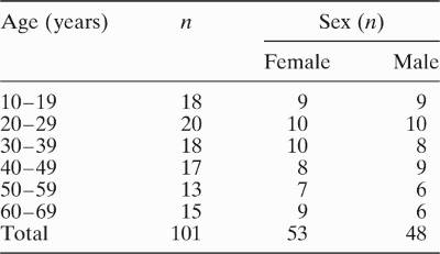
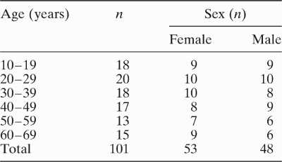
Our study group comprised 53 women and 48 men diagnosed within the audiology and speech pathology section of the department of otolaryngology, Hacettepe University. Subjects were aged 10–69 years, with a mean age of 36.37 ± 16.72 years, and were divided into six age ranges (10–19, 20–29, 30–39, 40–49, 50–59 and 60–69 years) (Table I). All had normal otoscopy findings. For the present study, social status was not taken into consideration. Subjects' age ranges reflected the fact that, within the study population, no subject older than 70 years with normal hearing could be found. According to the age groups, sex was not considered in the subject selection. Testing was performed bilaterally. A total of 202 ears was evaluated.
Subjects' age and sex
During the selection process, any individuals with the following were excluded: hearing impairment, otological disorder, metabolic disease, noise exposure and ototoxic medication. Included individuals were required to have: hearing thresholds better than 25 dB hearing level (HL) at octave intervals from 0.25 to 6.0 kHz; speech recognition scores better than 88 per cent; no cochlear sensitivity; middle-ear pressure between the limits of ±50 mm H2O; and good detectable transient evoked otoacoustic emissions in both ears.
All subjects underwent pure tone hearing threshold measurements between 125 and 6000 Hz, according to International Standards Organiation (ISO) 1964 standards. Air and bone conduction hearing thresholds, speech tests and masking level difference tests were conducted within soundproof rooms, using an AC-40 audiograph (Interacoustics, Assens, Denmark). Subjects' air conduction hearing thresholds were measured, using standard earphones (TDH-39, Interacoustics, Assens, Denmark) with MX41/AR covers (Interacoustics, Assens, Denmark) at 125–6000 Hz. Bone conduction hearing thresholds were measured using 60273 vibrators (Oticon, Smørum, Denmark) at 0.5–4 kHz. Audiometers were calibrated using 4152 artificial ears with a Brüel-Kjaer (Nærum, Denmark) sound level meter.
Subjects' speech reception thresholds were assessed using a three-syllable word list. Speech recognition was tested using a monosyllable, phonetically balanced word list developed in the Hacettepe University audiology unit. The uncomfortable loudness level was also determined.
For the impedancemetric tests, middle-ear pressure and acoustic reflex measurements were made, using Interacoustics AT-22, AZ-7 and AZ-3 impedancemeters and TDH-39 earphones.
Masking level difference test
A pure tone signal (S) of 500 Hz and narrowband noise (N) of 500 Hz was transmitted to both ears at SoNo and SπNo states.Reference Mueller, Bright and Katz10, Reference Bellis11 While the noise level was fixed at 50 dB HL (which is the efficient masking level for both conditions), the threshold was determined first for the SoNo state, then for the SπNo state at 1 dB levels.Reference Mueller, Bright and Katz10 The masking level difference was obtained by subtracting the antiphasic state threshold (SπNo) from the homophasic state threshold (SoNo) (SπNo = Sπ; antiphasic signal, No; homophasic noise = antiphasic state and SoNo = So; homophasic signal, No; homophasic noise = homophasic state).
Speech recognition in noise test
The speech recognition in noise test was applied with a signal-to-noise ratio of +10 dB.Reference Baran, Musiek, Musiek and Rintelmann9, Reference Beattie, Barr and Roup12 Phonetically balanced word lists were transmitted at 40 dB sensation level (SL) and white noise at 30 dB SL to subjects' ipsilateral test ear, at the same time. The word lists were presented to subjects via an adapted CD player (Sony, New York, United States). Four different word lists comprising 25 syllables were presented. The results of the last word list were included in the statistical analysis, in order to reduce the effect of learning.
Transient evoked otoacoustic emissions
The initial part of the experimental session included an audiological evaluation to determine candidacy. Transient evoked otoacoustic emission measurement was conducted with a transient evoked (TE) full menu. The eliciting stimuli were conventional, nonlinear clicks of 80-microsecond duration, delivered at a peak sound pressure level of 83 ± 3 dB.Reference de Ceulaer, Yperman, Daemers, van Driessche, Somers, Offeciers and Govaerts13, Reference Parthasarathy14 The signal-to-noise ratio in at least three of the five highest frequency bands (i.e. 1, 2, 3, 4 and 5 kHz) exceeded 3 dB.Reference Hall and Hall3, Reference Hood, Berlin, Hurley, Cecola and Bell15
Probe fit for each subject was assessed by closely examining the stimulus spectrum in the ear canal and then adjusting the probe to produce minimal ringing and a flat frequency response. The response was produced using the standard nonlinear differential averaging technique in order to minimise stimulus and other artefacts. An average of two transient evoked otoacoustic emission waveforms, composed of 260 accepted click trains, was automatically cross-correlated and used to determine the reproducibility of the measured transient evoked otoacoustic emissions; V5, 6Y software (Otodynamics, Herts, United Kingdom) was used. The transient evoked otoacoustic emissions measurement was included for further analysis if both the whole wave reproducibility and the stimulation stability was 70 per cent or higher.Reference Hall and Hall3, Reference Parthasarathy14, Reference Hood, Berlin, Hurley, Cecola and Bell15
Contralateral suppression of transient evoked otoacoustic emissions
The transient evoked otoacoustic emissions recording was first conducted without noise stimulation from the opposite direction. It was then repeated by applying 40 dB SL contralateral speech noise.Reference de Ceulaer, Yperman, Daemers, van Driessche, Somers, Offeciers and Govaerts13 The intensity of the contralateral stimulation was less than that presented to the ear in which otoacoustic emissions were being recorded, and less than that needed to create an acoustic stapedial reflex.Reference Hall and Hall3 The reported suppression values were derived by subtracting the emission levels with contralateral speech noise from the emissions levels without contralateral speech noise, at the five highest frequency bands (1, 2, 3, 4 and 5 kHz).Reference de Ceulaer, Yperman, Daemers, van Driessche, Somers, Offeciers and Govaerts13–Reference Hood, Berlin, Hurley, Cecola and Bell15
An ILO88DP OAE V5,6y DP Echoport (Otodynamics, Herts, United Kingdom) and an adult transient evoked otoacoustic emissions probe were used. The probe was calibrated with a 1 cm3 acoustic calibration cavity. Contralateral speech noise was supplied by an Interacoustics AC-5 clinical audiometer, using an Ear Tone 3A insert earphone (Interacoustics, Assens, Denmark).Reference de Ceulaer, Yperman, Daemers, van Driessche, Somers, Offeciers and Govaerts13
All the findings were evaluated statistically using the Statistical Package for the Social Sciences version 11.0 for Windows software (SPSS, Chicago, Illinois, USA). The relationship between the results for the contralateral supression of transient evoked otoacoustic emissions tests, the masking level difference tests and the speech recognition in noise tests was then determined. Kruskall–Wallis variance analysis was used for statistical evaluation of hearing thresholds, transient evoked otoacoustic emission amplitudes and contralateral transient evoked otoacoustic emission supression values. One way analysis of variance was used to statistically evaluate masking level difference and speech recognition in noise test results. The paired t-test was used to compare the right and left ear results for the speech recognition in noise test. Spearman's rank correlation was used to analyse inter-variable relations.
All subjects gave informed consent for the study. This study was approved by the ethical committee of the Hacettepe University Institute of Health Sciences.
Results
Subjects' hearing thresholds were evaluated by age. Subjects' hearing thresholds decreased with age, within normal limits (p < 0.001). All subjects had lower hearing thresholds at 6000 Hz than at other frequencies (p < 0.05).
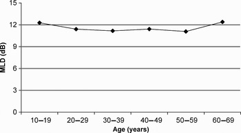
A statistically significant increase was seen in the average masking level difference test results of subjects in the 10–19 and 60–69 year age ranges, compared with other age ranges (p < 0.05) (Figure 1). Masking level difference averages generally decreases with age in the literature, but this decrease was not seen in the 60–69 year age range in our study. Masking level difference averages were found to be high in the 60–69 year age range as in the 10–19 year age range.
Subjects' masking level difference (MLD) averages, by age.

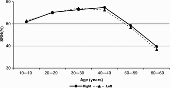
We found a statistically significant difference in speech recognition in noise scores at different ages (Figure 2). These scores decreased with the age, and score averages in the 60–69 year age range were lower than those in other age ranges (p < 0.01). There was no statistically significant difference between left ear and right ear speech recognition in noise scores (p < 0.05).
Subjects' speech recognition in noise (SRN) test scores, by age.

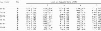
Transient evoked otoacoustic emission amplitudes reduced with age (Table II). These amplitudes were significantly lower in the 50–59 and 60–69 year age ranges, compared with other age ranges (p < 0.05). Transient evoked otoacoustic emission amplitudes in the 4 and 5 kHz frequency bands were significantly lower than those in the 1, 2 and 3 kHz frequency bands (p < 0.001).
Average TEOAE by test frequency and subject age

TEOAE = transient evoked otoacoustic emissions; SD = standard deviation; R = right; L = left
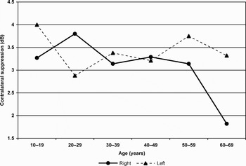
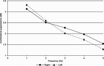
We evaluated contralateral transient evoked otoacoustic emission suppression with respect to age and test frequency band (Figures 3 and 4). The degree of supression was found to decline with age; however, this decline was not statistically significant (p > 0.05). The degree of supression was greater at 1 kHz than at other frequency bands, but, again, this was not statistically significant (p > 0.05). No statistically significant difference was obtained for contralateral transient evoked otoacoustic emission suppression values between the right and left ears (p > 0.05).
Subjects' contralateral transient evoked otoacoustic emission suppression values, by age.

Subjects' contralateral transient evoked otoacoustic emission suppression values, by test band frequency.

When the results for contralateral transient evoked otoacoustic emission suppression and masking level difference were compared, we observed a negative correlation, such that the degree of supression decreased as the masking level difference values increased. This association was found to be statistically significant at 1 kHz (for the right ear, p < 0.01; for the left ear, p < 0.05).
When the results for contralateral transient evoked otoacoustic emission supression and speech recognition in noise testing were compared, we observed a positive correlation, such that the speech recognition in noise scores decreased as the suppression amounts decreased. This association was statistically significant at 1 kHz for the right and left ears (p < 0.01) and at 2 kHz for the left ear (p < 0.01).
When the results for masking level difference and speech recognition in noise scores were compared, we observed a negative correlation, such that the masking level difference decreased as the speech recognition in noise score increased. This association was found to be statistically significant (for the right ear, p < 0.05; for the left ear, p < 0.01).
Discussion
In daily life, one must continuously detect sound signals against background noise. Using both ears facilitates this function. For all the subjects in our study, the masking level difference values obtained were between 10 and 15 dB. These masking level difference values indicate normal hearing and are comparable with other published findings.Reference Jiang, McAlpine and Palmer6, Reference Schoeny, Talbott and Katz16
From the literature, we expected that masking level difference averages would decrease with age.Reference Pichora-Fuller and Schneider17 We did find such a decrease; however, we also found that the masking level difference averages were high in the 60-69 year age range. Therefore, there was no direct association between age and masking level difference averages. We cannot explain this increase in masking level difference averages in the 60–69 year age range.
The elderly experience more difficulty in understanding daily speech compared with the young, although they do not have considerable hearing loss. Such age-related speech comprehension difficulties are due to high level cognitive functions (such as language ability, memory and attention) and also to low level cognitive functions (such as perceptive and sensory functions). Studies show that an age-related decline in general performance, reduced memory capacity and high frequency hearing loss considerably affects speech recognition scores.Reference Barrenäs and Wikström18–Reference Salvi20
We observed that subjects' speech recognition in noise scores decreased with age. These scores dropped in the 60–69 year age range. This was considered to be very important, and it was concluded that ageing decreases the ability to recognise speech against background noise. Our findings are supported by those of von Wedel et al. Reference von Wedel, von Wedel and Streppel21 These authors inferred from their study that the ability to recognise speech against background noise stabilises at about 40 years of age, decreases gradually after 50 years and decreases significantly after 70 years. They theorised that this was possibly because of the reduced capacity for frequency, intensity and time resolution in the ageing hearing organ's peripheral and central pathways. In contrast, some published studies do not support our findings. In these studies, where hearing is normal, age does not affect speech recognition in noise scores.Reference Barrenäs and Wikström18, Reference Poulsen and Keidser22
Fışıloğlu and KatzReference Fışıloğlu and Katz23 studied 14 children with central auditory processing disorders, 14 children with normal hearing and 12 adults, and found mean speech recognition in noise scores of 41.1, 54 and 67.7 per cent, respectively, at 10 dB signal/noise (S/N). They explained that the SRN scores obtained from the children's group with central auditory processing disorder decreased significantly while compared with the 14 children and 12 adults have normal hearing.
In subjects in the 10–69 year age range, we found mean speech recognition in noise scores of 51.96 ± 13.97 per cent for the right ear and 51.60 ± 14.23 per cent for the left ear. One reason for low speech recognition in noise scores may be central auditory processing problems. We concluded that this situation should be investigated in detail.
We observed an age-related drop in both transient evoked otoacoustic emission amplitudes and hearing thresholds. The former may result from a decrease in auditory sensitivity, as stated by other authors.Reference Laukli, Molde, Solem and Stenklev24, Reference Stenklev and Laukli25
We found that the degree of contralateral transient evoked otoacoustic emission suppression decreased with age, but it was not statistically significant. Suppression of transient evoked otoacoustic emissions was found to be greater at 1 kHz than at other frequency bands, but this difference was not statistically significant.
Kim et al. Reference Kim, Frisina and Frisina26 found that contralateral distortion product otoacoustic emission (DPOAE) suppression levels were much lower in the middle-aged and elderly than in younger subjects, and stated that this finding supported the presence of a functional disorder in the medial olivocochlear system. Others have also observed an age-related loss of medial olivocochlear suppressive function.Reference Castor, Veuillet, Morgon and Collet27, Reference Jacobson, Kim, Romney, Zhu and Frisina28
When we evaluated the relations of the tests to each other, independent of age, it was observed that, as masking level difference values dropped, speech recognition in noise scores and contralateral transient evoked otoacoustic emission suppression values increased. Masking level difference values and speech recognition in noise scores were inversely related; however, there is no physiological explanation for this correlation. As Musiek reported, Lionell Collet et al. compared masking level difference results with the contralateral supression effect (as the masking level difference involves the superior olivary complex anatomically), and concluded that there was an inverse relationship between them.Reference Musiek29 In this study, in which both measurement methods were compared, it was determined that the smaller contralateral supression effect was seen in the group whose masking level difference value was bigger.
In our study, a directly proportional relationship was found between speech recognition in noise scores and contralateral transient evoked otoacoustic emission suppression. It was observed that, as speech recognition in noise scores decreased, so too did the degree of contralateral suppression (statistically significant at 1 kHz). As a decrease in the amount of contralateral transient evoked otoacoustic emission suppression indicates a medial olivocochlear system disorder, such a drop is considered to be associated with a disorder of medial efferent system function, and thus of the central auditory system.Reference Veuillet, Khalfa and Collet5, Reference Hood30 Better central auditory function is usually associated with better speech recognition in noise scores and greater contralateral suppression of transient evoked otoacoustic emissions.
ParthasarathyReference Parthasarathy14 indicated that contralateral wide band noise can activate the medial olivocochlear system and consequently suppress transient evoked otoacoustic emission amplitudes, as a function of age. This author explained that recognizing speech in noise worsens with age as the functional degeneration of the efferent control of the outer hair cells with age.
In the evaluation of neural disorders, physiological measurements are more sensitive, more objective and less variable than behavioural tests such as pure tone thresholds and speech recognition. Recognising speech in noise is more sensitive for neural disorders; however, it may be affected by other factors, including age, language function and the state of the patient. In order to allow evaluation of the medial olivocochlear system, it is first necessary to have extant otoacoustic emissions, hearing sensitivity better than 30–35 dB HL and a normally functioning middle ear.Reference Hood30 With stimulation of the medial olivocochlear system, signal finding in noise improves, cochlear signal noise rate increases and the outer hair cells are protected against excessive acoustic stimulation.Reference Aran, Pajor, de Sauvage and Erre31–Reference Sahley, Nodar and Musiek34
In our study, we evaluated the effect of age on: speech recognition in noise; masking level differences; transient evoked otoacoustic emission amplitudes; and contralateral transient evoked otoacoustic emission suppression results. All test parameters showed decreased performance with age, except for the masking level difference test. In light of the relationships between these tests, the present study represents a step toward more detailed investigation of the relationships between medial olivocochlear system functioning and psychophysical performance in humans.
• Understanding the efferent system may aid patients with disorders of auditory processing of background noise and help develop new treatment approaches
• This may benefit: children experiencing auditory perception difficulties in noisy classrooms; children and adults with central auditory noise processing disorders, who have little or no peripheral hearing loss and high frequency hearing loss; and elderly people with presbycusis experiencing communication difficulties due to background noise
• Research on the efferent auditory system may help develop hearing aids to improve speech recognition in noise or may help to improve the cochlear implantation method