Introduction
Brain development is a complex process regulated by genes and sculpted by environmental experiences (Lenroot & Giedd, Reference Lenroot and Giedd2008). Although experiential influences affect brain structure and function throughout the lifespan, childhood experience is particularly crucial with early stress adversely affecting the nature and trajectory of normal brain development (Pechtel & Pizzagalli, Reference Pechtel and Pizzagalli2011).
Childhood maltreatment, which includes physical, sexual and emotional abuse and neglect, is common in the UK with paediatric prevalence rates of 7–10% (NSPCC, 2011). It has been associated with a host of adverse consequences, such as low IQ, abnormal error processing (Lim et al. Reference Lim, Hart, Mehta, Simmons, Mirza and Rubia2015), impaired attention, inhibition, emotion and reward processing (Pechtel & Pizzagalli, Reference Pechtel and Pizzagalli2011; Hart & Rubia, Reference Hart and Rubia2012; De Bellis & Zisk, Reference De Bellis and Zisk2014). Large-scale epidemiological studies found that childhood maltreatment is significantly associated with first onsets of various psychiatric disorders, such as depression and post-traumatic stress disorder (PTSD) (Green et al. Reference Green, McLaughlin and Berglund2010).
The psychopathological outcomes associated with childhood maltreatment may be mediated by the disruption of neural underpinnings (Bremner & Vermetten, Reference Bremner and Vermetten2001). Structural MRI studies show that, relative to non-maltreated controls, individuals exposed to childhood maltreatment have grey matter volume (GMV) abnormalities in several relatively late-developing brain regions, such as the orbitofrontal cortex (OFC) (Hanson et al. Reference Hanson, Chung, Avants, Shirtcliff, Gee, Davidson and Pollak2010; Thomaes et al. Reference Thomaes, Dorrepaal, Draijer, de Ruiter, van Balkom, Smit and Veltman2010; Edmiston et al. Reference Edmiston, Wang, Mazure, Guiney, Sinha, Mayes and Blumberg2011; De Brito et al. Reference De Brito, Viding, Sebastian, Kelly, Mechelli, Maris and McCrory2013; Hodel et al. Reference Hodel, Hunt, Cowell, Van Den Heuvel, Gunnar and Thomas2015), insula (Edmiston et al. Reference Edmiston, Wang, Mazure, Guiney, Sinha, Mayes and Blumberg2011; Dannlowski et al. Reference Dannlowski, Stuhrmann, Beutelmann, Zwanzger, Lenzen, Grotegerd, Domschke, Hohoff, Ohrmann, Bauer, Lindner, Postert, Konrad, Arolt, Heindel, Suslow and Kugel2012; Lim et al. Reference Lim, Radua and Rubia2014), temporal lobes (Bremner et al. Reference Bremner, Randall, Vermetten, Staib, Bronen, Mazure, Capelli, McCarthy, Innis and Charney1997; De Bellis et al. Reference De Bellis, Keshavan, Frustaci, Shifflett, Iyengar, Beers and Hall2002; Tomoda et al. Reference Tomoda, Sheu, Rabi, Suzuki, Navalta, Polcari and Teicher2011) and cerebellum (De Bellis & Kuchibhatla, Reference De Bellis and Kuchibhatla2006; Bauer et al. Reference Bauer, Hanson, Pierson, Davidson and Pollak2009; Walsh et al. Reference Walsh, Dalgleish, Lombardo, Dunn, Van Harmelen, Ban and Goodyer2014). Volumetric abnormalities in subcortical regions such as the hippocampus and amygdala have been mainly observed in adults but not children/adolescents exposed to childhood maltreatment (Woon & Hedges, Reference Woon and Hedges2008). Recent studies also reported reduced visual cortex GMV in childhood maltreatment (Tomoda et al. Reference Tomoda, Navalta, Polcari, Sadato and Teicher2009; Reference Tomoda, Polcari, Anderson and Teicher2012; Edmiston et al. Reference Edmiston, Wang, Mazure, Guiney, Sinha, Mayes and Blumberg2011). Several reviews of childhood maltreatment have consistently reported structural deficits in several stress-susceptible brain regions including the OFC, limbic, insula and cerebellar regions (McCrory et al. Reference McCrory, De Brito and Viding2011a ; Hart & Rubia, Reference Hart and Rubia2012; Lim et al. Reference Lim, Radua and Rubia2014; Nemeroff, Reference Nemeroff2016; Teicher et al. Reference Teicher, Samson, Anderson and Ohashi2016), with the late-developing OFC and cerebellum being particularly vulnerable to the effects of early stress (Hanson et al. Reference Hanson, Chung, Avants, Shirtcliff, Gee, Davidson and Pollak2010; Pechtel & Pizzagalli, Reference Pechtel and Pizzagalli2011), and the insula is known to be involved in regulating the glucocorticoid effect (Fornari et al. Reference Fornari, Wichmann, Atucha, Desprez, Eggens-Meijer and Roozendaal2012). Our meta-analysis also showed that childhood maltreatment is associated with GMV reduction in OFC–limbic–temporal regions and inferior frontal cortices that mediate top–down affect and cognitive control, respectively; and with GMV reduction in pre-/postcentral gyri that mediate sensory functions (Lim et al. Reference Lim, Radua and Rubia2014).
Cortical volume (CV) is determined by two separable cortical indices, cortical thickness (CT) and surface area (SA), which are genetically (Panizzon et al. Reference Panizzon, Fennema-Notestine, Eyler, Jernigan, Prom-Wormley, Neale, Jacobson, Lyons, Grant, Franz, Xian, Tsuang, Fischl, Seidman, Dale and Kremen2009) and phenotypically (Winkler et al. Reference Winkler, Kochunov, Blangero, Almasy, Zilles, Fox, Duggirala and Glahn2010) independent with differing developmental trajectories (Panizzon et al. Reference Panizzon, Fennema-Notestine, Eyler, Jernigan, Prom-Wormley, Neale, Jacobson, Lyons, Grant, Franz, Xian, Tsuang, Fischl, Seidman, Dale and Kremen2009). Studies examining CT, SA and CV may be more sensitive to individual differences than considering volume alone (Hutton et al. Reference Hutton, Draganski, Ashburner and Weiskopf2009). However, as most of the earlier structural studies on childhood maltreatment examined abuse-related volumetric abnormalities, examining group differences in volume in the current study thus allows for comparison with the existing literature. Volume measurements are also useful for subcortical structures where CT/SA measurements are unavailable. Therefore, it is important to explore these brain measures to better understand the structural correlates of childhood maltreatment.
To date, few studies on childhood maltreatment examined whole-brain differences in CV, CT and SA within the same sample. Compared with healthy controls, maltreated young people had significantly reduced CT in right OFC (Kelly et al. Reference Kelly, Viding, Wallace, Schaer, De Brito, Robustelli and McCrory2013; Reference Kelly, Viding, Puetz, Palmer, Samuel and McCrory2016; Gold et al. Reference Gold, Sheridan, Peverill, Busso, Lambert, Alves, Pine and McLaughlin2016), and reduced SA in left middle temporal and lingual regions (Kelly et al. Reference Kelly, Viding, Wallace, Schaer, De Brito, Robustelli and McCrory2013). Children who experienced psychosocial deprivation exhibited widespread CT reductions in lateral OFC, precuneus, insula, parietal and lingual gyri, which were furthermore associated with inattention and impulsivity (McLaughlin et al. Reference McLaughlin, Sheridan, Winter, Fox, Zeanah and Nelson2014). In adults, childhood sexual abuse was associated with reduced CT in left somatosensory cortex, while emotional abuse was associated with reduced CT in bilateral precuneus and left somatosensory cortex (Heim et al. Reference Heim, Mayberg, Mletzko, Nemeroff and Pruessner2013). Individuals exposed to domestic violence during childhood had reduced CT in bilateral lingual (Tomoda et al. Reference Tomoda, Polcari, Anderson and Teicher2012).
Given that childhood maltreatment is associated with the development of psychiatric complications (Sugaya et al. Reference Sugaya, Hasin, Olfson, Lin, Grant and Blanco2012; Herrenkohl et al. Reference Herrenkohl, Hong, Klika, Herrenkohl and Russo2013; MacMillan et al. Reference MacMillan, Tanaka, Duku, Vaillancourt and Boyle2013), it is crucial to control for these in order to disentangle the effects of maltreatment from the psychiatric comorbidities (McCrory et al. Reference McCrory, De Brito and Viding2011a ; Hart & Rubia, Reference Hart and Rubia2012; Lim et al. Reference Lim, Radua and Rubia2014). Only two prior structural studies in childhood maltreatment controlled for psychiatric disorders. However, they examined CV alone in specific disorders, such as psychosis (Sheffield et al. Reference Sheffield, Williams, Woodward and Heckers2013) and depression (Chaney et al. Reference Chaney, Carballedo, Amico, Fagan, Skokauskas, Meaney and Frodl2014), which limits the generalizability of their findings to other psychiatric comorbidities. The majority of patients in the two studies were also on psychotropic medications (e.g. chlorpromazine, SSRIs), which are known to affect brain structure and function (Murphy, Reference Murphy2010).
Therefore, the aim of this study was to control for the limitations of earlier studies by conducting both region-of-interest (ROI) and whole-brain structural (CV, CT, SA) analyses in medication-naïve, drug-free youth exposed to documented childhood physical abuse and in healthy controls. To assess the specificity of the association with childhood abuse, we included a third group of psychiatric controls that was matched with the abuse group on psychiatric comorbidities. Sexual abuse was excluded because it has different effects on brain structure (Heim et al. Reference Heim, Mayberg, Mletzko, Nemeroff and Pruessner2013) and different behavioural and psychiatric consequences (Teicher et al. Reference Teicher, Ito, Glod, Andersen, Dumont and Ackerman1997; Weierich & Nock, Reference Weierich and Nock2008; Lopez-Castroman et al. Reference Lopez-Castroman, Melhem, Birmaher, Greenhill, Kolko, Stanley, Zelazny, Brodsky, Garcia-Nieto, Burke, Mann, Brent and Oquendo2013; Lewis et al. Reference Lewis, McElroy, Harlaar and Runyan2016). For instance, both childhood physical abuse and neglect, but not sexual abuse, were associated with alterations in regional corpus callosum size (Teicher et al. Reference Teicher, Ito, Glod, Andersen, Dumont and Ackerman1997) and with GMV reduction in a distributed corticostriatal–limbic system (Edmiston et al. Reference Edmiston, Wang, Mazure, Guiney, Sinha, Mayes and Blumberg2011). Furthermore, childhood sexual abuse is associated with experiences unique to sexual victimization relative to other abuse experiences; for example, traumatic sexualization, stigmatization, attributions of responsibility as well as feelings of guilt and shame may impact sexual abuse victims differently than victims of other abuse experiences (Finkelhor & Browne, Reference Finkelhor and Browne1985; Feiring et al. Reference Feiring, Taska and Lewis1996). For these reasons, and in order to obtain a more homogenous group, we only included youth exposed to childhood physical abuse. Nevertheless, it is unrealistic to separate physical abuse from typically co-occurring emotional abuse and neglect (Claussen & Crittenden, Reference Claussen and Crittenden1991; Edwards et al. Reference Edwards, Holden, Felitti and Anda2003) since psychological maltreatment would be present in almost all cases of physical maltreatment (Claussen & Crittenden, Reference Claussen and Crittenden1991).
Since childhood maltreatment is consistently associated with structural deficits in several stress-susceptible brain regions including the OFC, limbic, insula and cerebellar regions (McCrory et al. Reference McCrory, De Brito and Viding2011a ; Hart & Rubia, Reference Hart and Rubia2012; Lim et al. Reference Lim, Radua and Rubia2014; Nemeroff, Reference Nemeroff2016; Teicher et al. Reference Teicher, Samson, Anderson and Ohashi2016), we hypothesized that the abuse group, relative to both healthy and psychiatric controls, would have structural abnormalities particularly in the OFC, insula and cerebellum. We also investigated abnormalities outside our priori defined ROIs with a whole-brain analysis.
Methods and materials
Participants
Seventy (23 abuse, 20 psychiatric controls, 27 healthy controls) right-handed, medication-naïve, drug-free and age- and gender-matched youth (aged 13–20) were assessed by a child psychiatrist (KM) using the Development and Well-Being Assessment (DAWBA) (Goodman et al. Reference Goodman, Ford, Richards, Gatward and Meltzer2000), which was designed to generate International Classification of Diseases, 10th Edition (ICD-10) and Diagnostic and Statistical Manual of Mental Disorders, Fourth Edition (DSM-IV) psychiatric diagnoses. The Strengths and Difficulties Questionnaires (SDQ) (Goodman, Reference Goodman1997) and Beck's Depression Inventory (BDI) (Beck et al. Reference Beck, Steer and Carbin1988) were also used to provide symptom scores on psychopathology. IQ was assessed using the Wechsler Abbreviated Scale of Intelligence (WASI) (Wechsler, Reference Wechsler1999). The Childhood Trauma Questionnaire (CTQ) (Bernstein & Fink, Reference Bernstein and Fink1998) was used to measure the severity of childhood physical, sexual and emotional abuse, and physical and emotional neglect. Socioeconomic status (SES) was measured by two non-sensitive items (on housing tenure and room occupancy) from the Family Affluence Scale (FAS) (Currie et al. Reference Currie, Elton, Todd and Platt1997).
Exclusion criteria for all participants were childhood sexual abuse, drug abuse, learning disability, neurological abnormalities, epilepsy, IQ < 70 and MRI contraindications. Urine screening for recent drug use was conducted with 10-panel urine drug test integrated cups (T-Cup; http://www.testfield.co.uk). All participants or their guardians, if they were under the age of 18, provided written informed consent to participate in the study. The study was approved by the National Research Ethics Service Committee London – Fulham.
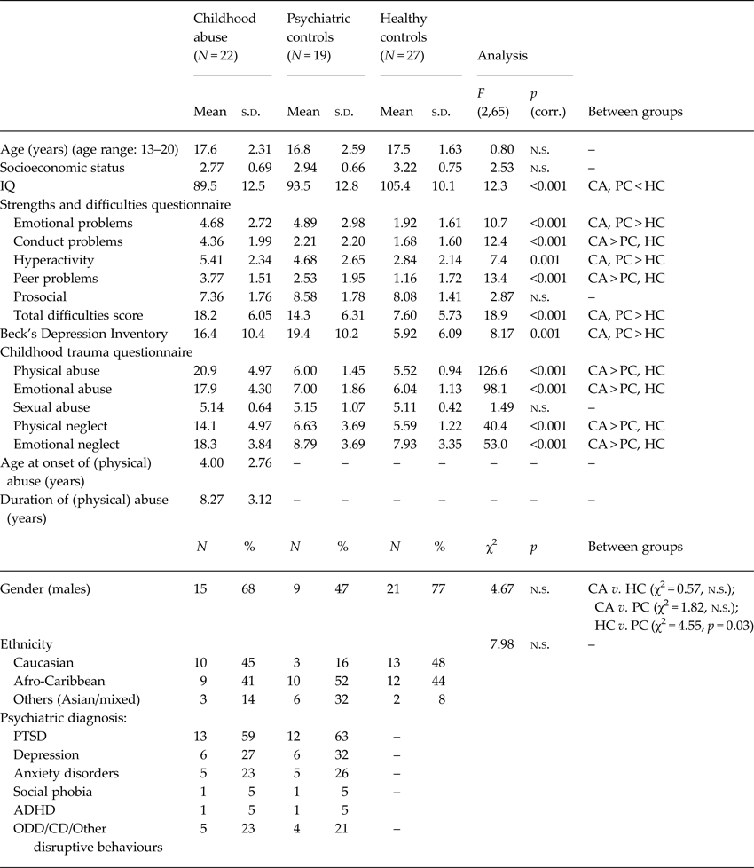
The 23 youth who experienced physical abuse before the age of 12 were first recruited through social services and psychiatric clinics. They or their guardians were first asked to provide signed permission to contact their social services for written confirmation of official records of physical abuse. The Childhood Experience of Care and Abuse (CECA) interview (Bifulco et al. Reference Bifulco, Brown and Harris1994) was used to corroborate the CTQ and provide information on the age of onset and duration of abuse. The participants scored ⩾ 13 (i.e. the cut-off for severe/extreme physical abuse) (Bernstein & Fink, Reference Bernstein and Fink1998) on the CTQ physical abuse subscale and information from the CECA interview and the CTQ were consistent with the official records. The common psychiatric comorbidities included PTSD, depression, anxiety and conduct disorder (Table 1). One participant was excluded due to MRI motion artefacts, leaving a final sample of 22 participants.
Demographic characteristics of 22 young people exposed to childhood abuse, 19 psychiatric controls and 27 healthy controls

CA, childhood abuse group; PC, psychiatric controls; HC, healthy controls; corr, Bonferroni corrected; ADHD, attention deficit hyperactivity disorder; PTSD, post-traumatic stress disorder; ODD, oppositional defiant disorder; CD, conduct disorder; n.s., non-significant.
The 20 psychiatric patients that were matched with the abuse group on psychiatric comorbidities but with no history of childhood maltreatment (scoring below the cut-offs for the respective CTQ subscales) (Bernstein & Fink, Reference Bernstein and Fink1998) were recruited through psychiatric clinics and social services (Table 1). PTSD patients experienced non-abuse-related trauma (e.g. witnessed a murder, experienced a car accident or experienced the death of a loved one). One participant was excluded due to motion artefacts, leaving a final sample of 19 patients.
The 27 healthy controls with no history of psychiatric illness and childhood maltreatment (scoring below the same cut-offs as above) were recruited through advertisements in the same geographic areas of South London to ensure similar SES (Table 1).
MRI acquisition and analysis
The MRI acquisition procedures are described in the online Supplementary Materials.
Image preprocessing and analyses were carried out using FreeSurfer version 5.3.0 (http://surfer.nmr.mgh.harvest.edu). After preprocessing (online supplementary materials), whole-brain between-group differences in CV, CT and SA were investigated within the QDEC surface-based group analysis. For each hemisphere, the General Linear Model was computed vertex-by-vertex for analysis of each cortical morphometric measure (CV, CT and SA), with group as a between-subjects factor and IQ, age and a total brain measure (total brain volume for CV, mean CT for CT and total SA for SA) included as covariates. Although there were no significant group differences in age, it was included given the relatively wide age range of the current sample. Cortical maps were smoothed with a full width at half maximum Gaussian kernel of 10 mm. Between-group differences were corrected for multiple comparisons with a Monte Carlo z-field simulation at p < 0.05 (two-tailed).
For group differences in the hypothesized ROIs (i.e. OFC, insula, cerebellum), analysis of variance with group (abuse v. healthy controls; abuse v. psychiatric controls) as a between-subject factor and covariates outlined above were used on the cortical measures of these regions generated during the automated segmentation and parcellation process (Fischl et al. Reference Fischl, van der Kouwe, Destrieux, Halgren, Segonne, Salat, Busa, Seidman, Goldstein, Kennedy, Caviness, Makris, Rosen and Dale2004). Given that a limited number of studies have aimed at specifying surface-based brain indices in relation to abuse exposure (Kelly et al. Reference Kelly, Viding, Puetz, Palmer, Samuel and McCrory2016), the stringent Bonferroni multiple comparisons correction was not applied in this analysis to limit potential type II errors.
Tests for normality were conducted in SPSS using the Kolmogorov–Smirnov and Shapiro–Wilk tests. None of the volume measurement distributions deviated significantly from normality.
Finally, we also conducted three preliminary analyses. First, we explored if gender influenced the impact of maltreatment on brain measures at the whole-brain level in QDEC with age, IQ and a total brain measure included as covariates. Second, the significant clusters were extracted for exploratory Pearson correlational analysis with the clinical measures within each group and with the abuse measures within the abuse group only. Lastly, we explored if the groups differed on hippocampus volume (online Supplementary Materials).
Results
Subject characteristics
The groups did not differ significantly on age, gender, ethnicity nor SES (p > 0.05), but differed on IQ (p < 0.001), which was expected as this is typical for the population (Mills et al. Reference Mills, Alati, O'Callaghan, Najman, Williams, Bor and Strathearn2011; Young & Widom, Reference Young and Widom2014; Geoffroy et al. Reference Geoffroy, Pinto Pereira, Li and Power2016) (Table 1). Although we selected participants with severe childhood physical abuse, they also experienced severe emotional abuse and neglect (Table 1), which typically co-occur with physical abuse; hence, we consider this group representative of the childhood abuse population (Edwards et al. Reference Edwards, Holden, Felitti and Anda2003; Trickett et al. Reference Trickett, Kim and Prindle2011).
ROI analysis
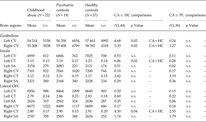
Relative to healthy controls only, the abuse group showed significantly reduced CV in left [F(1,44) = 4.68, p = 0.03] and right [F(1,44) = 5.33, p = 0.02] cerebellum, and reduced CT in left insula [F(1,44) = 6.06, p = 0.02] and right lateral OFC [F(1,44) = 4.30, p = 0.04]. The abuse and psychiatric groups did not differ significantly (Table 2). There were no significant group differences on hippocampus volume (online Supplementary Materials).
Group differences in the cortical measures of the regions of interest among 22 young people exposed to childhood abuse, 19 psychiatric controls and 27 healthy controls

CA, childhood abuse group; HC, healthy controls; PC, psychiatric controls; OFC, orbitofrontal cortex; CV, cortical volume (mm3); CT, cortical thickness (mm); SA, surface area (mm2); n.s., non-significant; s.d., standard deviation.
Whole-brain analysis
Cortical volume
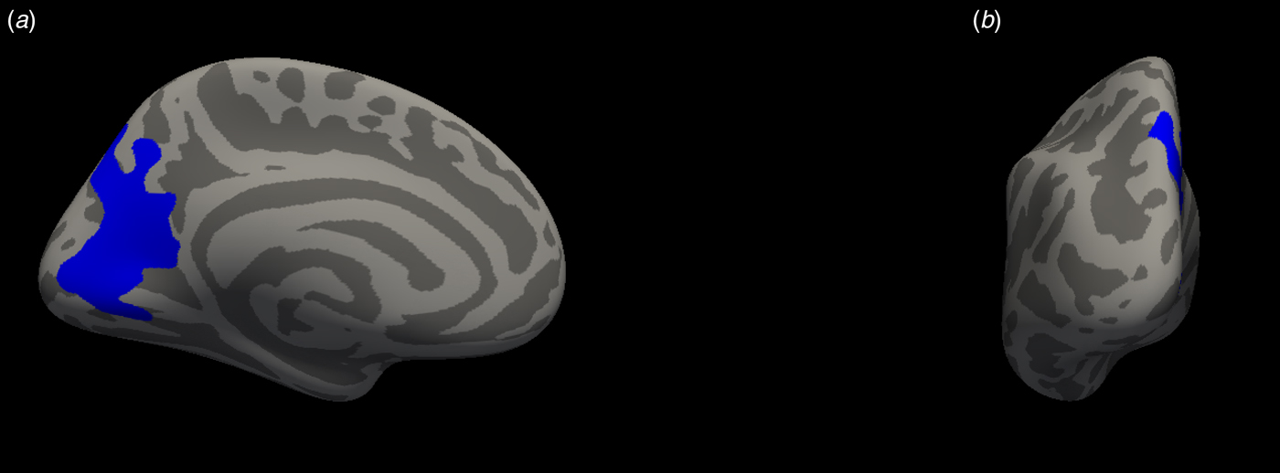
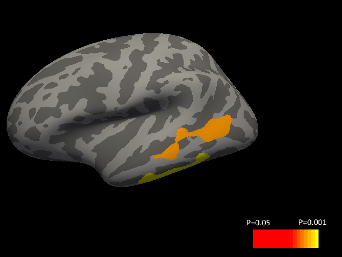
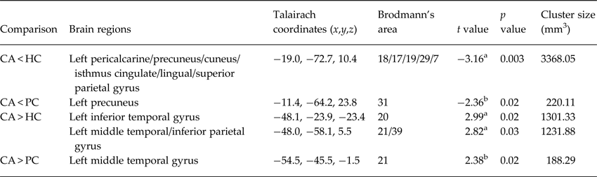
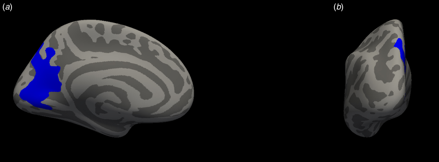
Compared with healthy controls, the abuse group had significantly reduced CV in a left-hemispheric posterior cluster comprising lingual, pericalcarine, precuneus, cuneus, isthmus cingulate and superior parietal gyri (Table 3, Fig. 1; cluster-corrected p < 0.05). They had larger CV in two left-hemispheric clusters: inferior temporal gyrus, along with middle temporal and inferior parietal gyri (Table 3, Fig. 2; cluster-corrected p < 0.05). Two of these regional differences, reduced CV in left precuneus (t = −2.36, p < 0.05) and larger CV in left middle temporal gyrus (t = 2.38, p < 0.05), were also significant relative to psychiatric controls at an uncorrected level; suggesting that the CV abnormalities in these two regions could be abuse-specific. Healthy and psychiatric controls did not differ significantly from each other. There was no significant maltreatment by gender interaction.
Significant cortical volume cluster projected onto the inflated surface of the left hemisphere in (a) medial and (b) tilted anterior views. The significant cluster shows reduced cortical volume in the childhood abuse group compared with healthy controls, and survived cluster correction for multiple comparisons using Monte Carlo simulation, p < 0.05.
Significant cortical volume clusters projected onto the inflated surface of the left hemisphere in lateral view. The significant clusters show increased cortical volume in the childhood abuse group compared with healthy controls, and survived cluster correction for multiple comparisons using Monte Carlo simulation, p < 0.05.

Regions with significant group differences in cortical volume among 22 young people exposed to childhood abuse, 19 psychiatric controls and 27 healthy controls

CA, childhood abuse group; HC, healthy controls; PC, psychiatric controls.
a The t value at which the test statistic is significant at p < 0.05, corrected for multiple comparisons with a Monte Carlo z-field simulation.
b The t value at which the test statistic is significant at p < 0.05, uncorrected for multiple comparisons.
Cortical thickness
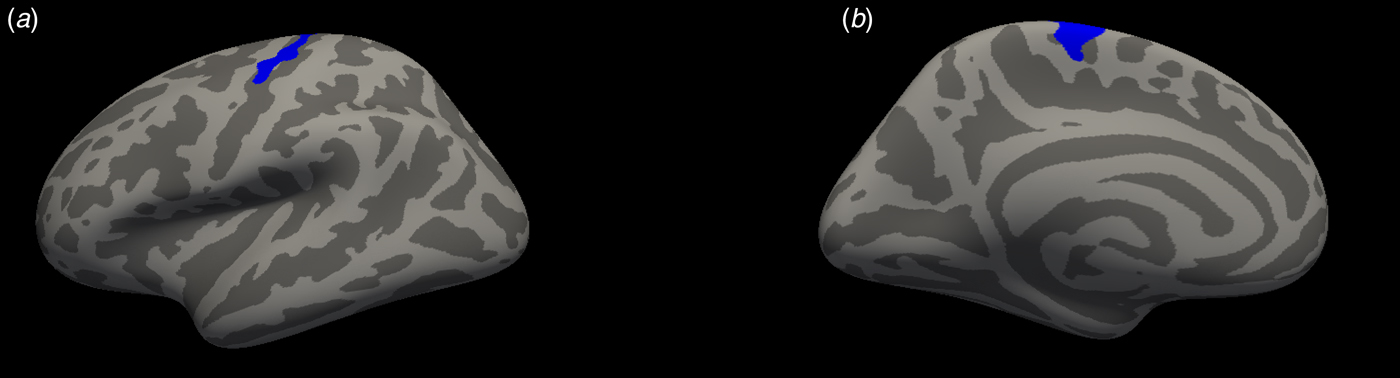
The abuse group had significantly reduced CT in left precentral, postcentral and paracentral gyri (Table 4, Fig. 3; cluster-corrected p < 0.05) relative to healthy controls, and significantly reduced left precentral CT (t = −2.18, p < 0.05) relative to psychiatric controls at an uncorrected level, suggesting that the precentral deficit could be abuse-specific. Healthy and psychiatric controls did not differ significantly from each other. There was no significant maltreatment by gender interaction.
Significant cortical thickness cluster projected onto the inflated surface of the left hemisphere in (a) lateral and (b) medial views. The significant cluster shows reduced cortical thickness in the childhood abuse group compared with healthy controls, and survived cluster correction for multiple comparisons using Monte Carlo simulation, p < 0.05.

Regions with significant group differences in cortical thickness among 22 young people exposed to childhood abuse, 19 psychiatric controls and 27 healthy controls

CA, childhood abuse group; HC, healthy controls; PC, psychiatric controls.
a The t value at which the test statistic is significant at p < 0.05, corrected for multiple comparisons with a Monte Carlo z-field simulation.
b The t value at which the test statistic is significant at p < 0.05, uncorrected for multiple comparisons.
Surface area
There were no significant group differences or maltreatment by gender interaction in SA.
Correlational analysis
The significant clusters were correlated with the SDQ and abuse measures within each group, controlling for IQ and age. Lower CV in the lingual–pericalcarine–precuneus cluster was significantly associated with higher CTQ physical abuse (r = −0.45, p < 0.05) and total score (r = −0.46, p < 0.05) in the abuse group, and with higher SDQ total score (r = −0.49, p < 0.05) and peer problems (r = −0.56, p < 0.05) in the healthy controls. Reduced CT in the pre-/postcentral cluster was also significantly associated with higher CTQ total score (r = −0.46, p < 0.05) in the abuse group.
Discussion
To our knowledge, this is the first structural study on childhood abuse that examined group differences in CV, CT and SA within the same sample in a group of medication-naïve, drug-free youth that also controlled for psychiatric comorbidities by inclusion of a psychiatric control group. Both are crucial to elucidate the effects of abuse independent from effects associated with psychiatric comorbid conditions or medication and drug abuse (McCrory et al. Reference McCrory, De Brito and Viding2011a ; Hart & Rubia, Reference Hart and Rubia2012; Lim et al. Reference Lim, Radua and Rubia2014).
For the ROI, the abuse group had significantly reduced CV in bilateral cerebellum and reduced CT in left insula and right lateral OFC, compared with healthy controls only. At the whole-brain level, relative to healthy controls, the abuse group showed significantly reduced CV in a cluster comprising left lingual, pericalcarine, precuneus and superior parietal regions, along with reduced CT in left pre-/postcentral and paracentral regions, which were furthermore significantly associated with greater abuse severity. Lower lingual–pericalcarine–precuneus CV was associated with greater SDQ total score and peer problems in the healthy controls, thereby suggesting possibly detrimental effects particularly in terms of peer problems, at least in the general healthy population. The abuse group also had increased CV in left inferior and middle temporal regions compared with healthy controls. Abnormalities in the precuneus, middle temporal and precentral regions were abuse-specific relative to psychiatric controls, albeit at a more lenient level.
The OFC receives strong inputs from the limbic system and is involved in emotion regulation, social behaviour and reward-related decision making (Rempel-Clower, Reference Rempel-Clower2007). It also receives inputs from the visual and somatosensory regions, and the lateral OFC is activated when viewing aversive pictures (Nitschke et al. Reference Nitschke, Sarinopoulos, Mackiewicz, Schaefer and Davidson2006) and experiencing unpleasant touch (Rolls et al. Reference Rolls, O'Doherty, Kringelbach, Francis, Bowtell and McGlone2003). The current finding of a thinner right lateral OFC is consistent with previous studies that found thinner right (lateral) OFC in children who experienced severe early-life deprivation and childhood abuse (Kelly et al. Reference Kelly, Viding, Wallace, Schaer, De Brito, Robustelli and McCrory2013; McLaughlin et al. Reference McLaughlin, Sheridan, Winter, Fox, Zeanah and Nelson2014; Gold et al. Reference Gold, Sheridan, Peverill, Busso, Lambert, Alves, Pine and McLaughlin2016), and extends findings of our meta-analysis (Lim et al. Reference Lim, Radua and Rubia2014) and other volumetric studies that found significantly reduced OFC CV in children (Hanson et al. Reference Hanson, Chung, Avants, Shirtcliff, Gee, Davidson and Pollak2010; Edmiston et al. Reference Edmiston, Wang, Mazure, Guiney, Sinha, Mayes and Blumberg2011; De Brito et al. Reference De Brito, Viding, Sebastian, Kelly, Mechelli, Maris and McCrory2013; Hodel et al. Reference Hodel, Hunt, Cowell, Van Den Heuvel, Gunnar and Thomas2015) and adults (Thomaes et al. Reference Thomaes, Dorrepaal, Draijer, de Ruiter, van Balkom, Smit and Veltman2010) exposed to childhood maltreatment.
The insula plays a key role in interoceptive awareness and emotion regulation (Goldin et al. Reference Goldin, McRae, Ramel and Gross2008) and together with the somatosensory, motor and prefrontal cortices, forms part of the neural circuitry of pain (Tracey & Mantyh, Reference Tracey and Mantyh2007). It is also part of the salience network that detects threat (Pichon et al. Reference Pichon, de Gelder and Grezes2012), where it integrates information about salience into perceptual decisions about pain (Wiech et al. Reference Wiech, Lin, Brodersen, Bingel, Ploner and Tracey2010). Previous structural studies have found thinner insula in children who experienced severe early-life derivation (McLaughlin et al. Reference McLaughlin, Sheridan, Winter, Fox, Zeanah and Nelson2014), as well as reduced insula CV in children (Edmiston et al. Reference Edmiston, Wang, Mazure, Guiney, Sinha, Mayes and Blumberg2011) and adults (Dannlowski et al. Reference Dannlowski, Stuhrmann, Beutelmann, Zwanzger, Lenzen, Grotegerd, Domschke, Hohoff, Ohrmann, Bauer, Lindner, Postert, Konrad, Arolt, Heindel, Suslow and Kugel2012) exposed to physical abuse and childhood maltreatment, respectively.
The cerebellum is vulnerable to the effects of early stress (Pechtel & Pizzagalli, Reference Pechtel and Pizzagalli2011). It plays a crucial role in emotion processing and fear conditioning via its connection with limbic structures and the hypothalamic–pituitary–adrenal axis (Schutter & van Honk, Reference Schutter and van Honk2005), and is a key region in many cognitive processes, particularly attention and timing functions (Stoodley & Schmahmann, Reference Stoodley and Schmahmann2009; Arnsten & Rubia, Reference Arnsten and Rubia2012). Cerebellar deficit has also been reported in previous studies of childhood abuse (De Bellis & Kuchibhatla, Reference De Bellis and Kuchibhatla2006; Bauer et al. Reference Bauer, Hanson, Pierson, Davidson and Pollak2009; Edmiston et al. Reference Edmiston, Wang, Mazure, Guiney, Sinha, Mayes and Blumberg2011), and may possibly underlie the affective and cognitive deficits in this population.
Childhood maltreatment has been associated with abnormal development of the sensory systems that relay adverse sensory experiences. For instance, studies reported reduced lingual CV in women who experienced childhood sexual and physical abuse (Tomoda et al. Reference Tomoda, Navalta, Polcari, Sadato and Teicher2009), reduced lingual CT in children who experienced psychosocial deprivation (McLaughlin et al. Reference McLaughlin, Sheridan, Winter, Fox, Zeanah and Nelson2014) and in young adults who witnessed domestic violence during childhood (Tomoda et al. Reference Tomoda, Polcari, Anderson and Teicher2012), as well as reduced lingual SA in maltreated children (Kelly et al. Reference Kelly, Viding, Wallace, Schaer, De Brito, Robustelli and McCrory2013). Women exposed to childhood sexual and emotional abuse also had reduced CT in left somatosensory cortex (Heim et al. Reference Heim, Mayberg, Mletzko, Nemeroff and Pruessner2013). Thus, the current findings of reduced left lingual CV and motor–somatosensory CT in the abuse group are consistent with these earlier studies and our meta-analysis finding of a smaller left motor–somatosensory CV in childhood maltreatment (Lim et al. Reference Lim, Radua and Rubia2014). Together, these findings support the suggestion that the sensory systems that process and interpret adverse sensory inputs may be altered by the abuse experience, reflecting an adaptive response of the developing brain to protect the child from highly hostile environmental conditions by gating sensory experiences and processing related to the abuse (Heim et al. Reference Heim, Mayberg, Mletzko, Nemeroff and Pruessner2013). Given that painful stimulation decreases blood flow in the somatosensory cortex (Tommerdahl et al. Reference Tommerdahl, Delemos, Vierck, Favorov and Whitsel1996), severe and painful punishments during the critical time of synapse formation and development in childhood may possibly reduce the number of synapses leading to a thinner somatosensory cortex. Moreover, the association between abuse and deficits in the sensory regions is further underpinned by the current findings of significant negative correlations between them.
The finding of a possibly abuse-specific reduced precuneus CV corroborates earlier findings of a negative association between precuneus CV and abuse severity (Dannlowski et al. Reference Dannlowski, Stuhrmann, Beutelmann, Zwanzger, Lenzen, Grotegerd, Domschke, Hohoff, Ohrmann, Bauer, Lindner, Postert, Konrad, Arolt, Heindel, Suslow and Kugel2012), as well as reduced CT (Heim et al. Reference Heim, Mayberg, Mletzko, Nemeroff and Pruessner2013; McLaughlin et al. Reference McLaughlin, Sheridan, Winter, Fox, Zeanah and Nelson2014) and network centrality of the precuneus (Teicher et al. Reference Teicher, Anderson, Ohashi and Polcari2014) in individuals exposed to childhood maltreatment. The precuneus plays a critical role in self-referential processing (Cavanna & Trimble, Reference Cavanna and Trimble2006). Childhood maltreatment has been associated with an increase in negative self-associations, which are postulated to further enhance negative bias when engaged in new situations, leading to the development and maintenance of affective disorders after exposure to childhood maltreatment (van Harmelen et al. Reference van Harmelen, de Jong, Glashouwer, Spinhoven, Penninx and Elzinga2010). Hence, the abuse-specific deficit in the precuneus may possibly be related to disturbances in self-referential processing in victims of childhood abuse, making them more vulnerable to the development and maintenance of psychopathology.
The possibly abuse-specific increased middle temporal CV is also novel. The middle temporal lobe is involved in moral function and intention attributions, and its dysfunction is often implicated in violent psychopathy (Sommer et al. Reference Sommer, Sodian, Döhnel, Schwerdtner, Meinhardt and Hajak2010; Fumagalli & Priori, Reference Fumagalli and Priori2012). Boys with callous-unemotional conduct problems had greater middle temporal CV than healthy controls (De Brito et al. Reference De Brito, Mechelli, Wilke, Laurens, Jones, Barker, Hodgins and Viding2009), while thicker middle temporal cortex correlated with higher concurrent psychopathic traits and psychopathic tendencies in adolescents (Yang et al. Reference Yang, Wang, Baker, Narr, Joshi, Hafzalla, Raine and Thompson2015). Thus, the abuse-specific increase in middle temporal CV may possibly serve as a biomarker for the development of psychopathic propensity in later life.
The OFC–limbic–cerebellar structural deficits may possibly underlie the neuropsychological deficits in emotion and reward processing (Pine et al. Reference Pine, Mogg, Bradley, Montgomery, Monk, McClure, Guyer, Ernst, Charney and Kaufman2005; Weller & Fisher, Reference Weller and Fisher2013) and attention (Pollak et al. Reference Pollak, Nelson, Schlaak, Roeber, Wewerka, Wiik, Frenn, Loman and Gunnar2010) observed in childhood maltreatment. This relationship is further supported by findings of fMRI studies of childhood maltreatment of abnormal OFC–limbic–cerebellar activation during emotion processing. For instance, increased activation in the insula (McCrory et al. Reference McCrory, De Brito, Sebastian, Mechelli, Bird, Kelly and Viding2011b ; Garrett et al. Reference Garrett, Carrion, Kletter, Karchemskiy, Weems and Reiss2012) and cerebellum (McCrory et al. Reference McCrory, De Brito, Kelly, Bird, Sebastian, Mechelli, Samuel and Viding2013) relative to controls to angry faces has been reported in maltreated children; together with lower OFC activation to angry faces in severely deprived children (Tottenham et al. Reference Tottenham, Hare, Millner, Gilhooly, Zevin and Casey2011) and healthy adults exposed to childhood physical abuse (Taylor et al. Reference Taylor, Eisenberger, Saxbe, Lehman and Lieberman2006), suggesting a deficit in their emotion-regulation abilities.
Finally, although there were no significant differences in the ROIs between the abuse group and psychiatric controls or between the psychiatric and healthy controls, the brain measurements of the psychiatric controls were in between those of the abuse group and healthy controls. This suggests that the abuse group, by nature of the abuse experience and the psychiatric comorbidities, was more adversely impaired than the psychiatric controls.
Among the strengths of this study are that all participants were medication-naïve and drug-free, and their abuse experience was carefully assessed and corroborated by social service records. We also included a psychiatric control group to determine the specificity of childhood abuse in our findings. The inclusion of a childhood abuse group without any psychiatric disorders would have provided a more robust means of determining abuse-specific abnormalities; however, such a ‘pure’ group would not be representative of the general childhood abuse populations, as large-scale epidemiological and longitudinal studies have consistently reported that childhood maltreatment is linked developmentally to psychiatric disorders (Sugaya et al. Reference Sugaya, Hasin, Olfson, Lin, Grant and Blanco2012; Herrenkohl et al. Reference Herrenkohl, Hong, Klika, Herrenkohl and Russo2013; MacMillan et al. Reference MacMillan, Tanaka, Duku, Vaillancourt and Boyle2013), and a meta-analysis further reported a causal relationship between non-sexual childhood maltreatment and a range of mental disorders (Norman et al. Reference Norman, Byambaa, De, Butchart, Scott and Vos2012). It is unclear to what extent pubertal development, malnutrition, prenatal drug exposure and presence of current life stressors may have influenced the findings. The SES measure used is limited, as it does not provide information on parents’ income and education; however, youth often have difficulties in reporting this information (Currie et al. Reference Currie, Elton, Todd and Platt1997). Although we recruited participants exposed to childhood physical abuse, it is unrealistic to separate physical abuse from typically co-occurring emotional abuse and neglect (Edwards et al. Reference Edwards, Holden, Felitti and Anda2003; Trickett et al. Reference Trickett, Kim and Prindle2011).
In summary, using medication-naïve, drug-free, carefully assessed age- and gender-matched groups of youth exposed to childhood abuse and psychiatric controls matched on psychiatric comorbidities, we found that childhood abuse is associated with widespread structural abnormalities in the OFC–limbic, cerebellar, parietal, temporal and sensory regions; which likely underlie the abnormal affective, motivational and cognitive functions typically observed in this population.
Supplementary Material
The supplementary material for this article can be found at https://doi.org/10.1017/S0033291717002392
Acknowledgements
This study, LL and HH were supported by the Reta Lila Weston Trust for Medical Research and the Kids Company. LL was also supported by the National Medical Research Council. AS and KR were supported by the UK Department of Health via the National Institute for Health Research (NIHR) Biomedical Research Centre (BRC) for Mental Health at South London and the Maudsley NHS Foundation Trust and Institute of Psychiatry, Psychology and Neuroscience, King's College London. The views expressed are those of the authors and not necessarily those of the NHS, the NIHR or the Department of Health. The authors thank all the individuals and their families who participated in this study. They also thank the staff of Kids Company London for their help with recruitment.
Financial Disclosure/Declaration of Interest
KR has received speaker's honoraria from Lilly and Shire. MM has acted as a consultant for Cambridge Cognition and Lundbeck and has received fees from Shire for contribution towards education. KM has received research and educational grants from Glaxo Smith Kline and Shire pharmaceuticals and has served on the advisory boards of Janssen, Eli Lily and Shire pharmaceuticals. KM has also received honoraria for speaking at conferences organized by Janssen, Eli Lilly and Shire pharmaceuticals. L.L., H.H., A.W. and A.S. reported no financial interests or potential conflicts of interest.