INTRODUCTION
Nematode parasites of the gastrointestinal tract are one of the most prevalent and economically important pathogens in livestock the world over, particularly in animals that are grazed outdoors for significant parts of the year (Fitzpatrick, Reference Fitzpatrick2013; Charlier et al. Reference Charlier, van der Voort, Kenyon, Skuce and Vercruysse2014). Control of these parasites relies almost exclusively upon regular treatment with synthetic anthelmintic drugs. The limitations of this approach include the on-going threat of drug resistance, which has already reached crisis levels in small ruminant production (Sargison, Reference Sargison2012), and rising consumer demand for organic animal products produced with minimal usage of synthetic drugs (Roepstorff et al. Reference Roepstorff, Mejer, Nejsum and Thamsborg2011). One approach that has received renewed attention in recent years is the identification of bioactive forages which contain natural compounds with direct anti-parasitic activity, such as plant secondary metabolites (PSM).
Whilst a number of studies have investigated the anthelmintic effects of PSM on nematode parasites of ruminants (Hoste et al. Reference Hoste, Martinez-Ortiz-De-Montellano, Manolaraki, Brunet, Ojeda-Robertos, Fourquaux, Torres-Acosta and Sandoval-Castro2012; Novobilský et al. Reference Novobilský, Stringano, Hayot Carbonero, Smith, Enemark, Mueller-Harvey and Thamsborg2013), less well studied are effects on swine parasites. In developed countries, pig production is normally now concentrated in large, industrial indoor farms. Consequently, pig parasites have received less attention due to these indoor conditions being generally considered unfavourable for transmission of most helminth parasites (Roepstorff et al. Reference Roepstorff, Mejer, Nejsum and Thamsborg2011). However, outdoor pig production is also an important (and increasing) contributor to the pork sector in many countries due to animal welfare concerns with intensive indoor production, and is still the norm in developing countries. In these systems, helminths pose a very real threat to sustainable and profitable production, more so because many outdoor or low-input farms are also classified as organic and do not routinely treat animals with anthelmintic drugs (Thamsborg et al. Reference Thamsborg, Roepstorff, Nejsum and Mejer2010).
Nematodes commonly found in pigs include Ascaris suum, the large roundworm that lives in the small intestine and is closely related to the human parasite Ascaris lumbricoides, and the large intestine-dwelling Oesophagostomum spp. (O. dentatum and O. quadrispinulatum), which are generally considered less pathogenic but can also significantly reduce animal growth and performance due to the chronic nature of infections arising from very slow and incomplete acquired immunity by the host (Stewart and Hale, Reference Stewart and Hale1988; Thamsborg et al. Reference Thamsborg, Nejsum, Mejer and Holland2013). These nematodes are prevalent not only in outdoor systems but, particularly for A. suum, are also found in high numbers in intensive production systems, e.g. in farms with loose-herded sows, and thus novel and sustainable control options need to be investigated (Haugegaard, Reference Haugegaard2010).
Chicory (Cichorium intybus) is a perennial herb that has been used for grazing in ruminants due to its high nutritive value, and has also been investigated as forage for outdoor pig production. Chicory also may have anthelmintic properties; sheep grazing chicory have been shown to have reduced worm burdens (Tzamaloukas et al. Reference Tzamaloukas, Athanasiadou, Kyriazakis, Huntley and Jackson2006; Kidane et al. Reference Kidane, Houdijk, Athanasiadou, Tolkamp and Kyriazakis2010), and extracts prepared from chicory leaves can directly kill eggs of parasites such as Haemonchus contortus in vitro (Foster et al. Reference Foster, Cassida and Turner2011). The active compounds responsible for these anthelmintic effects have not been definitively identified, although it is hypothesized that abundant sesquiterpene lactones (SL) such as lactucin in chicory leaves may play a role (Foster et al. Reference Foster, Cassida and Turner2011). The potential of forage chicory to be used as an alternative worm-control option in pigs remains to be investigated.
The overall objective of the current work was to prepare purified extracts from forage chicory and investigate direct anthelmintic effects in vitro against A. suum and O. dentatum, with the hypothesis that incubation of parasites in SL-containing extracts would directly compromise their survival. In addition, we also determined whether different life-cycle stages of the parasites were equally susceptible to these effects.
MATERIALS AND METHODS
Chicory
Forage chicory cultivar (cv.) Spadona and cv. Puna II were sown at the experimental farm of the University of Copenhagen in May 2013. Leaves from both cultivars were hand-picked in July 2013, during the first-year vegetation stage, and then stored in the dark at −20 °C before extraction.
Extraction and analysis of sesquiterpene lactones
The extraction and analysis procedures are described in detail by Peña-Espinoza et al. (Reference Peña-Espinoza, Boas, Williams, Thamsborg, Simonsen and Enemark2015). Briefly, leaves were freeze dried, pulverized and then extracted for 10 min in bath sonication at room temperature with methanol: water (4:1) containing 2% formic acid. After centrifugation, the supernatant was evaporated using rotary evaporation and the resulting extract was freeze-dried overnight. Subsequently, the extract was re-suspended in methanol and dissolved in a cellulase enzyme (Aspergillus niger, Sigma-Aldrich) solution to release glycoside-bound SL. After 2 h incubation at 40 °C, total SL were extracted three times with ethyl acetate. Sesquiterpene lactones were further purified from other plant compounds by solid-phase extraction (Supelclean LC-Si SPE tubes, Supelco). The resulting purified SL-containing extracts were dissolved in either methanol for ultra-high liquid chromatography-mass spectrometry (UPLC-MS) analyses or in Dimethyl sulfoxide (DMSO) for anthelmintic assays. Two additional extractions were performed with the same plant material from both cultivars to account for variation in the yield of isolated SL using the above described method.
For identification of SL in the purified extracts, UPLC-MS was performed with a Shimadzu Nexera X2 and a Bruker MicrOTOF-Q III using an Ascentis Express Peptide ES-C18 column (Supelco, 2·7 µm, 160 Å) and a 1 mL min−1 linear gradient from 0 to 100% buffer B over 10 min [buffer A comprised 0·025% trifluroacetic (TFA) acid in 10% aqueous acetonitrile (MeCN) and buffer B comprised 0·025% TFA in 90% aqueous MeCN]. Compounds were identified by comparison of detected fragment ions (m/z) with previously published reports. To quantify individual SL, UPLC was carried out a wavelength where the UV absorption of the different SL were approximately the same (i.e. the isobestic point) and concentrations determined by a standard curve derived from a commercially available lactucopricin standard (Extrasynthese 3813).
Ethical considerations
All animal experimentation was conducted under the guidelines and with approval of the Danish Animal Experimentation Inspectorate (Licence number 2010/561-1914).
Parasites
Adult A. suum worms were collected from a local slaughterhouse and eggs were isolated from the uteri and embryonated as described (Oksanen et al. Reference Oksanen, Eriksen, Roepstorff, Ilsøe, Nansen and Lind1990). Eggs were hatched by stirring with 2 mm diameter glass beads for 30 min at 37 °C. A pure suspension of viable third-stage larvae (L3) was obtained in a two-step process, by firstly collecting larvae after 4 h incubation in a Baermann apparatus, and then incorporation of the larvae into 0·5% agar. Larvae that migrated from the agar after overnight incubation were collected and placed into RPMI 1640 medium supplemented with L-glutamine (2 mm), penicillin (100 U mL−1), streptomycin (100 µg mL−1) and amphotericin B (0·5 µg mL−1). Larvae were maintained at 37 °C in 5% CO2 in air. Fourth-stage larvae (L4) were recovered from the small intestine of pigs 14 days after experimental infection with 10 000 A. suum eggs, washed and then suspended in culture medium as described above. Oesophagostomum dentatum eggs were obtained from the faeces of infected donor pigs, and L3 stages produced by copro-culture. L4 stages were produced by in vitro culture of exsheathed L3 as described previously (Williams et al. Reference Williams, Ropiak, Fryganas, Desrues, Mueller-Harvey and Thamsborg2014b ). Briefly, L3 were seeded at 1000 larvae mL−1 in LB broth supplemented with 10% inactivated porcine serum, penicillin (100 U mL−1), streptomycin (100 µg mL−1) and amphotericin B (1 µg mL−1), cultured for 21–24 days, and the L4 purified by sedimentation. Adult worms were collected from the large intestine of donor pigs after slaughter at 35 days post-infection. For the anthelmintic assays, all 3 O. dentatum stages (L3, L4 and adult) were suspended in supplemented RPMI1640 medium as described above.
Anthelmintic assays
To test the viability of newly hatched A. suum, L3 were added to 96-well flat-bottomed plates (100 larvae per well) and SL-containing extracts were added at appropriate concentrations (DMSO concentration never exceeded 1%). All assays were performed in triplicate and included 1% DMSO as a negative control. The larvae were then incubated at 37 °C and observed after 16 and 36 h. Larvae were considered alive if they were moving and displayed a characteristic coiled appearance, whilst immobile and straight larvae were considered to be dead (Masure et al. Reference Masure, Vlaminck, Wang, Chiers, Van den Broeck, Vercruysse and Geldhof2013). The viability of A. suum L4 was assessed essentially as described previously (Williams et al. Reference Williams, Fryganas, Ramsay, Mueller-Harvey and Thamsborg2014a ). Briefly, 5–6 worms were placed into triplicate wells on a 48-well plate, incubated overnight and the motility scored by a single observer on a 0–5 scale where 0 is no movement and 5 is vigorous movement (i.e. identical to the beginning of the incubation period). To test the viability of O. dentatum L3, a migration inhibition assay was performed. Larvae were exsheathed (Williams et al. Reference Williams, Ropiak, Fryganas, Desrues, Mueller-Harvey and Thamsborg2014b ) and then added (100 L3 per well) to triplicate wells on a 96-well plate. Purified chicory extracts were added (final DMSO concentration 0·5%), and all plates included a negative control of 0·5% DMSO and a positive control of ivermectin (50 µg mL−1). Larvae were incubated overnight before the addition of agar to 0·8%. The agar was allowed to set and then media was added to the top of the gel and incubated for a further 3–4 h. The number of larvae that migrated from the agar was counted by light microscopy. The motility of O. dentatum L4 and adults was assessed as described above for A. suum L4, at 12-h intervals. Experiments were performed in triplicate using 5–6 L4 per well and 3 adults per well.
Transmission electron microscopy (TEM)
Worms were washed well in phosphate-buffered saline, and then fixed in 2% glutaraldehyde in 0·05 m phosphate buffer. The worms were then post-fixed, dehydrated and embedded and sectioned as described previously (Williams et al. Reference Williams, Fryganas, Ramsay, Mueller-Harvey and Thamsborg2014a ). TEM was performed on a Phillips CM100 microscope. Images were captured with an Olympus Veleta camera and processed using ITEM software. At least 10 individual larvae were examined for each treatment.
Data analysis and statistics
EC50 values were calculated using non-linear least squares regression (Williams et al. Reference Williams, Fryganas, Ramsay, Mueller-Harvey and Thamsborg2014a ). Differences between the EC50 of the two chicory cultivars were assessed by extra sum-of-squares F-test assuming a null hypothesis of equal EC50. For analysis the O. dentatum larval migration assay, one-way ANOVA and Bonferroni post-hoc testing was used. All analyses were conducted in GraphPad Prism (v6·00, GraphPad Software, La Jolla, California, USA, www.graphpad.com).
Results
Chemical analyses of purified chicory extracts
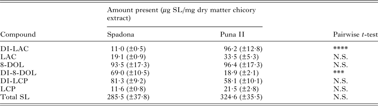
Detailed analysis of the two different cultivars is presented elsewhere (Peña-Espinoza et al. Reference Peña-Espinoza, Boas, Williams, Thamsborg, Simonsen and Enemark2015), and is summarized here briefly in Table 1. Three SL were detected – lactucin (LAC), lactucopricin (LCP), and 8-deoxylactucin (8-DOL), as well as their corresponding 11,13 dihydro (DI) derivatives. Of the 6 compounds, the concentration of only 2 differed significantly between cultivars; DI-LAC was more abundant in Puna II (P < 0·001) and DI-8-DOL was found in higher concentrations in Spadona (P < 0·001).
Identification and quantification of sesquiterpene lactones present in purified extracts from two forage chicory cultivars, Puna II and Spadona (also reported in Peña-Espinoza et al. (Reference Peña-Espinoza, Boas, Williams, Thamsborg, Simonsen and Enemark2015))

SL, sesquiterpene lactones; LAC, Lactucin; DI-LAC, 11, 13-dihydrolactucin; 8-DOL, 8-deoxylactucin; DI-8-DOL, 11, 13-dihydro-8-deoxylactucin; LCP, Lactucopicrin; DI-LCP, 11, 13-dihydro-lactucopicrin.
N.S.: P > 0·05; ***P = 0·01; ****P < 0·0001.
Purified chicory extracts reduce survival of Ascaris suum third-stage larvae
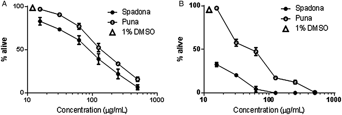
The activity of the purified chicory extracts was first assessed against A. suum L3 that were obtained by the in vitro hatching of embryonated eggs. L3 that were incubated in control media (RPMI1640 + 1% DMSO) remained alive and fully motile over the course of 36 h (Fig. 1). In contrast, L3 exposed to extracts from either Spadona or Puna II chicory exhibited a reduction in survival that was both dose and time dependent. After 16 h incubation, more than 90% of larvae were dead at concentrations of ⩾250 µg mL−1 of both extracts (Fig. 1A). The Spadona extract was more potent than the Puna II extract, as evidenced by a significantly lower EC50 value (81 µg mL−1 vs 146 µg mL−1, P < 0·001). When the larvae were observed again after 36 h, here there were clear differences between the two cultivars. For the L3 incubated in Spadona extract, all larvae were dead at concentrations of ⩾125 µg mL−1 and greater than 50% of larvae were dead at concentrations as low as 15 µg mL−1. In contrast, only the highest concentration of Puna II extract (500 µg mL−1) resulted in 100% larval mortality, and at lower concentrations most larvae remained alive and motile (Fig. 1B).
Survival of Ascaris suum third-stage larvae (L3) after exposure for 16 h (A) or 36 h (B) to varying concentrations of extracts from Spadona or Puna II varieties of chicory, or 1% DMSO (control). Experiments were conducted in triplicate and performed on three independent occasions. Results are the mean and s.e.m. of the pooled replicates from the three experiments.

Purified chicory extracts reduce motility of Ascaris suum fourth-stage larvae
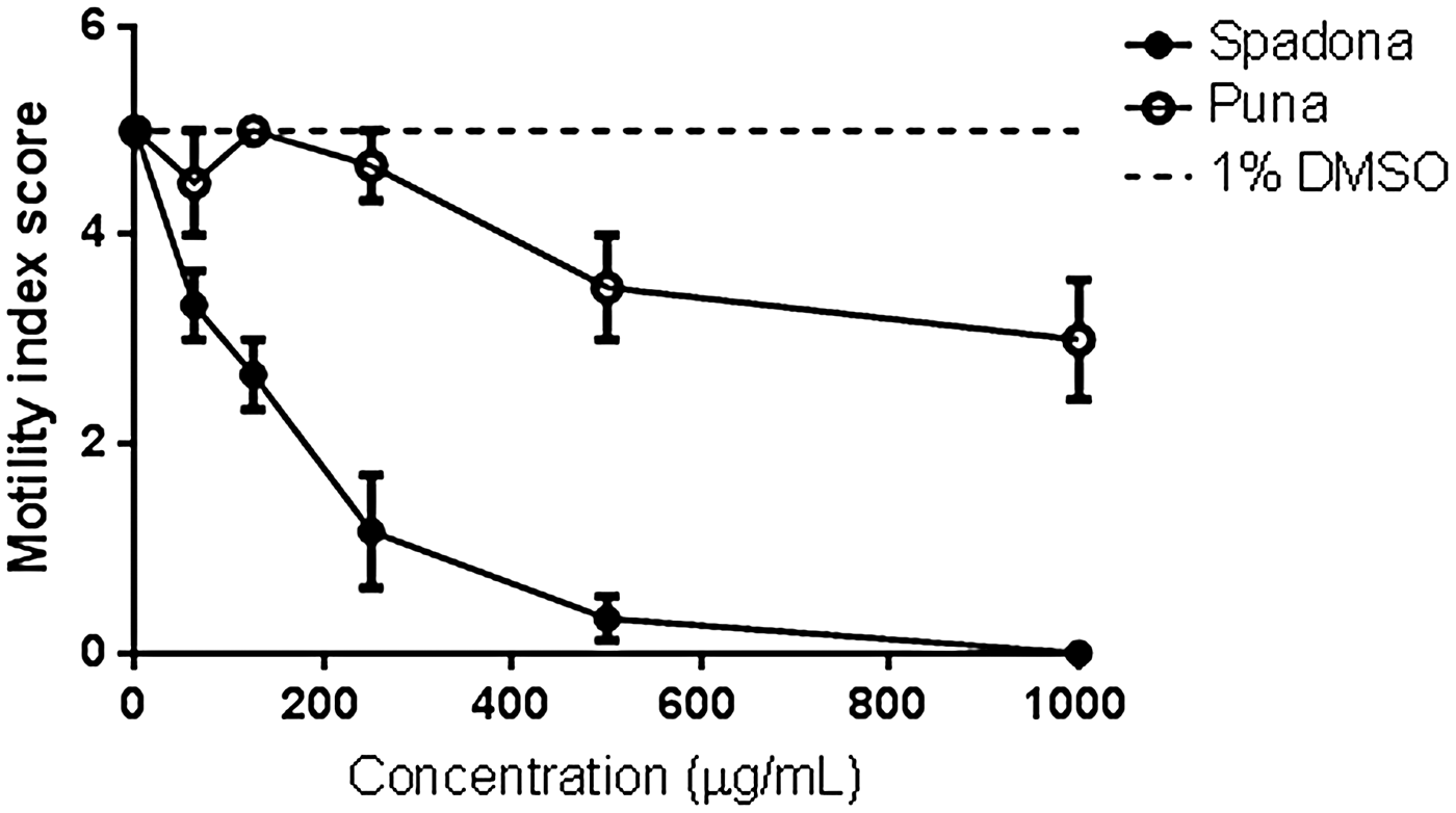
To confirm the anthelmintic activity of the SL-containing extracts, and also to ascertain whether different larval stages were equally susceptible to this activity, we obtained A. suum L4 from the intestine of experimentally infected pigs and exposed these to the extracts in vitro. Exposure of the L4 to the Spadona extract exhibited a significant loss in motility (Fig. 2). Concentrations of 250 or 500 µg mL−1 resulted in near-total paralysis of the larvae, and no movement was observed in L4 exposed to the top concentration of 1000 µg mL−1 despite extended observation. In contrast, L4 exposed to the Puna II extract exhibited only a minor loss of motility even at concentrations as high as 1000 µg mL−1 (Fig. 2).
Motility of Ascaris suum fourth-stage larvae after exposure for 12 h to varying concentrations of extracts from Spadona or Puna II varieties of chicory, or 1% DMSO (control). Results are the mean of two independent experiments, each performed in triplicate. Error bars indicate the inter-replicate s.e.m.

Ultrastructural analysis of A. suum exposed to Spadona extracts
To gain insight into the possible anthelmintic mechanism(s), A. suum L4 exposed to either 1000 µg mL−1 of Spadona extract or media only for 12 h were examined by transmission-electron microscopy. In all the parasites exposed to the Spadona extract, the cuticle was notably different in appearance to control larvae, being paler in colour with slight erosion of the epicuticle and a less pronounced delineation between the basal layer overlying the hypodermis and the medial zone (Fig. 3A and B). In addition, some lesions were visible in the muscular tissue underlying the hypodermis (Fig. 3C and D), however there were localized and infrequent, and no differences were noted in the internal digestive tissues (data not shown). Thus, despite the complete paralysis and apparent death of the parasites, only subtle ultrastructural changes were apparent.
Transmission electron micrographs of Ascaris suum fourth-stage larvae (L4) exposed for 12 h to 1% DMSO only (A,C) or 1000 µg mL−1 of Spadona chicory extract (B,D). Note the differing appearance of the basal and medial layers of the cuticle (cu) in A and B (marked by arrows), and the appearance of lesions (arrows) in the muscular layer (mu) of chicory-exposed L4 (D), compared with the control (C). For all images scale bar indicates 2 µm.

Anthelmintic activity of purified chicory extracts against O. dentatum
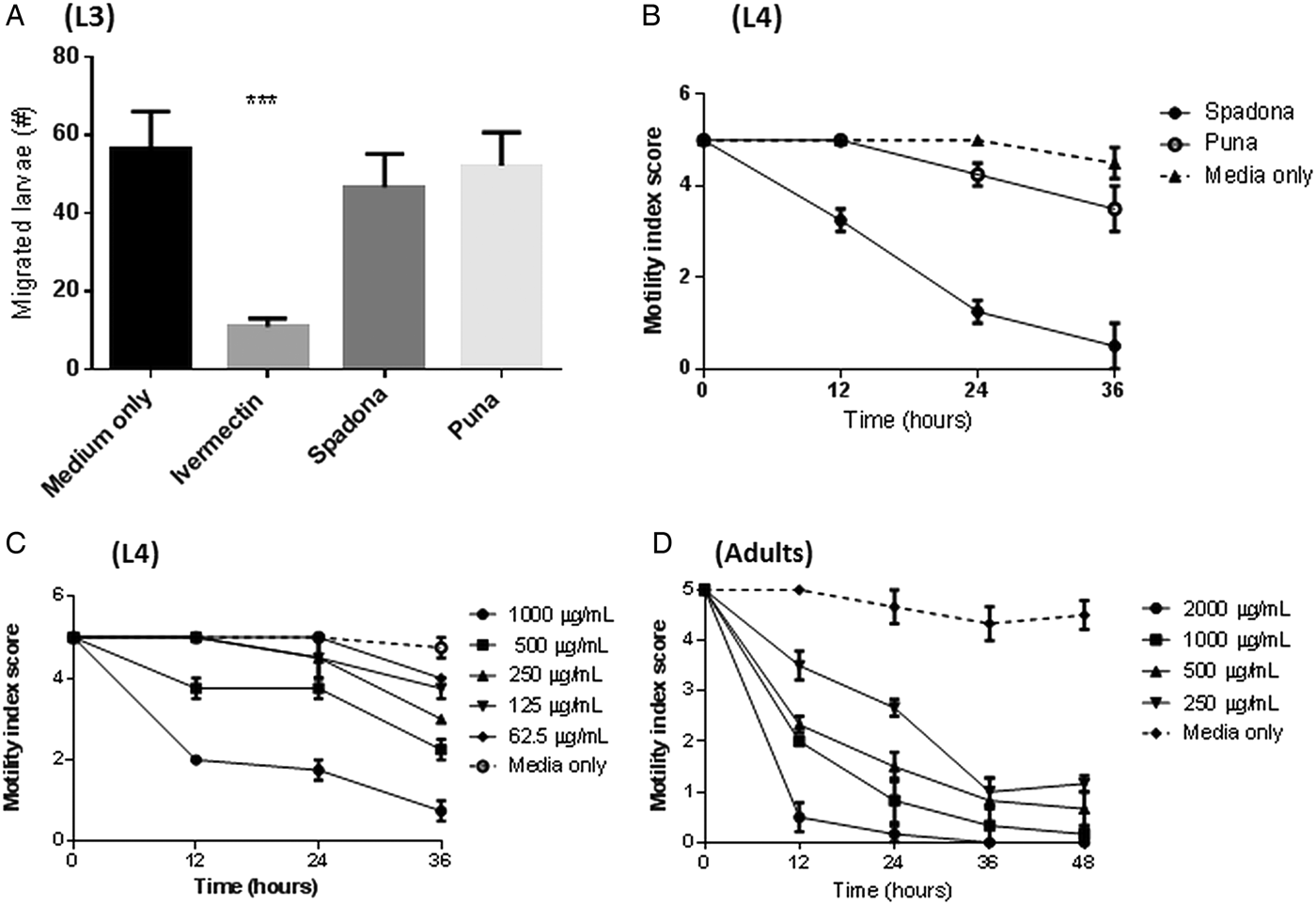
Having shown clear anthelmintic activity against A. suum, we next assessed whether the extracts were also active against O. dentatum. Initially, the extracts were tested against exsheathed L3 stages using a larval migration assay. In contrast to ivermectin, which inhibited migration by more than 80%, neither Spadona or Puna II extracts inhibited larval migration (Fig. 4A). However, when the extracts were tested at a concentration of 1000 µg mL−1 against L4 stages, the Spadona extract had strong anthelmintic activity, with a time-dependent inhibition of larval motility that resulted in almost total paralysis after 36 h incubation. Moreover, a complete cessation of pharyngeal pumping was observed in the Spadona-exposed worms. In contrast, no significant effect of the Puna II extract was apparent at this concentration (Fig. 4B). A subsequent experiment confirmed the activity of the Spadona extract against O. dentatum L4, and demonstrated that the effect was dose-dependent, with strong reductions in motility apparent at concentrations of 500 and 1000 µg mL−1 and lesser activity at concentrations ⩽250 µg mL−1 (Fig. 4C). Finally, we assessed whether the Spadona extract also had activity against adult stages of O. dentatum. Similar to the results with L4, incubation of adults in the SL-containing extract resulted in a reduction in motility that was both time and dose-dependent (Fig. 4D). These results show that the tested Spadona extract had strong activity against O. dentatum L4 and adult stages, but not L3, whilst Puna II extract seemed to have little effect against this parasite.
Anthelmintic effects of chicory extracts against Oesophagostomum dentatum. (A) Numbers of third-stage larvae (L3) that migrated after overnight incubation in ivermectin (50 µg mL−1) or extracts from Spadona or Puna II chicory (both 1000 µg mL−1). Results are the mean of two independent experiments, each performed in triplicate. Error bars indicate inter-replicate s.e.m. ***P < 0·001. (B) Motility of fourth-stage larvae (L4) exposed to only 0·5% DMSO (culture media) or 1000 µg mL−1 of Spadona or Puna II chicory extracts over 36 h. Results are the mean of two independent experiments, each performed in triplicate. Error bars indicate inter-replicate s.e.m. (C) Motility of L4 exposed to only 0·5% DMSO (culture media) or varying concentrations of Spadona chicory extract over 36 h. Results are from a single experiment performed in triplicate. Error bars indicate inter-replicate s.e.m. (D) Motility of adult worms exposed to only 0·5% DMSO (culture media) or varying concentrations of Spadona chicory extract over 48 h. Results are from a single experiment performed in triplicate. Error bars indicate inter-replicate s.e.m.

DISCUSSION
The identification of natural anti-parasitic compounds in livestock forages is likely to play a crucial role for the adoption of integrated parasite-control methods that have a reduced reliance on prophylactic drug treatment. Here we have demonstrated that extracted SL from chicory are able to directly kill larvae of A. suum and O. dentatum, two of the most prevalent and economically important nematode parasites of pigs.
We found that A. suum L3 exposed to the extracts rapidly died in a dose-dependent manner, whilst L4 also exhibited a rapid reduction in motility that, in the case of the Spadona extract, resulted in a complete cessation of movement after 12 h incubation. This suggests that diets containing chicory may be able to reduce the establishment of incoming A. suum larvae, and/or remove established larval infections from the gut. We also observed anthelmintic efficacy of the Spadona extract (but not the Puna II extract) against O. dentatum, although there were notable differences compared to the activity against A. suum, namely the lack of efficacy against L3 stages. This is consistent with recent results from our group which have demonstrated potent activity of the same purified chicory extracts against free-living and adult stages of the cattle nematode Ostertagia ostertagi, but not against the L3 stages (Peña-Espinoza et al. Reference Peña-Espinoza, Boas, Williams, Thamsborg, Simonsen and Enemark2015). Moreover, we have previously observed that O. dentatum L3 appear to be less susceptible to the effects of plant-derived condensed tannins than A. suum L3, suggesting that the L3 stage of O. dentatum may have a generalized lack of susceptibility to anthelmintic PSM (Williams et al. Reference Williams, Fryganas, Ramsay, Mueller-Harvey and Thamsborg2014a , Reference Williams, Ropiak, Fryganas, Desrues, Mueller-Harvey and Thamsborg b ). Recent results from Caenorhabditis elegans, which indicate that the adult and L4 stages are also more susceptible to anthelmintic plant extracts than L3 stage parasites (Kumarasingha et al. Reference Kumarasingha, Palombo, Bhave, Yeo, Lim, Tu, Shaw and Boag2014), suggest that this phenomenon may be a more generalized feature of helminths that have a free-living larval stage, which A. suum does not. Overall, despite this lack of effect against O. dentatum L3, it is clear that SL from chicory have broad-spectrum anthelmintic activity against swine nematodes in vitro.
The clear differences in potency we observed between the two cultivars, with Spadona significantly more active than Puna II, may be related to the differing SL profiles. In addition to our current data, we also have observed that Spadona extracts are substantially more active than Puna II against O. ostertagi in vitro (Peña-Espinoza et al. Reference Peña-Espinoza, Boas, Williams, Thamsborg, Simonsen and Enemark2015). Thus, it appears that the enhanced activity of the tested Spadona extract is conserved across multiple helminth species from different host species. Six major SL were identified in extracts from both chicory cultivars: LAC, LCP, 8-DOL, and their DI derivatives, all which have been previously reported in chicory leaves (Foster et al. Reference Foster, Cassida and Turner2011; Ferioli and D'Antuono, Reference Ferioli and D'Antuono2012). The relative abundances differed only subtly between the cultivars, with only the concentration of DI-8-DOL significantly elevated in Spadona as compared with Puna II. The major mechanism of biological activity of SL is proposed to involve a Michael-addition reaction between the α-methylene functional group on the γ-lactone ring of the SL and nucleophiles such as thiol groups on cysteine-containing amino acids (Simonsen et al. Reference Simonsen, Weitzel, Christensen, Ramawat and Merillon2013). However, the mechanism of DI-8-DOL may involve a different process, as the DI derivatives lack this α-methylene moiety, and thus are unable to form Michael-additions with nucleophiles. Notwithstanding, these derivatives can still be biologically active – e.g. insecticidal activities of SL lacking the α-methylene moiety have previously been reported (Bardón et al. Reference Bardón, Popich, Valdés and Catalán1999). However, it is not yet clear from our results whether the anthelmintic activity can be ascribed to discrete compounds or whether it represents a synergistic effect of a number of compounds within the extract. Indeed, whilst SL are likely candidates for the anthelmintic activity, it cannot be ruled out that other unidentified compounds in the extract may be responsible. Experiments to identify the active compound(s) and the mechanism of anthelmintic activity are on-going in our laboratory. It is interesting to note, however, that TEM of A. suum L4 exposed to high concentrations of Spadona extract showed only subtle structural damage – in clear contrast to previous experiments with other anthelmintic PSM such as condensed tannins which cause a generalized, toxic degradation of the parasite's external and internal structure (Martínez-Ortíz-de-Montellano et al. Reference Martínez-Ortíz-de-Montellano, Arroyo-López, Fourquaux, Torres-Acosta, Sandoval-Castro and Hoste2013; Williams et al. Reference Williams, Fryganas, Ramsay, Mueller-Harvey and Thamsborg2014a ). The lack of such a generalized necrotic effect with SL suggests that the potential mechanism may involve inhibition of specific key molecules involved in parasite metabolism, although this is only speculative at this stage and needs further investigation.
If these in vitro results can be confirmed in in vivo experiments, than the use of forage chicory may represent a novel option for control of helminths in outdoor pigs. Chicory has received renewed attention as a forage source for pigs, due in part to its high mineral content and favourable effects on gut health and microbiota due to its fibre content (Ivarsson et al. Reference Ivarsson, Liu, Dicksved, Roos and Lindberg2012; Liu et al. Reference Liu, Ivarsson, Lundh and Lindberg2013). Chicory roots have also been investigated as a bioactive feed supplement for organically-reared outdoor pigs, and in fact it has been shown that feeding chicory roots to young pigs can reduce A. suum and O. dentatum burdens and faecal egg excretion (Mejer, Reference Mejer2006; Jensen et al. Reference Jensen, Mejer, Mølbak, Langkjær, Jensen, Angen, Martinussen, Klitgaard, Baggesen, Thamsborg and Roepstorff2011). As chicory roots also contain SL (Cavin et al. Reference Cavin, Delannoy, Malnoe, Debefve, Touché, Courtois and Schilter2005), our current results may offer a mechanistic explanation to these observed results; however, chicory roots also contain high amounts of inulin which is rapidly fermented in the large intestine resulting in increased production of short-chain fatty acids, and these physio-chemical changes have a known antagonistic effect on helminths residing in the large intestine such as O. dentatum (Petkevičius et al. Reference Petkevičius, Murrell, Bach Knudsen, Jørgensen, Roepstorff, Laue and Wachmann2004). As A. suum L3 hatch in the caecum and penetrate the mucosa of the large intestine to begin their migratory phase (Murrell et al. Reference Murrell, Eriksen, Nansen, Slotved and Rasmussen1997), it may be that that this inulin-related effect is also partly or mainly responsible for the observed anthelmintic effects of chicory roots against A. suum. Therefore, further experiments with chicory forage, which contain only low levels of these easily fermentable fructans (Ernst et al. Reference Ernst, Chatterton and Harrison1995; Milala et al. Reference Milala, Grzelak, Król, Juśkiewicz and Zduńczyk2009), will be necessary to clearly demonstrate the in vivo efficacy of chicory SL.
In vivo trials will also provide an opportunity to examine the pharmacokinetics and possible changes to the SL structures during digestion that may affect the anthelmintic activity of these compounds when they reach the predilection site of A. suum (small intestine) and O. dentatum (large intestine). Factors to be considered include the possible degradation and absorption of SL in the gut, as well as decreased biological availability of total SL as a result of being bound to other molecules e.g. glycosides. Whilst our in vitro experiments were conducted with extracts that had been digested with cellulase (resulting in release of glycoside-bound SL), we also showed that this digestion was not necessary for in vitro anthelmintic activity as extracts prepared with the omission of the cellulase step showed comparable anthelmintic efficacy (data not shown). Therefore, consumption of whole forage chicory should provide a source of available and functionally active SL in pigs, which unlike ruminants lack the ability to digest cellulose.
In conclusion, we have shown for the first time that SL-containing extracts from chicory have direct anthelmintic effects against nematode parasites of swine. This effect was, however, dependent on cultivar, with extracts from Spadona displaying enhanced activity compared to extracts from Puna II, a difference which may be attributable to differing SL compositions in the two varieties. Multiple larval stages of A. suum were susceptible to extracts from Spadona, and O. dentatum L4 and adult stages were also susceptible to Spadona extracts whilst O. dentatum L3 were not. Future experiments will focus on in vivo efficacy, further purification of the active compounds and determination of the mechanism(s) of the anthelmintic effect.
ACKNOWLEDGEMENT
The authors are grateful to Lise-Lotte Christiansen for assistance with infection of pigs.
FINANCIAL SUPPORT
This work was funded by the Danish Council for Independent Research (Technology and Production Sciences; Grant no. 12-126230). MPE, HLE and SMT acknowledge support of the EMIDA ERA-NET project ‘Coping with anthelmintic resistance in ruminants’ (CARES; 3405-11-0430/32).