Transcatheter balloon dilation for an occluded aortopulmonary shunt is an optional treatment to avoid high-risk reoperation among children with cyanotic CHD. Reference Gillespie and Rome1 Although the success rate of balloon dilatation has been reported to be as high as 80–90%, Reference Bonnet, Petit and Lambert2–Reference Wang, Wu, Chang, Chiu and Lue4 this procedure may be associated with major complications, including intimal injury of the pulmonary artery, thrombotic occlusion in the peripheral pulmonary arteries, thromboembolic stroke, and death. Reference Bonnet, Petit and Lambert2–Reference Wang, Wu, Chang, Chiu and Lue4 Younger age is associated with failure of this procedure. We herein present the case of a 2-year-old girl who presented catastrophic haemoptysis due to an arterio-bronchial fistula after transcatheter balloon dilatation for an occluded aortopulmonary shunt. She was successfully treated with coil embolisation for the fistula, while haemoptysis was controlled with the left bronchial block ventilation and haemostatic balloon occlusion of the left subclavian artery.
Case report
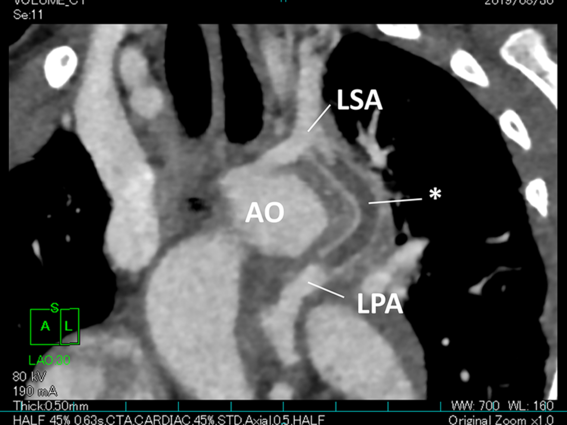
A 2-year-old girl was admitted to our hospital because of the development of cyanosis. The patient had been diagnosed with severe Epstein’s malformation and pulmonary atresia. She underwent Starnes operation with an aortopulmonary shunt (in the diameter of 3.5 mm) from the ascending aorta to the main pulmonary trunk at the age of 11 days, and the placement of a left modified Blalcok–-Taussig shunt (in the diameter of 4 mm) at the age of 24 months. She also underwent transcatheter balloon dilatation for narrowing aortopulmonary shunts twice at age of 22 and 27 months and was treated with oral aspirin of 5 mg/kg/day. She had visited the emergency department at the last month because of bronchitis and gastroenteritis. On admission, her systemic oxygen saturation decreased from 83% to 68%. Contrasted CT showed that the left aortopulmonary shunt was completely occluded due to thrombosis (Fig 1), and thereby urgent cardiac intervention was attempted.
Contrasted CT shows that a left aortopulmonary shunt is completely occluded due to thrombosis (asterisk). AO: aorta, LSA: left subclavian artery, LPA: left pulmonary artery.

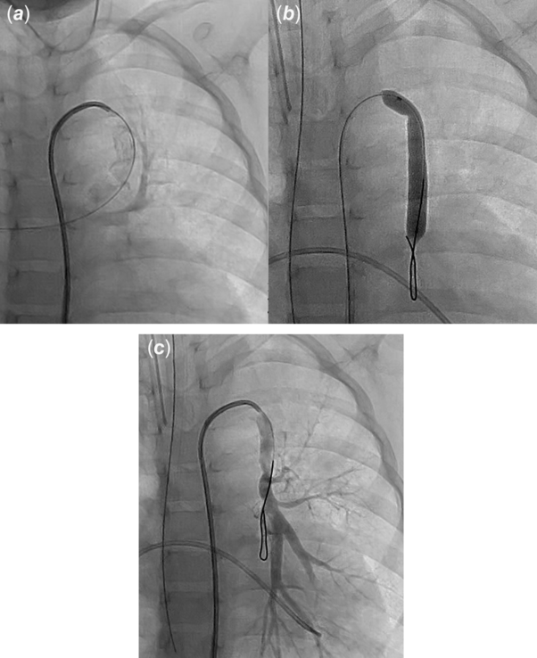
Under the sedation with thiamylal, a 4-French sheath was introduced from the right femoral artery. Intravenous heparin of 100 units/kg was administrated before the procedure. Angiography at the left subclavian artery showed the complete occlusion of the graft. We engaged the occluded shunt using the right Judkins catheter and recanalised it using a 2.7-French microcatheter (Progreat™, Terumo Corp., Tokyo, Japan). Urokinase of 4,000 units/kg was slowly and cautiously injected via a microcatheter into the thrombus. However, she suddenly developed haemoptysis just after taking an angiogram at the left aortopulmonary shunt (Fig 2a; Supplemental Movie 1). Soon, she was intubated and administrated with intra-tracheal epinephrine. After the confirmation of haemostasis and haemodynamic stabilisation, we subsequently performed transcatheter balloon dilation for the occluded shunt. After a 0.018-inch guidewire (Thruway™, Boston Scientific Japan, Tokyo, Japan) was placed through the shunt into the distal left pulmonary artery, a balloon catheter in the diameter of 5 mm and the length of 20 mm (Sterling™, Boston Scientific Japan, Tokyo, Japan) was introduced and dilated (Fig 2b). Then, the shunt was successfully recanalised, and the procedure was finished (Fig 2c). She continued to be on admission to observe systemic saturation and haemostasis.
( a ) An angiogram at the left subclavian artery shows the opacification of the left bronchioles following haemoptysis (also shown in Supplemental Movie 1). ( b ) Balloon dilatation of an occluded shunt was performed using Sterling™ balloon. ( c ) The final angiogram shows the shunt is recanalised.

On the 6th day after the procedure, she suddenly developed haemoptysis following haemorrhagic shock on the ward. She was transferred to the ICU following conservative treatments with transfusion and mechanical ventilation under sedation. However, she repetitively developed haemoptysis five times for 2 weeks, which required transfusion. Contrasted CT obtained on the 9th day after the procedure showed that blood flow through the shunt was kept adequate and no apparent causative lesion of haemoptysis was detected. Bronchoscopy also failed to show any origin of haemoptysis. On the 19th day after the procedure, she developed catastrophic haemoptysis following haemorrhagic shock again, and therefore we underwent urgent catheter intervention for haemostasis.
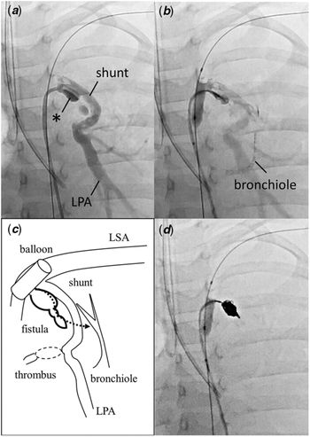
Under general anesthesia, tracheal bleeding was aspirated using a bronchoscope, while a bronchial blocker (Arndt®, 5 French Cook Japan, Tokyo, Japan) was placed for single lung ventilation. A 4-French sheath was introduced from the femoral artery, and a balloon catheter (Sterling™, 5 mm × 20 mm) was placed in the left subclavian artery for haemostasis. Another 4-French sheath was introduced from the femoral artery, and a 4-French right Judkins catheter was introduced. An angiogram at the left subclavian artery showed an arterio-bronchial fistula originating from the left subclavian artery adjusted to the left aortopulmonary shunt into the left bronchiole. The fistula looked like an aneurysm in the diameter of 4 mm × 13 mm (Fig 3a–c). We introduced a 2.7-French microcatheter into the fistula and embolised it using neurovascular coils (Target XL360 Soft™, Stryker Japan Corp., Tokyo, Japan) (Fig 3d, Supplemental Movie 2). The final angiogram confirmed the complete occlusion of the fistula. After then, she never developed haemoptysis. On the 56th day of admission, cardiac catheterisation showed that the fistula was completely occluded, and that the left aortopulmonary shunt was patent with the mean left pulmonary artery pressure of 13 mmHg. She was discharged with arterial oxygen saturation of 85%. Nowadays, she is waiting for Glenn operation at the age of 3 years.
( a ) An angiogram at the left subclavian artery conjunct with the aortopulmonary shunt shows an arterio-bronchial fistula (asterisk). ( b ) A selective angiogram in the fistula shows that contrast media run down into the left bronchiole. ( c ) The anatomical scheme was drawn. ( d ) The fistula is embolised using a total of six coils (also shown in Supplemental Movie 2).

Discussion
This is a notable case of a child who developed catastrophic haemoptysis due to an arterio-bronchial fistula after transcatheter balloon dilatation of an aortopulmonary shunt. To the best of our knowledge, this is the first case of a patient with an arterio-bronchial fistula as a complication of catheter intervention for CHD. Arterio-bronchial fistula is known as a rare but serious complication after tracheotomy and occasionally results in haemoptysis. Previous reports have shown that an arterio-bronchial fistula after tracheotomy has the mortality as high as 15% despite the occurrence less than 1%. Reference Bobka, Sukato, Kohli, Worku, Gulkarov and D’Ayala5 There were two cases of patients after radiochemotherapy and tracheotomy for laryngeal cancer who developed an arterio-bronchial fistula between the subclavian artery and the trachea. Reference Bobka, Sukato, Kohli, Worku, Gulkarov and D’Ayala5,Reference Hizette, Horn, Lemaitre and Segers6 They two presented haemoptysis as the first symptom, which were similar to our present case. It is speculated that compression of the tracheal wall by a tracheostomy tube elicits necrosis or erosion of the innominate artery resulting in the formation of an arterio-bronchial fistula. However, there is no previous report about paediatric arterio-bronchial fistula associated with CHD, although there is one patient with an aneurysmal formation at the anastomosis site of a shunt graft after transcatheter balloon dilatation for a stenotic aortopulmonary shunt. Reference Wang, Wu, Chang, Chiu and Lue4 As we usually dig and pass through an occluded aortopulmonary shunt using a guidewire during transcatheter balloon dilatation, it is possible that a subclinical injury may occur at the site of the anastomosis of a shunt graft, leading to the development of an arterio-bronchial fistula. In our present case, contrasted CT failed to clearly show the presence of an arterio-bronchial fistula. Thereby, we consider that angiography should be necessary when haemoptysis unknown origin occurs during or after catheter intervention for CHD.
We successfully embolised the arterio-bronchial fistula using additional haemostatic techniques. Firstly, we performed single lung ventilation using a bronchial blocker. This method is used for not only single lung ventilation during thoracotomy but also a temporary haemostatic treatment for haemoptysis. Reference Kathuria, Hollingsworth, Vilvendhan and Reardon7,Reference Radchenko, Alraiyes and Shojaee8 It allows us to prevent aspiration into the contralateral lung and to stabilise respiratory and haemodynamic conditions during procedures. An Arndt™ bronchial blocker can be available among small infant because of the variable sizes. Winch et al recently reported the feasibility of a bronchial blocker for pulmonary haemorrhage during catheter intervention in children with CHD. Reference Winch, Tumin, Moore, Vizzini, Berman and Naguib9 Although an endotracheal balloon or an endotracheal embolisation with silicon sponges may be alternative to a bronchial blocker, Reference Kathuria, Hollingsworth, Vilvendhan and Reardon7,Reference Radchenko, Alraiyes and Shojaee8 we consider that these haemostatic methods are not suitable in the present case because of the size limitation and technical problems. Secondly, we performed haemostatic occlusion of the left subclavian artery using another angioplasty balloon. This method is feasible for haemostasis when bleeding is active during procedures, especially portal venous haemorrhage during liver transplantation. Reference Seal, Bohorquez and Battula10 The balloon remodelling technique is commonly used for coil embolisation of intracranial aneurysms or coronary arteriovenous fistulae. Reference Pierot, Cognard, Spelle and Moret11 A non-detachable balloon catheter is guided into the front of a lesion and temporarily inflated to prevent a coil to migrate. Scepter C™ (Microvention Inc., CA, USA) is a occlusion balloon system for this technique with a dual lumen catheter which accommodates a 0.014-inch guidewire Reference Spiotta, Miranpuri, Hawk, Chaudry, Turk and Turner12 , which might be available in our present case.
Conclusion
We present the case of a 2-year-old girl who presented catastrophic haemoptysis due to an arterio-bronchial fistula that is a rare complication of transcatheter balloon dilatation for an occluded aortopulmonary shunt. We successfully embolised the fistula using coils, while haemoptysis was controlled with the left bronchial block ventilation and haemostatic balloon occlusion of the left subclavian artery.
Acknowledgement
The authors thank the patient’s guardians for the written informed consent.
Financial Support
This research received no specific grant from any funding agency, commercial or not-for-profit sectors.
Conflicts of Interest
None.
Ethical Standards
This work does not involve any human/animal experimentation.
Supplementary Material
To view supplementary material for this article, please visit https://doi.org/10.1017/S1047951120002681