Introduction
Laryngeal squamous cell carcinoma (SCC) is the 11th most common form of cancer with a male predominance, and the second most common malignancy of the head and neck.Reference Chu and Kim1 The major prognostic factors for laryngeal SCC are tumour–node–metastasis (TNM) stage, clinical stage, cartilage invasion, differentiation, perineural invasion and lymphatic invasion.Reference Yilmaz, Gedikoglu and Gursel2 Studies of laryngeal SCC have investigated the effect of genetic and epigenetic modification, alteration in tumour environment, and immune evasion, in order to better understand the tumour's development and course.Reference Chu and Kim1, Reference Loyo and Pai3 Protein-based alterations in tumour cells may also contribute to carcinogenesis.Reference Loyo and Pai3
Fascin is a 55-kDa, actin-binding protein. Actin bundle proteins are concentrated in cell membrane protrusions, and these protrusions provide motility for the cell. These actin bundle proteins are rearranged by fascin proteins.Reference Adams4, Reference Kureishy, Sapountzi, Prag, Anilkumar and Adams5 During the neoplastic transformation of cells, the development of motility and migratory capacity is important for the tumour's invasive and metastatic potential. A relationship between fascin overexpression and an unfavourable prognosis has been demonstrated for many malignant tumours.Reference Chen, Zhang, Ren, Tao, Shen and Lv6–Reference Lee, Poon, Man, Guan, Ma and Liu9 However, the relationship between fascin expression and laryngeal SCC aggressiveness has not previously been reported, to the best of our knowledge.
In this study, we aimed to investigate the relationship between fascin expression and age, sex, tumour localisation, TNM stage and tumour differentiation, in laryngeal SCC patients.
Materials and methods
The surgical specimens of 30 patients who had undergone surgical treatment for laryngeal SCC at the Gulhane Military Medical Academy between 1995 and 2007 were randomly retrieved for immunohistochemical study.
We chose a representative block from each tumour for immunohistochemical analysis. A 5-mm section from each formalin-fixed, paraffin-embedded tumour tissue block (which consisted almost entirely of tumour tissue) was automatically immunostained (Autostainer-480-LV1; Labvision, Fremont, California, USA) using a primary antibody against fascin (FCN01, 1:80, mouse monoclonal antibody; Neomarkers, Fremont, California, USA), employing the avidin-biotin peroxidase technique. The sections were incubated with the primary antibody at room temperature for 60 minutes.
All immunohistochemically stained slides for each case were reviewed by two pathologists, using a previously described fascin staining and scoring method, as follows.Reference Gunal, Onguru, Safali and Beyzadeoglu10 Cytoplasmic staining was accepted as positive. The percentage of positively stained cells was assessed semiquantitatively and scored from one to five, as follows: one = 0–5 per cent, two = 6–25 per cent, three = 26–50 per cent, four = 51–75 per cent and five = 76–100 per cent. The intensity of immunohistochemical staining was also scored, as either one (=weak; Figure 1a), two (=moderate; Figure 1b) or three (=strong; Figure 1c). Overall immunohistochemical scores for fascin expression were calculated for each case by multiplying the scores for staining percentage and staining intensity; possible overall fascin scores ranged from one to 15. Finally, we categorised the overall fascin scores into two groups: eight and below, and more than eight.Reference Gunal, Onguru, Safali and Beyzadeoglu10
Photomicrographs showing tissue sections with (a) weak, (b) moderate and (c) strong fascin immunohistochemical staining intencity (×200).

The overall immunohistochemical fascin score was compared with the age, sex, localisation, TNM stage and differentiation status of the cases. Statistical significance was assessed by the chi-square test. All analyses were performed using the Statistical Package for the Social Sciences version 11.0 software program (SPSS Inc, Chicago, Illinois, USA).
Results
Patients' ages ranged from 29 to 79 years. There were 28 men and two women.
Tumours were supraglottic in 11 cases, transglottic in 10, glottic in four, supraglottic with glottic extension in three and glottic with subglottic extension in two. Four of the cases were well differentiated, 21 moderately differentiated and five poorly differentiated. Nine cases had one to four metastatic lymph nodes. No lymph node metastasis was found in 21 cases.
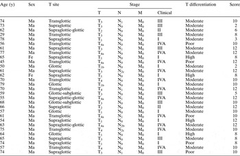
Fascin expression of varying degrees was found in all tissue samples. The overall fascin score was 12 in nine cases, 10 in 10 cases, eight in six cases and six or less in five cases (Table I).
Cases' clinicopathological variables and fascin expression scores

Y = years; T = tumour; N = node; M = metastasis; score = overall immunohistochemical fascin expression score; Ma = male; Fe = female
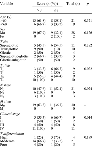
There was no significant relationship between the cases' age or sex and the overall fascin score (p = 0.571 and 0.126, respectively).
An overall fascin score of more than eight was found in five of the 11 supraglottic cases, nine of the 10 transglottic cases, two of the four glottic cases, two of the three supraglottic with glottic extension cases, and one of the two glottic with subglottic extension cases. Although most transglottic laryngeal SCC cases had high overall fascin scores, we found no statistically significant relation between cases' overall scores and their tumour localisation (p = 0.282).
The overall fascin score was more than eight in one of the four well differentiated cases, 14 of the 21 moderately differentiated cases, and four of the five poorly differentiated cases. There was no statistically significant relationship between tumour differentiation and overall fascin score (p = 0.199) (Table II).
Cases' clinicopathological variables by overall fascin expression score
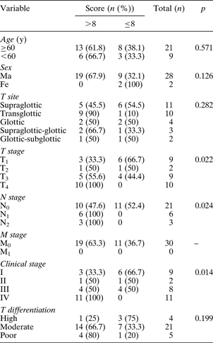
Score = overall immunohistochemical fascin expression score; y = years; Ma = male; Fe = female; T = tumour; N = node; M = metastasis
Since none of the cases in our series had distant metastasis, we were not able to evaluate the significance of fascin expression for this parameter.
All T4, N1 to N2, and stage IV cases had high overall fascin scores. Half of stage II and III cases had high overall fascin scores, whereas the remainder had low scores. Only 33 per cent of stage I cases had high overall fascin scores. Similar results were observed for T staging. Ten of the 21 N0 cases had low overall fascin scores. There was a statistically significant relationship between cases' T stage (p = 0.022), N stage (p = 0.024) and clinical stages (p = 0.014) and their overall fascin score (Table II).
Discussion
Despite the availability of new surgical techniques and organ preservation protocols, laryngeal SCC is still a serious health problem, with low survival rates especially for patients with advanced cancers. Prevention and early diagnosis of laryngeal carcinoma is the most effective way of maximising cure rates and preserving function. In addition, a better understanding of the steps involved in malignant transformation may assist the development of novel diagnostic, prognostic and therapeutic strategies.
In this study, we investigated the relationship between fascin expression and clinicopathological prognostic parameters in laryngeal SCC. We demonstrated that fascin expression was greater in more aggressive tumours.
Fascin expression was first reported in dendritic cells, and then in Hodgkin lymphoma, glial, neuron and muscle cells.Reference Adams4, Reference Kureishy, Sapountzi, Prag, Anilkumar and Adams5 It is only expressed in the basal layer of the epidermis within normal epithelium. The fascin protein may be related to tumour invasion and distant metastasis, due to its association with motility. It has been studied in cancers of the breast, oesophagus, stomach, lung, colon, pancreatic and biliary tract, ovary, and bladder, and an association with an unfavourable prognosis has been demonstrated.Reference Grothey, Hashizume, Ji, Tubb, Patrick and Yu11–Reference Tong, Yee, Chiriboga, Hernandez and Waisman18
Tumoural protein expression is known to play a role in carcinogenesis, in addition to genetic and epigenetic factors. Various studies have addressed the relationship between laryngeal SCC prognosis and epidermal growth factor receptor, dysadherin, cyclooxygenase-2 (COX-2) and vascular endothelial growth factor C.Reference Demiral, Sarioglu, Birlik, Sen and Kinay19–Reference Kyzas, Stefanou and Agnantis21
Epidermal growth factor receptor is a transmembrane tyrosine kinase receptor. It plays an important role in cell survival and proliferation. Overexpression has been demonstrated in more than 90 per cent of head and neck SCC cases. Overexpression in early glottic carcinomas has been reported to be associated with an unfavourable prognosis.Reference Demiral, Sarioglu, Birlik, Sen and Kinay19
Dysadherin is another cancer-associated cell membrane glycoprotein which has recently been defined and characterised. Kyzas et al. have demonstrated a relationship between high expression of dysadherin in head and neck region SCC and higher clinical stage and presence of lymph node metastasis at the time of diagnosis.Reference Kyzas, Stefanou, Batistatou, Agnantis, Nakanishi and Hirohashi20
Similarly, it has been shown that high expression levels of COX-2 and vascular endothelial growth factor C are correlated with the presence of lymph node metastasis at the time of diagnosis.Reference Kyzas, Stefanou and Agnantis21
There are no previous studies investigating the relationship between fascin expression and laryngeal SCC aggressiveness. However, fascin has been studied in oral cancers and SCC of the oesophagus.
In 2006, Zhang et al. studied a total of 102 sections, comprising tissue from normal regions of cancerous oesophageal tissue, dysplastic cases and SCC cases.Reference Zhang, Xu, Xiao, Xie, Zeng and Cai12 They also evaluated 49 fresh cases of oesophageal SCC. They found that high fascin expression levels were associated with high levels of cellular proliferation and lymph node metastasis.
In 2007, Chen et al. studied 129 oral SCC cases, and found more fascin expression in tumour tissue than in normal tissue.Reference Chen, Yang, Li, Nieh, Lin and Fu22 They also found an association between fascin overexpression and tumour diameter, lymph node metastasis, distant metastasis and clinical stage.
Similarly, we found a high degree of fascin expression to be associated with tumour stage and lymph node metastasis, in laryngeal SCC cases. The overall immunohistochemical fascin score was usually high in cases of advanced stage tumour; however, it is not clear which causes which. Despite this, in both of these situations, knowing a tumour's fascin expression level may assist tumour management, for the following reasons. The fascin protein has been shown to play a role in the carcinogenesis of many cancer types. It does this by forming actin-based structures, changing cellular motility and causing migratory changes.Reference Yamashiro, Yamakita, Ono and Matsumura23 According to recent reports, tumours which overexpress fascin generally have markedly more lymph node metastases than tumours with low fascin expression. Lymph node metastasis is an important negative prognostic indicator for laryngeal SCC.Reference Yilmaz, Gedikoglu and Gursel2 We have found a statistically significant relationship between high fascin expression levels and lymph node metastasis.
• Fascin is an actin-binding protein which is expressed in the basal areas of healthy squamous epithelium
• This study aimed to investigate the relationship between fascin expression and tumour behaviour in 30 laryngeal squamous cell carcinoma (SCC) cases
• Results suggest that laryngeal SCC tumours with high levels of fascin expression may be more aggressive than those with low levels
We did not find a statistically significant relationship between tumour differentiation and overall fascin score. However, the percentage of cases with a high score tended to increase as cases become less differentiated (Table II). The reason for the statistical insignificance of this association may be the small number of patients in our series.
Conclusion
We found an association between a high fascin expression score and the T stage, N stage and clinical stage of our laryngeal SCC cases. This may indicate that tumours with high levels of fascin expression behave more aggressively. In light of recent studies, fascin could be considered as a candidate molecular target for cancer therapy. However, studies employing larger series and evaluating survival versus fascin expression are needed, in order to fully understand the significance of fascin expression for patients with laryngeal SCC.