Introduction
Alzheimer's disease (AD) is a multifactorial neurodegenerative disease which affects more than 20 million people worldwide. Hypotheses based on amyloid formation, inflammation and cholesterol metabolism have been suggested. Large-scale epidemiological studies have shown that women have a higher prevalence of AD, and estrogen replacement therapy has been reported as reducing the risk of developing AD (Henderson et al., Reference Henderson, Benke, Green, Cupples and Farrer2005) and delay the onset of AD (Tang et al., Reference Tang1996; Waring et al., Reference Waring1999). Some studies have also suggested that estrogen might have pleiotropic effects on the inflammatory response as shown, for example, in the estrogen decreased inflammation in animal models of arthritis (Badger et al., Reference Badger1999).
Estrogen is a steroidal hormone produced in the ovaries. Its physiological function extends beyond the role in reproduction. It readily passes through the blood-brain-barrier and exerts physiological effects in the central nervous system. Estrogen has been shown to act as a neuroprotectant and neurotrophic agent by increasing the non-amyloidogenic processing of amyloid precursor protein (APP) (Jaffe et al., Reference Jaffe, Toran-Allerand, Greengard and Gandy1994), up-regulating the apolipoprotein E (ApoE) expression (Srivastava et al., Reference Srivastava1997), improving cerebral blood flow, facilitating neuronal repair and stimulating glucose transport and metabolism (Birge, Reference Birge1997). Estrogen exerts its action through at least two receptors (estrogen receptor α or ESR1 and estrogen receptor β or ESR2). The ESR1 gene is expressed throughout the brain. Different studies examining the association of ESR1 gene polymorphisms and AD have reported inconsistent findings (Brandi et al., Reference Brandi1999; Ji et al., Reference Ji, Urakami, Wada-Isoe, Adachi and Nakashima2000; Maruyama et al., Reference Maruyama2000; Lambert et al., Reference Lambert2001; Lin et al., Reference Lin2003; Usui et al., Reference Usui2006). It should be noted that the ESR1 gene is a highly polymorphic gene containing more than 1000 single nucleotide polymorphisms (SNPs) over its genomic span of 295.7kb. Only two polymorphisms – rs2234693 (also known as PvuII or ESR+397 in the literature) and rs9340799 (commonly known as XbaI or ESR+351) – are widely studied in diseases such as breast cancer (Cai et al., Reference Cai2003) and AD (Brandi et al., Reference Brandi1999). In the present study, we attempted to tag SNPs of higher density and evaluate further the association between ESR1 gene polymorphisms and the risk of AD in the Chinese population.
Methods
Subject recruitment
Two-hundred-and-thirty-three Chinese AD patients (84.3%women; mean age 81.4 years, SD = 6.7, range 65–97 years) with NINCDS-ADRDA diagnosis (McKhann et al., Reference McKhann1984) for probable or possible AD were recruited from the psychogeriatric services of New Territories East Cluster Hospitals in Hong Kong. Another 245 Chinese control subjects (75.5% women; mean age 73 years, SD = 6.3, range 65–89 years) were recruited from local elderly social centers for comparison. All control subjects underwent cognitive assessment using the Cantonese version of the Mini-mental State Examination (Chiu et al., Reference Chiu, Lee, Chung and Kwong1994) and were evaluated by a specialist psychiatrist as cognitively intact before recruitment. Both the AD and control subjects originated from Guangdong Province, a major province of southern China. Age-at-onset (AAO) for AD patients was defined as the age at which the family caregivers and/or individual first noted cognitive and/or behavioral problems sufficient to interfere with independent daily activities. Informed consent was obtained and the project was approved by the institutional ethics committee.
Analysis of the linkage disequilibrium (LD) structure of ESR gene
ESR1 gene contains more than 1000 putative SNPs over its genomic span of 295.7kb (dbSNP). Genotyping data of Caucasians (CEU) and Chinese (CHB) were obtained from HapMap (www.hapmap.org; International HapMap Consortium, 2003) and were used to compare the linkage disequilibrium (LD) structure among the two populations.
Genotyping of ESR1 polymorphisms
Genomic DNA was extracted from peripheral blood samples using DNA extraction kit according to the manufacturer's instruction (Roche, U.S.A.). PCR-RFLP was performed as previously described to genotype the selected polymorphisms (Ma et al., Reference Ma, Tang, Lam and Chiu2005). To validate the genotyping results, genotyping experiments were repeated and direct sequencing was performed in 10% of the samples. The ApoE genotyping was performed as reported earlier (Ma et al., Reference Ma, Tang, Lam and Chiu2005).
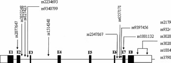
First, four commonly studied SNPs – rs2077647 (ESR + 30)(Dijkstra et al., Reference Dijkstra2006), rs2234693 (PvuII) (Wang et al., Reference Wang2007), rs9340799 (XbaI) (Yaffe et al., Reference Yaffe, Lui, Grady, Stone and Morin2002) and rs1801132 (ESR+975) (Kang et al., Reference Kang2002) – were selected and genotyped in a case-control setting. With the result of a pilot study showing significant association of rs1801132 and the risk of AD, Phase II study with 12 additional SNPs were selected and genotyped for the samples. SNPs in Phase II were selected according to available genotype information of 80 SNPs in the Phase I HapMap. Twelve tagSNPs were selected by a R2 algorithm among SNPs with minor allele frequencies of at least 5% in Asians (Figure 1) (Tang et al., Reference Tang, Pharoah, Ma and Easton2006a).
Positions of SNPs examined in this study. ESR1 contains 8 exons and extends over a distance of ∼300 kb in physical length. The coding regions are shown as solid boxes, and the 5’- and the 3’-untranslated regions are depicted as open boxes.

Statistical analysis
Deviations from the Hardy-Weinberg equilibrium for genotypes at individual loci were assessed by using Pearson χ2. Statistical analysis of genotype distribution and allele frequencies was performed by χ2 tests. Haplotypes were reconstructed by use of the PHASE software (Stephens et al., Reference Stephens, Smith and Donnelly2001). Logistic regression was performed to investigate the simultaneous effects of the ESR1 genotypes and ApoE ɛ4 allele status on the risk of AD (SPSS for Windows; 11.5). Cox regression or Kaplan-Meier was used to examine the effect of polymorphisms of interest to AAO of AD (Li et al., Reference Li1995; Maraganore et al., Reference Maraganore2006).
Results
Linkage disequilibrium (LD)
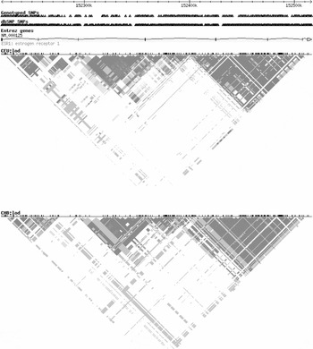
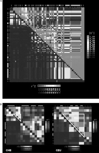
Four haploblocks were identified in Caucasians but only three haploblocks in Chinese (Figure 2). Genotyping data of SNPs at 3kb interval from 45 Chinese subjects (CHB) in HapMap project were obtained and the LD structure was analyzed (Zhang and Jin, Reference Zhang and Jin2003). The analysis showed a high degree of LD at the first two exons and the last exon. We also analyzed the LD structure of the 16 SNPs examined in this study. According to the genotyping data from 45 CHB subjects, we recapitulated the LD pattern of three haploblocks in Chinese. Comparing the LD structure of those 16 SNPs among the Caucasian (CEU) (genotyping data from 90 CEU subjects) and Chinese populations, the linkage disequilibrium measure (D′) between exon 2 to exon 7 was higher in Caucasians (Figure 3). Previous studies also suggested XX allele of XbaI was less frequent in Asians (Tang et al., Reference Tang2006b) and this decreased the tagging ability of this SNP. The selection of tagSNP would maximize the information provided by these SNPs in the association study.
LD structure of ESR1 gene in Caucasians (upper panel) and Chinese (lower panel).

Linkage disequilibrium measures with the ESR1 in Chinese, (A) with 90 SNPs (at 3kb interval), (B) 16 SNPs examined in this study (genotype data of 45 CHB subjects and 90 CEU subjects obtained from HapMap) (The International HapMap Consortium, 2003). The upper right triangle plot in each panel provides a measure of D’; the lower left triangle plot in each panel provides a measure of the correlation coefficient (Pearson's R2).

SNP analysis
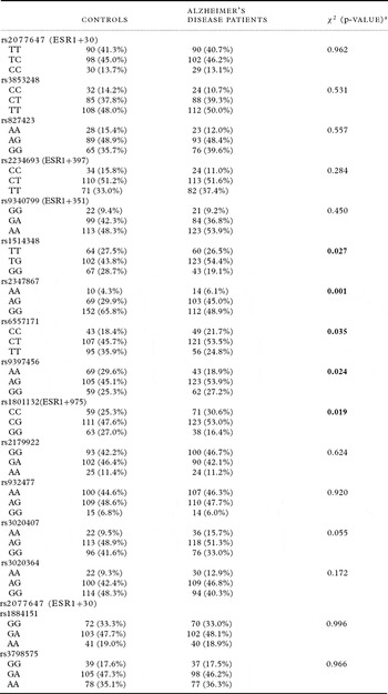
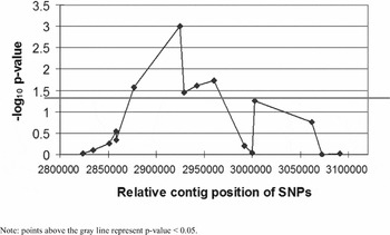
Among the four commonly studied SNPs, we found that rs2077647 (ESR+30), rs2234693 (PvuII) and rs9340799 (XbaI) were not associated with the risk of AD in the Chinese population; rs1801132 was associated with AD (p = 0.019). The genotyping results of the 12 additional tagging SNPs revealed that four SNPs were associated with the risk of AD (p-value ranging from 0.001 to 0.035; Table 1). From the graph showing the relative contig position of ESR1 genotyped SNPs (Figure 4) and the resulting p-value, those SNPs with significant association to the risk of AD were clustered and spanning a distance of 83 kb around intron 3 and intron 7.
The genotype frequencies of ESR1 polymorphisms

Graph showing the relationship of –log10 p-value and the relative contig position of ESR1 SNPs.

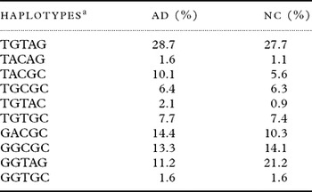
Three haploblocks were identified in our Chinese population. The five SNPs associated with the risk of AD were spanning on haploblock 2. Haplotypes of these five SNPs (rs1514348, rs2347867, rs6557171, rs9397456 and rs1801132) were reconstructed by the program PHASE (Stephens et al., Reference Stephens, Smith and Donnelly2001) (Table 2). Ten haplotypes with frequencies greater than 1% were obtained in our sample set. Comparing the haplotypes between AD patients and the normal control subjects, the frequencies of haplotypes GGTAG was significantly higher in the normal control group (p = 0.003) and TACGC was higher in the AD group (p = 0.07). The overall p-value estimated by PHASE was 0.01, which showed a significant association with the risk of AD. The haplotype GGTAG was composed of alternate alleles of haplotype TACGC. It is possible that haplotype GGTAG might be a protective haplotype while haplotype TACGC conferred an at-risk haplotype for AD. The frequency of ApoE ɛ4 carrier was more prevalent in the AD group and ɛ4+ genotypes were significantly associated with AD (p = 0.001). Previous studies suggested there was interaction effect between ApoE ɛ4 allele and ESR1 polymorphisms in AD (Brandi et al., Reference Brandi1999). Logistic regression analysis was performed to investigate the interaction effect between ApoE ɛ4 allele and ESR1 polymorphisms investigated in this study; such an interaction effect was not detected in this study (p = 0.243). In an additional female-only analysis, the same five SNPs were also significantly associated with the risk of AD.
Haplotypes for haploblock 2 of ESR1 polymorphisms

a Haplotype composed of five ESR1 polymorphisms on haploblock 2: rs1514348, rs2347867, rs6557171, rs9397456, rs1801132.
Note: Haplotypes with a negligible percentage were not shown and the overall p-value generated by PHASE was 0.01.
AD = Alzheimer's disease; NC = normal controls.
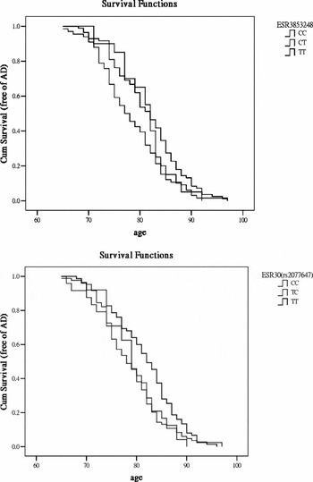
Two neighboring ESR1 SNPs (rs2077647 and rs3853248) were shown to be associated with the AAO in AD, while rs2077647 (ESR1+30) showed a delayed disease onset (3 years) for the T allele carrier (Breslow method p-value = 0.036). Another ESR1 SNP, rs3853248, which is about 70 kb away from rs2077647, also showed significant association to AAO in AD. A heterozygote of rs3853248 allele showed an intermediate but significantly earlier disease onset (2 years) (Breslow p-value = 0.011) (Figure 5).
Analysis the effect of rs3853248 and rs2077647(ESR+30) on age-at-onset(AAO) of AD. Event is defined as onset of AD.

Discussion
The possible role of estrogen and its receptors in etiology of AD was first suggested by epidemiology studies, which showed a higher prevalence of AD in women than men. Although clinical trials of estrogen replacement therapy in older women failed to demonstrate protective effects from development of dementia (Shumaker et al., Reference Shumaker2004), the exact clinical significance of estrogen treatment on maintenance of cognitive function in late life remains controversial. Studies showed that estrogen enhanced neurites outgrowth promoted the formation of dendrite spine and synapses, increased cerebral blood flow, modulated acetylcholine metabolism and decreased serum cholesterol and ApoE levels (Urabe et al., Reference Urabe1996; Slopien et al., Reference Slopien2003). In addition, estrogen increased soluble APP production and reduced Aβ42 and Aβ40 concentration (Yaffe et al., Reference Yaffe, Lui, Grady, Stone and Morin2002). These observations suggested that the ESR gene may be a candidate gene for AD susceptibility.
The ESR1 gene was a positional candidate gene for AD based on the evidence for linkage to 6q in a genome-wide study on AD (Myers et al., Reference Myers2002). Two adjacent polymorphisms in intron 2, rs2234693(PvuII) and rs9340799(XbaI), in ESR1 gene have been studied (Brandi et al., Reference Brandi1999; Lambert et al., Reference Lambert2001). A meta-analysis suggested these two polymorphisms were associated with the risk of AD in Caucasians but not in Asians (Bertram et al, Reference Bertram, McQueen, Mullin, Blacker and Tanzi2007). The association between other polymorphisms in ESR1 gene and the risk of AD has not been comprehensively studied. Any ethnic-specific association remained unclear. Based on the genotyping data from the HapMap project, our analysis on the LD structure among Caucasians and Chinese showed different patterns of haploblock, and the LD pattern also differs across ethnic groups. This may lead to the controversial results in the association study. Most study investigating the association of ESR1 polymorphisms and the risk of AD were focused on rs2234693 and rs9340799. Our findings did not support the association of these two ESR1 polymorphisms and the risk of AD in Chinese community. We found that five SNPs (rs1514348, rs2347867, rs6557171, rs9397456 and rs1801132) on the ESR1 gene were associated with the risk of AD. The SNP rs1801132 (ESR1+975 or P325P) is a synonymous polymorphism in exon 4 and the other four SNPs are located in introns of ESR1 gene. This silent SNP, rs1801132, has been associated with familial breast cancer (Roodi et al., Reference Roodi1995) and total cholesterol and LDL cholesterol levels (Zavratnik et al., Reference Zavratnik, Prezelj, Kocijancic and Marc2007) and is positioned within the hormone-binding domain of the ESR1 protein molecule. For the other four polymorphisms showing association to the risk of AD, they were located on introns of ESR1 gene (intron 3 to intron 7). Haplotype analysis of these five SNPs further suggested the association of the ESR1 polymorphisms with the risk of AD. Intronic polymorphisms may affect the splicing of pre-mRNA and result in alternative splicing. Alternate splicing had been reported downstream of exon 3 (Anandappa et al., Reference Anandappa2000) and SNPs in these introns may play a role in alternative splicing. Exon 4 to exon 8 of the ESR1 gene encode the ligand-binding domain of the ESR protein including determinants for heat-shock protein association in the cytoplasm, ligand-dependent receptor dimerization, activation function AF2 to promote gene transcription by recruiting coactivators, and estrogen and antiestrogen ligand binding (Garcia Pedrero et al., Reference Garcia Pedrero, Zuazua, Martinez-Campa, Lazo and Ramos2003). Splicing variants in this region might affect the property of the estrogen receptor and modulate the risk to AD.
The age of onset and clinical manifestation of disease may be modulated by genetic factors. It is possible that various disease susceptibility genotypes lead to differential rates of progression. Our study showed two SNPs (rs2077647 and rs3853248) in the ESR1 gene were associated with age of onset of clinical disease. Concurrent effects of gene polymorphisms on the risk of disease and AAO might explain the inconsistent results on association studies. It is possible that polymorphisms on the ESR1 gene affect both the risk and AAO of AD, and the interaction of these genotypes might further increase the risk of AD. To the best of our knowledge, this is the first time a high density of ESR1 polymorphisms have been studied for predisposition and AAO in AD.
It is well accepted that there are interactions between genetic and environmental factors in the risk of AD. In this study, only genetic factors were taken into consideration for associating the risk of AD. This is the major limitation of this study. We limited the subject recruitment criteria to reduce the possibility of variation in environmental factors. In addition, all subjects were recruited from our south Chinese community to avoid any bias between ethnic groups.
In summary, our study identified five SNPs in the ESR1 gene associated with the risk of AD and a further two SNPs associated with the age of onset. This lends support for a putative role of ESR1 in the pathogenesis of AD. Our result calls for more extensive functional studies to unravel the mechanism of ESR1 in modulating cognitive deterioration in late life.
Conflict of interest
None.
Descriptions of authors' roles
S. L. Ma, N. L. Tang and Y. P. Zhang were involved in genotyping and data analysis. L. C. Lam, C. W. Tam, V. W. Lui and F. K. Chiu were involved in design and data collection. S. L. Ma prepared the manuscript.
Acknowledgments
This work is supported by RGC Research Grant Direct Allocation from the Chinese University of Hong Kong (CRE-2003.1.072), CUHK Direct Grant and from the Mr. Lai Seung Hung and Mrs. Lai Chan Pui Ngong Dementia in Hong Kong Research Fund.