Introduction
There is a strong interrelationship between delirium and dementia, both pathophysiologically and clinically (Young and Inouye, Reference Young and Inouye2007). Delirium superimposed on dementia is common but frequently unrecognized, and these patients reportedly exhibit an accelerated decline in cognitive and functional abilities, a greater need for institutionalization, a greater re-hospitalization risk, and increased mortality (Fick et al., Reference Fick, Agostini and Inouye2002).
According to a recent review article, the prevalence of delirium in elderly individuals aged ≥65 years is 1–2%, and it increases up to 22% in elderly individuals with dementia (de Lange et al., Reference de Lange, Verhaak and van der Meer2012). The prevalence of delirium superimposed on dementia ranges from 22 to 89% in community and hospital populations (Fick et al., Reference Fick, Agostini and Inouye2002).
Although dementia is generally considered a major risk factor for delirium, the relationship between delirium and dementia remains unclear. We hypothesized that the frequency of delirium varies with each dementia type, and is associated with cerebrovascular disease (CVD) with neurodegenerative dementias. This study aimed to investigate the frequency of delirium superimposed on dementia in patients with each dementia type and examines the association between delirium and CVD in patients with neurodegenerative dementias.
Methods
All procedures followed the Clinical Study Guidelines of the Ethics Committee of Kumamoto Mental Health Hospital and were approved by the internal review board. A complete description of all procedures was provided to the patients, and written informed consents were obtained from all patients or their caregivers prior to participation.
This study was a prospective, dementia referral center-based, cohort study. The participants were 206 consecutive patients with dementia. These patients were defined at the time of initial assessment and selected on the basis of inclusion/exclusion criteria from a consecutive series of 261 patients who had undergone medical examination at the memory clinic of Kumamoto Mental Health Hospital from April 2010 to September 2011. All patients were examined by senior neuropsychiatrists (M. Ikeda and Y. Yatabe) using routine laboratory tests, computed tomography, and standard neuropsychological examinations, the results of which included Mini-Mental State Examination (MMSE; Folstein et al., Reference Folstein, Folstein and McHugh1975) and Physical Self-Maintenance Scale (PSMS; Lawton and Brody, Reference Lawton and Brody1969; Hokoishi et al., Reference Hokoishi2001) scores. Behavioral and psychological symptoms of dementia (BPSD) were assessed by the Neuropsychiatric Inventory (NPI; Cummings et al., Reference Cummings, Mega, Gray, Rosenberg-Thompson, Carusi and Gornbein1994; Hirono et al., Reference Hirono1997). In addition, we investigated antipsychotic and benzodiazepine use at the time of initial assessment. We also assessed delirium severity using the Delirium Rating Scale – Revised 98 (DRS-R98; Trzepacz et al., Reference Trzepacz, Mittal, Torres, Kanary, Norton and Jimerson2001; Kato et al., Reference Kato, Kishi, Okuyama, Trzepacz and Hosaka2010). The DRS-R98 severity scale score ranges from 0 to 39, with higher scores indicating more severe delirium and a cut-off score of ≥15 being consistent with a diagnosis of delirium (Meagher et al., Reference Meagher, Leonard, Donnelly, Conroy, Saunders and Trzepacz2010).
The diagnosis of delirium was based on the criteria for delirium outlined in the Diagnostic and Statistical Manual of Mental Disorders, fourth edition, text revision (DSM-IV-TR; American Psychiatric Association, 2000). All patients with delirium fulfilled the following core criteria: (a) disturbance of consciousness, (b) changes in cognition, (c) development of disturbances over a short period of time, with a fluctuating course during the day, and (d) evidence from the history, physical examination, or laboratory findings that the disturbance is caused by the direct physiological consequence of general medical condition or drug effect. The criteria for delirium state that the condition should not be better explained by a pre-existing dementia, whereas the criteria for dementia state that prior delirium should be excluded.
Therefore, dementia was diagnosed according to the Diagnostic and Statistical Manual of Mental Disorders, third edition-revised (DSM-III-R; American Psychiatric Association, 1987) after improvement of consciousness by clinical treatments. If dementia existed, each dementia type was diagnosed according to the international consensus criteria. Patients were divided into those with probable Alzheimer's disease (AD), defined according to the National Institute for Neurological and Communicative Disorders and Stroke and Alzheimer's disease and Related Disorders Association (NINCDS–ADRDA; McKhann et al., Reference McKhann, Drachman, Folstein, Katzman, Price and Stadlan1984); probable vascular dementia (VaD), defined according to the NINCDS and Association Internationale pour la Recherche et l’Enseignement en Neurosciences (NINDS–AIREN) criteria (Román et al., Reference Román1993); probable dementia with Lewy bodies (DLB), defined according to the consensus criteria for the clinical diagnosis of DLB, 2005 (McKeith et al., Reference McKeith2005); frontotemporal lobar degeneration (FTLD), defined according to the international collaborative workshop on FTLD, 1998 (Neary et al., Reference Neary1998); or other types. In patients where it was difficult to determine whether symptoms were due to delirium or DLB were diagnosed as DLB without delirium. Therefore, the prevalence of delirium in patients with DLB could have been underestimated in this study. The presence of CVD, including multiple lacunar infarcts and leukoencephalopathy, was determined by a senior neuropsychiatrist (M. Hashimoto), who was blinded to the clinical data. As suggested by a number of previous studies (Bennett et al., Reference Bennett, Wilson, Gilley and Fox1990; Román et al., Reference Román1993; Yoshitake et al., Reference Yoshitake1995; Leys et al., Reference Leys1999), we used a semi-standardized measure and classified CVD on the basis of computed tomography (CT) findings as follows (Ikeda et al., Reference Ikeda2001): multiple lacunar infarcts, leukoencephalopathy with or without lacunar infarcts, multiple infarcts, strategic single infarct, large cortical infarcts, cerebral hemorrhage, and subarachnoid hemorrhage.
Exclusion criteria for this study included the following: (a) absence of dementia according to DSM-III-R, (b) those with developmental abnormalities, serious psychiatric diseases, such as schizophrenia or major depression, or substance abuse before the onset of dementia, (c) absence of reliable informants, and (d) inability to obtain informed consent from patient and caregiver.
Statistical differences in age, education, and MMSE, PSMS, NPI total, and NPI subscale scores between patients with dementia and delirium and those with dementia without delirium were assessed by Student's t-tests. The χ 2 for independence test was conducted to compare sex; presence of CVD; use of donepezil, antipsychotics, or benzodiazepines; presence of CVD with neurodegenerative dementias; and the prevalence of delirium in patients with each dementia type. A significance level of 0.05 (two-tailed) was set for all analyses, which were conducted with SPSS for Windows, version 17.0 (IBM Corporation, Armonk, NY, USA). In addition, two-way analysis of variance (ANOVA) was used to analyze the effects of each diagnosis with or without delirium and their interaction effects on scores for individual items in the NPI. The significance level was set at 0.01 or less. Scheffe's post hoc test was performed to detect any significant differences between diagnoses.
Results
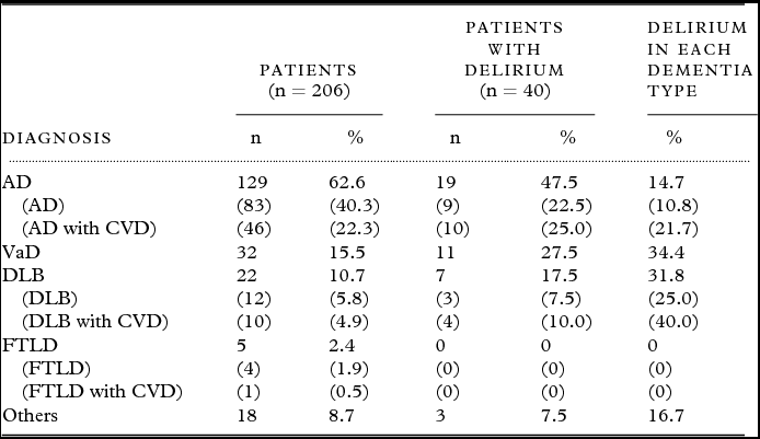
Among the 206 patients, there were 142 women and 64 men with a mean age of 81.4 years (standard deviation (SD), 6.0), a mean educational attainment period of 8.5 years (SD, 2.1), and a mean MMSE score of 15.9 points (SD, 6.0). AD was observed in 62.6% (129/206) patients, VaD in 15.5% (32/206) patients, DLB in 10.7% (22/206) patients, and FTLD in 2.4% (5/206) patients.
Forty patients (19.4%) suffered from delirium. The frequency of delirium varied with dementia type, as shown in Table 1. The proportion of patients who experienced episodes of delirium was 14.7% in the AD group, 34.4% in the VaD group, and 31.8% in the DLB group, whereas no patient with FTLD experienced these episodes, indicating that the prevalence of delirium was significantly diverse for each dementia type (p = 0.022). Furthermore, delirium was more frequent in patients with CVD than in those without (p = 0.006); 21.7% of AD with CVD and 40.0% of DLB with CVD developed delirium, while 10.8% of AD without CVD and 25.0% of DLB without CVD developed delirium.
Prevalence of delirium in patients with each dementia type

Notes: AD: Alzheimer's disease, CVD: cerebrovascular disease, VaD: vascular dementia, DLB: dementia with Lewy bodies; FTLD: frontotemporal lobar degeneration.
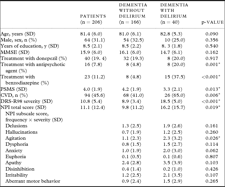
Demographic characteristics of the patients with and without delirium are shown in Table 2. There were no significant differences between the two groups with regard to age, sex, education, MMSE score, and use of donepezil, while patients with delirium more frequently used antipsychotics and benzodiazepines compared with those without delirium. PSMS scores were significantly lower for patients with delirium than for those without delirium (p = 0.013), while DRS-R98 severity was significantly higher for patients with delirium than for those without delirium (p < 0.001). With regard to NPI scores, NPI total scores (p = 0.019) and NPI agitation subscale scores (p = 0.026) were significantly higher for patients with delirium than for those without delirium.
Demographic characteristics of patients with dementia with or without delirium
Notes: *p < 0.05.
CVD: cerebrovascular disease, MMSE: Mini-Mental State Examination,
PSMS: Physical Self-Maintenance Scale, DRS-R98: Delirium Rating Scale-Revised 98, NPI: Neuropsychiatric inventory.
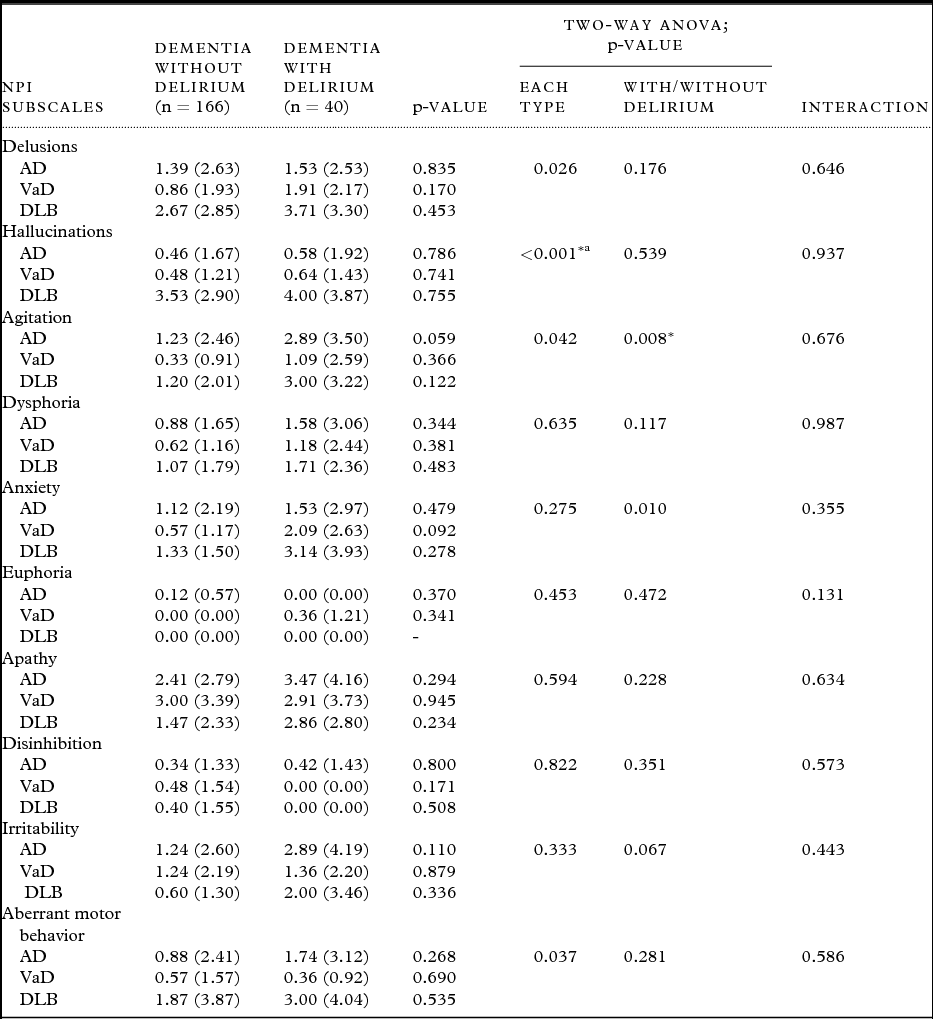
Table 3 shows scores for individual NPI subscales among patients with each dementia type with or without delirium. Two-way ANOVA revealed statistically significant differences in scores for hallucinations among patients with each dementia type (p < 0.001) and in scores for agitation between patients with and without delirium (p = 0.008). Scheffe's post hoc comparisons revealed that DLB patients with delirium exhibited significantly higher scores for hallucinations compared with AD or VaD patients with delirium (p < 0.01). Overall, no significant differences were observed in individual NPI scores between patients with each dementia type with or without delirium (p > 0.01).
Mean composite scores (frequency × severity) of individual Neuropsychiatric Inventory (NPI) items among patients with each dementia type with or without delirium
Notes: Values are presented as mean (SD).
AD: Alzheimer's disease; AD without delirium (n = 110), AD with delirium (n = 19).
VaD: Vascular dementia; VaD without delirium (n = 21), VaD with delirium (n = 11).
DLB: Dementia with Lewy bodies; DLB without delirium (n = 15), DLB with delirium (n = 7).
*p < 0.01; aDLB versus AD or VaD as per Scheffe's post hoc test.
Discussion
To the best of our knowledge, this is the first study to investigate the disease-specific frequency of delirium superimposed on dementia on an outpatient basis. Delirium was present in approximately 20% outpatients with dementia, although the prevalence of delirium was diverse for each dementia type: 14.7% in patients with AD, 34.4% in patients with VaD, 31.8% in patients with DLB, and 0% in patients with FTLD. The prevalence of delirium among AD and DLB patients with CVD was 1.6 times higher than that in AD and DLB patients without CVD. The findings suggest that delirium decreases activities of daily living (ADL) and exaggerates BPSD in patients with dementia.
In a study that examined patients (n = 175) admitted to a neuropsychiatric unit (Robertsson et al., Reference Robertsson, Blennow, Gottfries and Wallin1998), the overall prevalence of recurrent delirium was 37%: 57% in patients with late-onset AD, 14% in patients with early-onset AD, 19% in patients with frontotemporal dementia, and 40% in patients with VaD. Among patients with acute medical illness, delirium was observed in 26% patients with primary degenerative dementia and 52% patients with dementia associated with vascular changes, i.e., VaD (Erkinjuntti et al., Reference Erkinjuntti, Wikström, Palo and Autio1986). However, there have been no studies that have investigated the prevalence of delirium in patients with DLB. Among the patients with neurodegenerative dementias in this study, the prevalence of delirium was highest in the DLB group. It can be particularly difficult to distinguish delirium from DLB because some features such as hallucinations and symptom fluctuation are common to both (Young and Inouye, Reference Young and Inouye2007). In the current study, dementia was diagnosed immediately after it was determined that the symptoms of delirium had subsided. Moreover, DRS-R98 severity scores were significantly higher for DLB patients with delirium (mean, 19.6; SD, 5.2; range, 15–28) than for DLB patients without delirium (mean, 11.7; SD, 2.6; range, 5–14; p = 0.006). No DLB patient with delirium exhibited a score of <15, whereas no DLB patient without delirium exhibited a score of ≥15. DLB patients with delirium exhibited significantly higher scores for the hallucination subscale of the NPI compared with AD and VaD patients with delirium. However, overall differences in individual NPI scores among patients with each dementia type with or without delirium were not significant. Dementia researchers need to assess any delirium component more carefully and utilize instruments that capture characteristics that differentiate delirium and dementia disorders (Meagher and Trzepacz, Reference Meagher and Trzepacz2007).
In the 263 post-stroke patients of the Helsinki Stroke Aging Memory cohort study, patients with delirium were more likely to have pre-stroke cognitive decline compared with patients without delirium (28.0% vs. 4.2%), and the former group developed post-stroke dementia more frequently than the latter group (50.0% vs. 16.9%; Melkas et al., Reference Melkas2012). In addition, the findings of this study suggest that CVD in patients with neurodegenerative dementias, such as AD and DLB, are at an increased risk of delirium. However, the association of CVD with neurodegenerative dementia and delirium remains unclear. Delirium may be a disorder of corticostriatal loops, and a central cholinergic deficiency may be a major factor in its development (Robertsson et al., Reference Robertsson, Blennow, Gottfries and Wallin1998). In patients with CVD accompanied by neurodegenerative dementias, the cortex is probably partly disconnected from deeper structures, and this causes cortical and subcortical dysfunction and a decrease in cholinergic activity. Cerebrovascular disease may be one of the major risk factors causing vulnerability to delirium.
In this study, NPI scores, particularly agitation subscale scores, were higher in patients with delirium than in those without (“pure” dementia). In a previous study, the scores for a wide range of DRS-R98 non-cognitive items, such as sleep–wake cycles, perceptual abnormalities, affective liability, thought process abnormalities, motor agitation, and motor retardation, were more severe in patient groups with dementia and comorbid delirium than in patient groups with “pure” dementia (Meagher et al., Reference Meagher, Leonard, Donnelly, Conroy, Saunders and Trzepacz2010). Taking into account these findings, delirium has an additive impact on the phenomenological profile of patients with dementia. Delirium has been shown to accelerate the trajectory of cognitive decline during the follow-up course of memory-clinic patients with AD (Fong et al., Reference Fong2009). In addition, delirium is highly prevalent among hospitalized patients with AD and is associated with an increased rate of cognitive deterioration in these patients (Weiner, Reference Weiner2012). Moreover, delirium itself is a cause of long-term cognitive impairment. However, the key question of whether delirium is a risk factor for new-onset dementia remains unanswered (MacLullich et al., Reference MacLullich, Beaglehole, Hall and Meagher2009). In a true population-based study, delirium has been shown to be a strong risk factor for the incidence of dementia and cognitive decline, but the relationship did not appear to be mediated by the classic neuropathologies associated with dementia (Davis et al., Reference Davis2012). In a longitudinal future study, we should carefully investigate the relationship between delirium and onset of dementia.
A previous study demonstrated an increased risk of admission to long-term care facilities among patients with delirium superimposed on dementia than among patients with “pure” dementia or only delirium (Fick et al., Reference Fick, Agostini and Inouye2002). This study suggests that delirium superimposed on dementia exaggerates BPSD and decreases patients’ ADL. Therefore, delirium superimposed on dementia can become a major burden to long-term care services.
This study had some limitations. First, we did not have pathological confirmations of dementia subtype in our patients. Second, the results may have been biased because all patients were recruited from the dementia outpatient clinic of a mental hospital, and the prevalence of delirium varies depending on the care settings. Hospital admission is one of the major risk factors for delirium, and most previous studies have examined hospitalized patients. A well-defined dementia cohort in an outpatient clinic is the strength of our study. However, taking account of the number of people with delirium due to medical problems severe enough to preclude coming to the memory clinic, the prevalence of delirium with dementia in this study may estimate low. Third, in order to identify CVD, we used a visual assessment method using CT, which may not be as accurate as assessments conducted using magnetic resonance imaging (MRI). Fourth, our sample size with regard to patients with each dementia type was relatively small for analysis. We had to exclude FTLD and other types of dementia for statistical analysis. However, an advantage of our study was the relatively large study cohort. A future investigation similar to this one should be conducted with a larger number of patients.
In summary, the findings of this study suggest that the frequency of delirium varies among patients with different types of dementia. In addition, these findings suggest that delirium decreases ADL, exaggerates BPSD, and is associated with CVD in patients with neurodegenerative dementias.
Conflict of interest
None.
Description of authors’ roles
N. Hasegawa collected and analyzed the data and prepared the first draft of the paper. S. Yuuki, K. Honda, Y. Yatabe, and K. Araki assisted with the data collection and defining the diagnoses. M. Hashimoto evaluated the signs of CVD on CT scans. M. Ikeda conducted the literature review and assisted with the design of the study, data collection, definition of diagnoses, and writing of the paper.
Acknowledgments
The present study was undertaken with the support of grants that were provided by the Ministry of Health, Labor and Welfare (Research on dementia; H24-Dementia-General-001) to M. Ikeda. The authors gratefully acknowledge the assistance of the staff of Kumamoto Mental Health Hospital.