Introduction
The uncinate process plays an important role in internal nasal airflow and maxillary sinus ventilation.Reference Sirikci, Bayazit, Gümüsburun, Bayram and Kanlikama1, Reference Milczuk, Dalley, Wessbacher and Richardson2 It is one of the most important landmarks in functional endoscopic sinus surgery.Reference Erdem, Aktas, Erdem, Miman and Ozturan3 Developmental abnormalities such as agenesis or hypoplasia may lead to surgical difficulties and cause inflammatory changes such as chronic sinusitis.Reference Sirikci, Bayazit, Gümüsburun, Bayram and Kanlikama1, Reference Tasar, Cankal, Bozlar, Hidir, Saglam and Ors4 These variations can be detected by paranasal sinus computed tomography (CT); their identification is important for managing and treating sinusitis.Reference Bolger, Woodruff, Morehead and Parsons5 This study aimed to investigate the role of maxillary sinus hypoplasia and isolated agenesis of the uncinate process in sinusitis aetiology.
Materials and methods
In the present study, 685 paranasal sinus CT scans acquired between January 2012 and December 2013 at a tertiary referral centre were retrospectively reviewed. Three patients with isolated agenesis of the uncinate process and a total of 43 maxillary sinus hypoplasia variations in 27 patients (16 bilateral) were identified. Clinical features were evaluated by accessing patients' medical records and obtaining their medical histories by telephone. The frequency of sinusitis episodes over the previous five years and radiological findings associated with inflammation, such as mucosal thickening and maxillary opacification, were documented. The frequency of antibiotic treatment for sinonasal symptoms was compared between the isolated agenesis of the uncinate process group and each maxillary sinus hypoplasia subgroup. Statistical analysis was performed using a two-tailed t-test with SPSS version 20.0 for Windows software (SPSS, Chicago, Illinois, USA). Statistical significance was set at a p value of less than 0.05. Radiological features of maxillary sinus hypoplasia were categorised according to the maxillary sinus hypoplasia classification proposed by Bolger et al. in 1990.Reference Bolger, Woodruff, Morehead and Parsons5 Patients with a history of surgical or non-surgical trauma were excluded.
Results
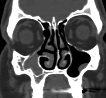
The 3 patients with unilateral isolated agenesis of the uncinate process were men aged 31, 44 and 53 years. Their major complaints were headache (3/3; 100 per cent), facial pain (3/3; 100 per cent) and purulent rhinorrhoea (2/3; 66 per cent). Over the previous five years, patients with isolated agenesis of the uncinate process underwent an average of seven antibiotic treatments per year for headache and facial pain symptoms. All three patients showed maxillary sinus opacification (100 per cent) on the affected side and a normal maxillary sinus on the contralateral side (Figure 1).
Coronal computed tomography image showing isolated agenesis of the uncinate process and maxillary sinus opacification on the right side.

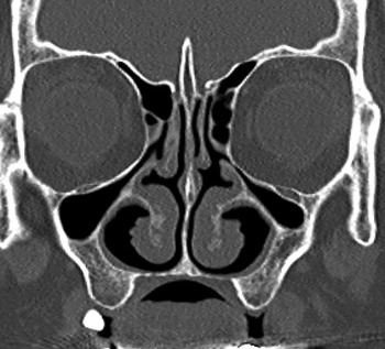
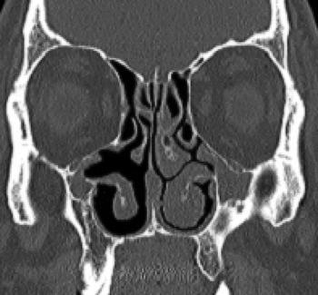
Twenty-seven patients with maxillary sinus hypoplasia were identified from 685 paranasal sinus CT scans (prevalence of 3.94 per cent). The mean age patient was 45.37 ± 2.3 years (range 19–75 years), and the male-to-female ratio was 2.4:1. According to Bolger and colleagues' maxillary sinus hypoplasia classification, 23 patients had type I variations (54 per cent), 13 had type II (30 per cent) and 7 had type III (16 per cent) (Figures 2 and 3). Bilateral maxillary sinus hypoplasia was identified in 16 patients, bilateral type I in 9, bilateral type II in 4 and bilateral type III in 2; 1 patient had type II on one side and type III on the other. Unilateral maxillary sinus hypoplasia was identified in 11 patients: of these, types I, II and III maxillary sinus hypoplasia variations were seen in 5, 5 and 1 patient, respectively. Patients with types I, II and III maxillary sinus hypoplasia had received 1.57, 3.22, 5.75 antibiotic treatments per year, respectively, for sinonasal complaints over the previous 5 years.
Coronal computed tomography image showing bilateral type I maxillary sinus hypoplasia.

Coronal computed tomography image showing type III maxillary sinus hypoplasia on the right side (cleft-like maxillary sinus) and type II maxillary sinus hypoplasia on the left side.

Antibiotic treatment frequencies were compared between patients with isolated agenesis of the uncinate process and those with each maxillary sinus hypoplasia subtype. The p values for differences between patient subgroups were 0.025 for isolated agenesis of the uncinate process vs type I maxillary sinus hypoplasia, 0.047 for isolated agenesis of the uncinate process vs type II maxillary sinus hypoplasia and 0.34 for isolated agenesis of the uncinate process vs type III maxillary sinus hypoplasia.
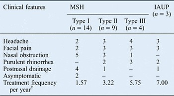
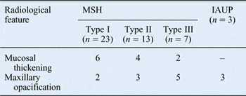
The clinical features of all patients with isolated agenesis of the uncinate process or maxillary sinus hypoplasia are summarised in Table I, and the radiological features are shown in Table II.
Clinical features of IAUP and MSH cases*

* Total n = 30. †Over the previous five-year period. IAUP = isolated agenesis of the uncinate process; MSH = maxillary sinus hypoplasia
Radiological features of IAUP and MSH cases*

* Total n = 46 sinuses. IAUP = isolated agenesis of the uncinate process; MSH = maxillary sinus hypoplasia
Discussion
Agenesis or hypoplasia of the uncinate process is a rare anatomical variation, and most reports in the literature have been associated with maxillary sinus hypoplasia. The overall prevalence of maxillary sinus hypoplasia is reported to be 4–10.4 per cent;Reference Erdem, Aktas, Erdem, Miman and Ozturan3, Reference Bassiouny, Newlands, Ali and Zaki6 the prevalence in this study was 3.94 per cent. Today, the maxillary sinus hypoplasia classification proposed by Bolger et al. in 1990 is most widely accepted. Type I maxillary sinus hypoplasia is characterised by mild hypoplasia of the maxillary sinus, with a well-developed infundibular passage and normal uncinate process. Type II maxillary sinus hypoplasia is characterised by significant hypoplasia of the maxillary sinus, with an absent or a pathological infundibular passage and a hypoplastic or absent uncinate process. Type III maxillary sinus hypoplasia is characterised by absence of the uncinate process and cleft-like maxillary sinus hypoplasia.Reference Bolger, Woodruff, Morehead and Parsons5
Isolated agenesis of the uncinate process is an extremely rare anatomical variation. To our knowledge, the three patients described in this study represent the largest reported case series diagnosed with an isolated agenesis of the uncinate process. In 1990, Aksungur et al. reported the case of a patient with unilateral aplasia of the uncinate process without maxillary sinus aplasia or hypoplasia.Reference Aksungur, Biçakçi, Inal, Akgül, Binokay and Aydogan7
It is important to identify isolated agenesis of the uncinate process or maxillary sinus hypoplasia before sinus surgery because there is an increased risk of orbital penetration when either condition is present. Thus, pre-operative detection of these anatomical variations may help avoid intra-operative damage to the medial orbital wall.Reference Erdem, Aktas, Erdem, Miman and Ozturan3, Reference Tasar, Cankal, Bozlar, Hidir, Saglam and Ors4, Reference Okur, Okur, Aksungur, Kiroğlu, Inal and Soyupak8 Paranasal sinus CT is an excellent imaging technique for diagnosing these anatomical variations and determining the extent of surgery.Reference Bolger, Woodruff, Morehead and Parsons5, Reference Earwaker9
The uncinate process forms part of the lateral nasal wall and ostiomeatal complex. In 2000, Sirikci et al. emphasised the important role played by the uncinate process in internal nasal airflow and maxillary sinus ventilation by directing expired air towards the sinus cavity.Reference Sirikci, Bayazit, Gümüsburun, Bayram and Kanlikama1 In 1993, Milczuk et al. noted that the distribution of nasal and paranasal air flow is related to uncinate process morphology and that absence of the uncinate process may impede mucociliary clearance, causing sinusitis.Reference Milczuk, Dalley, Wessbacher and Richardson2 In 2009, Jiang and XuReference Jiang and Xu10 and Xiong et al.Reference Jiang and Xu10 reported that the normal uncinate process permits the physiological commutation of air within the maxillary sinus, and that airflow patterns inside the maxillary sinus are inactivated by uncinate process excision in cadavers.
In the present study, symptoms related to sinus inflammation, such as facial pain and purulent rhinorrhoea, were mainly observed in type III maxillary sinus hypoplasia and isolated agenesis of the uncinate process cases. Maxillary sinus opacification was observed radiologically in all sinuses with isolated agenesis of the uncinate process and was much more frequent in maxillary sinus hypoplasia type III sinuses than in type I or II sinuses.
There was a significant difference in antibiotic treatment frequency between patients with isolated agenesis of the uncinate process and those with each maxillary sinus hypoplasia subtype, except for type III. Although there were no significant differences in treatment frequency and radiological abnormalities between type III maxillary sinus hypoplasia and isolated agenesis of the uncinate process patients, the values were higher in those with isolated agenesis of the uncinate process. Isolated agenesis of the uncinate process may cause ipsilateral maxillary sinusitis by disrupting airflow exchange between the nasal cavity and maxillary sinus, thus obstructing drainage pathways or impeding mucociliary clearance. In our case series, there was no evidence of inflammation of the contralateral maxillary sinus in any patient, suggesting that the uncinate process alone has a role in sinusitis aetiology. These findings indicate that isolated agenesis of the uncinate process plays a more important role than maxillary sinus hypoplasia in sinusitis aetiology.
• Isolated agenesis of the uncinate process is an extremely rare anatomical variation
• Isolated agenesis of the uncinate process or maxillary sinus hypoplasia can increase the risk of orbital penetration during sinus surgery
• Isolated agenesis of the uncinate process plays an important role in the pathophysiology of chronic sinusitis
Isolated agenesis of the uncinate process is a very rare condition, and the statistical analysis was limited by the small number of cases. Inclusion of more patients with isolated agenesis of the uncinate process would improve the statistical significance of our findings. In addition, information on the treatment frequency was obtained from both medical records and patient histories. Thus, patients' subjectivity in medical history recall could have reduced the data quality.
Conclusion
The uncinate process plays an important role in internal nasal airflow and maxillary sinus ventilation. Isolated agenesis of the uncinate process is an extremely rare anatomical variation and only a few cases have been reported to date. This study showed that agenesis may play a stronger role than maxillary sinus hypoplasia in the pathophysiology of chronic sinusitis.