The use of imaging modalities that require ionising radiation is rising, particularly in the setting of paediatric cardiology.Reference Brenner and Hall1–Reference Patel and Hijazi5 There are understandable concerns given that infants are 3 to 6 times more susceptible to the carcinogenic effects of ionising radiation in comparison to adults receiving an equivalent dose.6 This is due to the rapidly dividing cells in paediatric patients and the greater relative life expectancy over which radiation-induced malignancies may occur.6–10 In addition, children with congenital cardiac disease frequently undergo repeat imaging, with each examination adding to the cumulative lifetime risk of radiation-related sequelae. It is therefore essential that clinicians have sufficient knowledge of the radiation burden and corresponding risks associated with different imaging techniques in children. In this way, they can adopt “as low as reasonably achievable” radiation dose principles.Reference Strauss and Kaste11
Multi-detector computed tomography has emerged as a useful complementary diagnostic tool in congenital cardiac disease. It may be used to evaluate extra-cardiac structures such as the pulmonary arteries, veins, and the aortic arch, where echocardiography has failed to provide sufficient detail.Reference Leschka, Oechslin and Husmann12 Moreover, it offers good pre- and post-operative evaluation of cardiac structures, particularly in patients who experience a sudden deterioration.Reference Bean, Pannu and Fishman13 Despite the increasing use of computed tomography, we lack paediatric radiation dose data from the most contemporary dual source computed tomography scanners that incorporate multiple detector rows, electrocardiogram gating, and three-dimensional automatic tube current modulation. Also, despite a widespread change away from angiography to computed tomography, few dose comparisons between conventional fluoroscopic angiography and computed tomography exist. Notwithstanding that we appreciate the heterogeneous indications for cardiac computed tomography and diagnostic angiography, we believe it is important that clinicians are aware of the approximate radiation doses associated with each imaging modality when making decisions about the best diagnostic technique. There is evidence suggesting that clinicians, and in particular radiologists, are not always fully aware of the radiation burden associated with various imaging techniques.Reference Lee, Haims, Monico, Brink and Forman14 This study was performed in order to assess and compare the radiation dose data from contemporary cardiac dual-source computed tomography and diagnostic angiography performed in the catheter laboratory using the latest “state-of-the-art” equipment.
Materials and methods
Study setting and population
We conducted a retrospective survey of consecutive cardiac computed tomography examinations and conventional diagnostic angiograms carried out between January, 2008 and December, 2009, both inclusive, including all children with all congenital cardiac diseases aged less than 16 years. Procedures were carried out within a congenital cardiology tertiary referral centre at a large teaching hospital in the United Kingdom, serving a catchment population of approximately 5.25 million. Local ethics committee approval was sought but deemed unnecessary.
Cardiac computed tomography scans
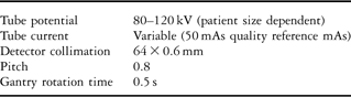
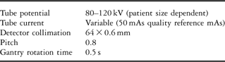
Computed tomography scans were performed on a 64-slice Siemens Somatom® Definition dual-source computed tomography scanner (Siemens Medical Solutions, Forchheim, Germany). Standard inbuilt presets for paediatric cardiac computed tomography examinations were used for all scans (Table 1). Automatic tube current modulation was employed: a dose-modifying system that optimises the computed tomography dose according to the shape and size of the patient to yield consistent image quality for all patients.Reference Goo15 All patients were sedated with chloral hydrate and received an intravenous bolus of Optiray®300 (Codali SA, Laboratoire Guerbert, Aulnay Sous Bois, France) totalling 2 millilitres per kilogram of body weight. A power injector was used to deliver the contrast at a rate of 2 millilitres per minute. Beta-blockers were not necessary in any of the patients; five cases required electrocardiogram-gating, using inbuilt retrospective electrocardiogram-gating protocols. Notwithstanding the use of two X-ray tubes for electrocardiogram-gated computed tomography scans, only one was used for non-gated scans.
Standard inbuilt paediatric thoracic computed tomography presets (Siemens Somatom® definition computed tomography scanner).
Diagnostic angiography
Angiography was performed using a biplane Siemens Axiom Artis dBC® system (Siemens Medical Solutions, Forchheim, Germany) with flat-panel detectors. The inbuilt paediatric protocol for this system uses 15 pulses per second for pulsed fluoroscopy and 30 frames per second for image acquisition. All patients were under general anaesthesia for the duration of the procedure. All interventional, electrophysiological, pacing, and hybrid procedures were excluded.
Data collection
Computed tomography and catheter laboratory logbooks were reviewed to generate a list of cases that met the inclusion criteria. Departmental databases were used to gather patient characteristics, radiation dose data, details of the patients’ diagnoses, and their congenital cardiac disease imaging history. Imaging history constituted the total number of lifetime cardiac computed tomography and catheterisation procedures. All data were entered onto a bespoke password-protected and encrypted Microsoft® Excel 2007 spreadsheet (Microsoft® Corporation, Redmond, Washington, United States of America) for reference purposes and statistical analysis.
Radiation dose measurements and conversion factors
With regard to the computed tomography dose, dose length product – the standard unit used is milliGray centimetre – is an indicator of the integrated radiation dose of an entire computed tomography examination.Reference Morin, Gerber and McCollough16 It incorporates the scan length and rises with increasing tube current and voltage, and the use of a lower pitch. Dose length product is the standard dose measurement reported on computed tomography scanner consoles and constituted our raw computed tomography dose data.
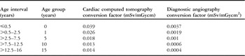
Similarly, the dose area product – the standard unit used is Gray-square-centimetre – reported in angiographic procedures indicates the total X-ray energy delivered to the patient as a result of fluoroscopy and cine-film sequences. It represents the radiation dose in the air at a given distance from the X-ray tube multiplied by the area of the beam at that distance.Reference Ait-Ali, Andreassi, Foffa, Spadoni, Vano and Picano17 This formed our raw angiography dose data. In order to estimate the risk of radiation-induced sequelae, the dose area product and dose length product were converted to another dose measurement, effective dose – the standard unit used is millisievert. This measurement accounts for the relative radiation risk of individual organs and also allows cross-modality comparisons of radiation dose. Published age-specific conversion factors that allow an estimation of effective dose were used (Table 2). We ensured that our angiographic imaging system closely matched that from which the conversion factors were derived.Reference Karambatsakidou, Sahlgren, Hansson, Lidegran and Fransson18 For computed tomography dose conversion, we used the generic factors published by Shrimpton.Reference Shrimpton19
Cardiac computed tomographyReference Shrimpton19 and diagnostic angiographyReference Karambatsakidou, Sahlgren, Hansson, Lidegran and Fransson18 age-specific dose conversion factors.

Statistical analysis
All statistical analysis was carried out using SPSS software version 18.0 (SPSS Inc®, Chicago, Illinois, United States of America). The Mann–Whitney U-test was used for non-parametric continuous variables, including effective dose. Categorical variables were measured using two-tailed Fisher's exact test and Chi-squared test. Spearman's rank correlation coefficients were used to determine the degree of dependence between two variables. For all analyses, a p-value of less than 0.05 was considered statistically significant.
Results
Frequency of imaging
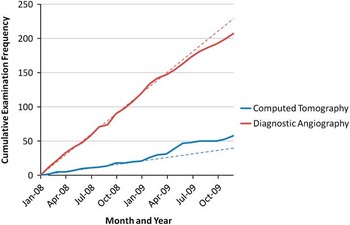
During the 2-year survey period, 62 cardiac computed tomography examinations were performed in 54 patients, whereas 210 diagnostic angiograms were carried out in 200 patients. There were 14 patients who had both a cardiac computed tomography and an angiogram during the survey period. There was a 95.2% increase in the number of computed tomography scans from 21 in 2008 to 41 in 2009 compared with a 26.4% decrease in the number of diagnostic catheters performed from 2008 – 121 procedures – to 2009 – 89 procedures. The median – range – number of computed tomography scans carried out per month in 2008 was 2.0 (0–4) compared with 3.5 (0–8) in 2009. The median number of monthly diagnostic angiograms that were performed during 2008 was 10.0 (3–16) compared with 7.5 (3–14) in 2009. Figure 1 shows the monthly cumulative examination frequencies for both of the imaging modalities.
Monthly cumulative examination frequency. Dashed lines indicate line of best fit based on 2008 trends.

Patient characteristics, cardiac lesions, and imaging history
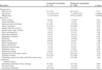
Patients undergoing cardiac computed tomography were on average younger and weighed less than patients having an angiogram (both p < 0.0001; Table 3). Sex differences were not significantly different between the two groups. There was a large spread of anatomical diagnoses among the two imaging groups (Table 3) with no significant difference between the groups. The most prevalent lesion was tetralogy of Fallot, including pulmonary atresia with ventricular wall defect, accounting for approximately one-third of all imaging procedures. In addition to the lesions specified in Table 3, there were single cases of cortriatriatum, absent pulmonary valve syndrome, aortopulmonary window, pulmonary vein stenosis, and hypoplastic aortic arch in the computed tomography group. There were six patients who underwent computed tomography for post-operative evaluation of the right ventricular outflow tract and pulmonary artery following a previous banding procedure. In addition to the cases in Table 3, the angiography group also contained three patients with diagnoses of Kawasaki disease, three cases of coronary fistulae, and single cases of Ebstein's anomaly, double aortic arch, tricuspid stenosis, dilated cardiomyopathy, hypoplastic aortic arch, and absent pulmonary valve syndrome.
Characteristics and cardiovascular lesions of patients undergoing diagnostic angiography and cardiac computed tomography.

Values are medians (range) for continuous variables and percentages (n) for categorical variables
In our study population of 240 patients, patients who had undergone multiple – more than one – lifetime diagnostic angiograms were more prevalent than those who had undergone multiple lifetime cardiac computed tomography scans (38.8% versus 3.4%, respectively; p<0.001). The median number of lifetime cardiac computed tomography scans was 0.0 (0–4), whereas the median number of diagnostic catheters was 1.0 (0–7). There were 39 patients (16.3%) who had undergone both a cardiac computed tomography and a diagnostic angiogram during their lifetime.
Radiation doses
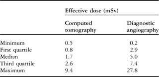
The median – range – dose length product for cardiac computed tomography was 28.0 (8.0–359.0) milliGray centimetres. This corresponded to an estimated median effective dose of 1.7 (0.5–9.5) millisieverts (Table 4). The correlation coefficients for dose length product with age and weight were ρ = 0.52 and 0.51, respectively (both p < 0.001). There was a strong positive correlation between dose length product and effective dose (ρ = 0.91; p < 0.001). Median effective dose was 1.4 (1.3–9.3) millisieverts for gated cardiac computed tomography and 1.9 (0.6–4.3) millisieverts for non-gated cardiac computed tomography (p = 0.64).
Effective dose quartiles from cardiac computed tomography and diagnostic angiography.
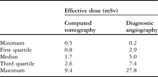
Regarding diagnostic angiography, the median fluoroscopy time was 7.5 (0.3–55.0) minutes. The median dose area product was 3.2 (0.2–21.0) Gray square centimetres. The respective correlation coefficients for dose area product with age, weight, and fluoroscopy time were ρ equal to 0.61, 0.58, and 0.50; all p's were less than 0.001. The estimated median effective dose was 5.0 (0.2–27.8) millisieverts (Table 4). There was a strong positive correlation between dose area product and effective dose (ρ = 0.87; p < 0.001).
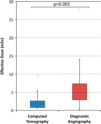
The distribution of effective dose in both imaging modalities exhibited high extreme values (Fig 2). The effective dose from cardiac computed tomography was significantly lower than that from angiography (p < 0.001; Fig 2).
Box and whisker plots of effective dose from cardiac computed tomography and diagnostic angiography. Boxes represent 25th-50th-75th centiles. Crosses demarcate extreme values.

Discussion
Our main findings were that, at our institution: diagnostic angiograms were performed over 3 times more often than cardiac computed tomography scans over a 2-year period; there was a trend towards reduction in the use of angiography and an increase in the use of cardiac computed tomography during the second year; multiple lifetime diagnostic angiography was more prevalent than multiple lifetime computed tomography examination; and radiation doses imparted by cardiac computed tomography were lower and more consistent in comparison to diagnostic angiography.
The finding that angiography was used more commonly than cardiac computed tomography is perhaps unsurprising given the intraluminal physiological measurements that can only be obtained during cardiac catheterisation. The finding that patients had undergone multiple diagnostic angiography procedures more frequently than multiple computed tomography examinations in their lifetimes is almost certainly due to the limited availability of cardiac computed tomography prior to the last decade. The trends demonstrated in our study are corroborated by 2004 data from the Great Ormond Street Hospital for Children, London, where the authors noted that the number of cardiac catheters far exceeded the number of cardiac computed tomography scans.Reference Puranik, Muthurangu, Celermajer and Taylor20 However, in this study by 2005 and 2006, the trend had reversed and cardiac computed tomography cases were two- and threefold, respectively, the number of angiograms performed. Similarly, other authors have reported that cardiac computed tomography has replaced conventional angiography in certain indications.Reference Ben Saad, Rohnean, Sigal-Cinqualbre, Adler and Paul21 Despite our data indicating a modest decline in the number of catheters mirrored by increasing numbers of computed tomography scans over time, we have not yet witnessed such a radical change in cardiac imaging. A potential reason may be a perception that the radiation burden of cardiac computed tomography is high.
Previously, thoracic computed tomography examinations using older scanners and protocols have yielded doses of between 1 and 50 millisieverts in children.Reference Dillman and Hernandez22 There are few studies in the literature reporting paediatric dose data from modern dual-source computed tomography scanners. A recent study of 110 infants, using the same dual-source scanner utilised in this study, reported mean effective doses of 0.5 and 1.3 millisieverts, respectively, for non-gated and gated examinations.Reference Ben Saad, Rohnean, Sigal-Cinqualbre, Adler and Paul21 Another study investigating the diagnostic accuracy of prospective electrocardiogram-triggered cardiac computed tomography in 35 children generated a median effective dose of 0.36 (0.25–0.58) millisieverts.Reference Cheng, Wang and Duan23 Kuettner et alReference Kuettner, Gehann and Spolnik24 have published effective doses ranging from 0.6 to 3.2 millisieverts in 12 paediatric gated cardiac computed tomography cases, using a dual source scanner. These are marginally lower doses than in this report and may reflect the lower tube voltage (80 kilovolts peak) used in all examinations. Nevertheless, it is reassuring that radiation doses from the latest computed tomography systems are broadly consistent among different centres, patients, and protocols.
Concerning radiation doses from diagnostic angiography, Bacher et alReference Bacher, Bogaert, Lapere, De Wolf and Thierens25 reported a median effective dose of 4.6 millisieverts with a range of 0.6 to 23.2 millisieverts in a group of 28 paediatric patients undergoing cardiac catheterisation. The authors’ recorded median fluoroscopy time of 4.35 minutes was shorter than ours, but their median dose area product was higher at 4.09 Gray-square-centimetres. Rassow et alReference Rassow, Schmaltz, Hentrich and Streffer26 calculated similar doses in infants undergoing diagnostic angiography ranging from 2 millisieverts – 25th centile – to 18 millisieverts – 90th centile. Both of these studies were conducted with older non-flatpanel fluoroscopic receivers making comparision with our data difficult.
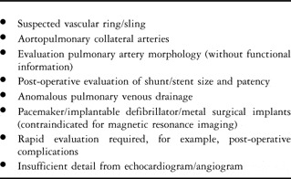
Radiation burden is only one consideration in selecting the best diagnostic imaging modality. Cardiac computed tomography may be selected as it precludes the need for general anaesthesia in many children, has fast image acquisition times, and is non-invasive in nature. Indeed, there are a growing number of indications in which computed tomography examination is warranted (Table 5). However, its primary disadvantages in comparison to angiography are the lack of intraluminal data and inability to undertake interventional procedures. Nevertheless, cardiac catheterisation is inherently limited by the risks of vascular and cardiac damage during instrumentation, and the need for multiple iodinated contrast media injections. The choice of examination, therefore, should result in the greatest yield of diagnostic information with minimal risk and radiation burden to the patient.
Common indications for cardiac computed tomography.

To put the radiation load of these examinations into perspective, our results suggest that the average cardiac computed tomography and angiographic examination are equivalent to almost 100 and 300 chest radiographs, respectively; a single chest radiograph = 0.02 millisievert. Furthermore, the United Kingdom average background radiation exposure is 2.2 millisieverts per year, which helps to contextualise the radiation doses to which we are subjecting patients.Reference Puranik, Muthurangu, Celermajer and Taylor20 Despite concerns that cardiac computed tomography may impart high radiation doses, we propose that cardiac computed tomography should be considered more frequently given its lower and more predictable radiation load than cardiac catheterisation.
Inevitably, there will be differences in the groups undergoing each investigation. This study was also limited by the use of dose conversion factors to estimate effective dose. Moreover, the authors of the angiography conversion factors that we used included both therapeutic and diagnostic catheters in the determination of these values, although we applied them to diagnostic catheters only.Reference Karambatsakidou, Sahlgren, Hansson, Lidegran and Fransson18 Dragusin et alReference Dragusin, Gewillig, Desmet, Smans, Struelens and Bosmans27 have also calculated conversion factors from therapeutic and diagnostic procedures using a flat-panel angiographic system identical to that used in our institution. Both sets of conversion factors were similar, and therefore we used those from Karambatsakidou et alReference Karambatsakidou, Sahlgren, Hansson, Lidegran and Fransson18 as the age bands used by them were consistent with the computed tomography age bands published by Shrimpton.Reference Shrimpton19 We are unaware of any flat-panel system conversion factors that exist in the literature for diagnostic procedures only. Despite our disregarding the number of projections used for angiography, some authors have recently shown that irradiation geometry has little effect on the overall estimation of effective dose.Reference Karambatsakidou, Sahlgren, Hansson, Lidegran and Fransson18 We also recognise the limited applicability of our findings as not all centres will use dual-source computed tomography and flat-panel detector angiography systems.
Conclusion
Our finding that the use of cardiac computed tomography is rising is probably due to an increased willingness to adopt this imaging technique for an initial diagnosis or pre- and post-surgical evaluation of congenital cardiac disease. Our data show that the radiation burden from this imaging modality is generally lower and more consistent than conventional angiography. Computed tomography will never completely replace other imaging modalities, and therefore it is clearly a useful complementary method for diagnosing complex congenital cardiac disease.
Acknowledgements
G.G. Gherardi is supported by The Royal College of Radiologists, Undergraduate Bursary and also by the University of Leeds’ Diagnostic Radiology Award.