INTRODUCTION
In recent years, potential dietary alternatives to antibiotics to promote the growth of broiler chicks have received increasing attention. Bacteriocins are a heterogeneous group of proteinaceous compounds lethal to bacteria other than the producing strain (Klaenhammer Reference Klaenhammer1993). A growing body of evidence indicates that bacteriocins modulate the metabolic processes of the host, including nutrient metabolism and immune activity (Nava et al. Reference Nava, Bielke, Callaway and Castañeda2005; Karimi Torshizi et al. Reference Karimi Torshizi, Moghaddam, Rahimi and Mojgani2010). The bacteriocin albusin B, isolated from rumen bacteria Ruminococcus albus 7, has been documented as an intestinal lactic acid bacteria growth promoter and pathogen inhibitor (Yang & Wang Reference Yang and Wang2008).
Nosiheptide (NHT) is produced by Streptomyces actuosus and is active against Gram-positive organisms. An early study with broiler chicks found NHT to be an effective growth promoter (McGinnis et al. Reference McGinnis, Johnson and Fox1978). However, a few studies have investigated the effects of NHT on broiler chicks’ gut fermentation and immunity. A more recent study by Li & Zou (Reference Li and Zou2005) reported that NHT displayed inhibitory effects on organs of the immune system and lymphocyte proliferation in peripheral blood and spleen.
Fermentation in the intestinal tract can be an indicator of microbial activity. The major products of undigested dietary compounds fermented by microflora are short-chain fatty acids (SCFA) such as acetate, propionate and butyrate (Williams et al. Reference Williams, Verstegen and Tamminga2001). The SCFA could provide an antibacterial effect, thereby preventing the establishment of pathogenic bacteria (Ricke Reference Ricke2003; Van Immerseel et al. Reference Van Immerseel, Ducatelle, De Vos, Boon, Van De Wiele, Verbeke, Rutgeerts, Sas, Louis and Flint2010). Because microflora can affect SCFA fermentation, some researchers have suggested that dietary supplementation with an antibiotic or its substitutes should be used to regulate the SCFA composition in the digestive tract (Louis & Flint Reference Louis and Flint2009).
Numerous studies have investigated the effect of bacteriocin or yeast products on broiler chick physiology. However, the literature contains a few reports on the use of yeast plus bacteriocin. Research focusing on NHT supplementation in broiler chicks is also scarce. Antibiotics or their substitutes have been proposed to regulate the microflora of the digestive tract and inhibit endemic subclinical infection to reduce the metabolic costs of the immune system (Dibner & Richards Reference Dibner and Richards2005; Niewold Reference Niewold2007). However, different antibiotics or their substitutes accomplish this through different mechanisms (Ferket Reference Ferket, Lyons and Jacques2004).
The commercial broiler is a stable experimental animal with smaller individual differences and variation in comparison with other farm animals (Hall et al. Reference Hall, Shirley, Bakalli, Aggrey, Pesti and Edwards2003). The key component of power analysis is the effect size. According to the effect size calculated by method of Roush & Tozer (Reference Roush and Tozer2004), effect sizes of 0·1, 0·4 and 0·8 can be viewed roughly as small, medium and large. Furthermore, the effect size value calculated from previous study data (Hsieh et al. Reference Hsieh, Wang, Chen and Chen2010) is large (0·6–0·8). Therefore, based on the effect size and in line with the ‘3Rs’ principle (reduce, refine and replace), a smaller-scale experiment was applied in the current study for a better understanding of the yeast with bacteriocin expression. The current study thus evaluated the effects of dietary supplementation yeast with bacteriocin expression on the performance, caecal flora, caecal fermentation and immunity of broiler chicks. NHT was used as an antibiotic positive control.
MATERIALS AND METHODS
Preparation of yeast with bacteriocin
Cloning of the structural gene and plasmid construction for expression in Saccaromyces cerevisiae and mass production of bacteriocin-expressing yeast were conducted as described by Wang et al. (Reference Wang, Yu, Hsieh, Chen, Chen and Chen2011). The albB gene (National Center for Biotechnology Information (NCBI) reference: AF469209) of the albusin B from R. albus 7 was applied for expression in S. cerevisiae DBY747 as a bacteriocin source. The albusin B-expressing yeast was mass-produced in the yeast extract peptone dextrose (YEPD) medium at pH 5·0 at 30 °C. After 48 h fed-batch fermentation, the yeast products were recovered by centrifugation, followed by spray drying for future use. The dried yeast contained 1 mg bacteriocin protein per gram of product yeast.
Animal treatments
A total of 180 one-day-old healthy Arbor Acres broiler chicks were obtained from a commercial hatchery. All chicks were wing-banded and weighed to obtain body weight at the first day. The 180 chicks were randomly assigned to nine groups of 20 birds (10 male and 10 female), kept in floor pens. Three groups were randomly assigned to one of the three dietary treatments: (1) control; (2) yeast with albusin B expression at a dose of 2·5 g/kg (equal to 2·5 mg/kg of albusin B activity); and (3) NHT 2·5 mg/kg (antibiotic control). The dose of additives was administered following Taiwanese drug feed additive standards (COA 2002) and previous studies (Hsieh et al. Reference Hsieh, Wang, Chen and Chen2010; Wang et al. Reference Wang, Yu, Hsieh, Chen, Chen and Chen2011).
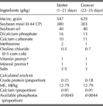
The experiment consisted of a two-stage nested design where the first stage is treatment and the second stage is chick group nested within treatment. Two basal diets, based on maize–soybean without fishmeal and which met the recommendations of the National Research Council (NRC 1994) (Table 1), were formulated for feeding during the starter (days 1–21) and grower (days 22–35) periods. All animal management was consistent with the recommendations of Arbor Acres Broiler Commercial Management Guide (Arbor Acres Farm 1996). Continuous lighting and access to feed and water were provided throughout the experiment. Room temperature was maintained between 26 and 28 °C for the first 4 weeks, and was thereafter gradually reduced according to normal management practices; additionally, a light bulb heater system was provided for the first 7 days. Ventilation was regulated by thermostat-controlled circulation and exhaust fans. The body weight of each bird was measured weekly and the feed consumption was recorded twice a day before feeding time (at 08.00 and 20.00 h). The feed efficiency of each group was calculated as g feed consumed/g body weight gained. During the experiment, 18 chicks/treatment (six chicks per group) were humanely slaughtered on days 21 and 35 for caeca and gallbladder collection.
Composition of the basal experimental diets

* Vitamin premix supplied per kg of diet: vitamin A, 12000 IU; vitamin D3, 3125 ICU; vitamin E, 37·5 IU; vitamine K3, 6·25 mg; vitamin B1, 3·75 mg; vitamin B2, 12·5 mg; vitamin B6, 10·0 mg; Ca-pantothenate, 18·8 mg; Niacin, 50 mg; Biotin, 0·06 mg; Folic acid, 1·25 mg and vitamin B12, 0·05 mg.
† Mineral premix supplied per kg of diet: Cu (CuSO4·5H2O, 0·25 Cu), 6 mg; Fe (FeSO4·7H2O, 0·20 Fe), 50 mg; Mn (MnSO4·H2O, 0·32 Mn), 40 mg; Zn (ZnO, 0·80 Zn), 60 mg; Se (NaSeO3,0·45 Se) and 0·075 mg.
Analysis of immunity
Blood samples were collected on days 21 and 35 from the wing vein of 18 chicks/treatment (six chicks per group); the serum was harvested and then stored (−20 °C) for immunoglobin analysis. After appropriate dilution, serum IgG, IgA and IgM were measured by double-antibody sandwich enzyme-linked immunosorbent assay (ELISA) quantification kits (Bethyl Lab Inc., USA). Bile samples were collected from the gallbladder after slaughter, and IgA concentrate was determined using the ELISA kit as described above.
Caecal microbial population and SCFA composition
Caecal samples for microbial population and SCFA determination were collected individually on days 21 and 35 from the fresh caeca of 18 chicks/treatment (six chicks per group). Approximately 1 g of fresh caecal content was weighed and transferred, within 2 min of extraction, to 9 ml of pre-reduced sterile dilution blank solution. It was then homogenized for c. 1 min before series dilution. The caecal microbial populations were determined by serial dilution (10−4–10−7) of caecal samples in anaerobic diluents (Bryant & Robinson Reference Bryant and Robinson1968) before inoculation onto Petri dishes of sterile agar. The plate media used were Rogosa Selective Lactobacillus (SL) agar (Difco™, Franklin Lakes, NJ, USA) for lactobacilli, eosin–methylene blue (EMB) agar (Difco™) for Escherichia coli, membrane-Enterococcus indoxyl-β-d-glucoside (m-EI) agar (Difco™) for Enterococcus, Reinforced Clostridial agar (Difco™) for Clostridium and Salmonella Shigella (SS) agar (Difco™) for Salmonella. Inoculated agar plates for lactobacilli and Clostridium were incubated anaerobically (0·97 CO2, 0·03 H2) at 37 °C. Enterococcus and E. coli were incubated aerobically at 37 °C. The contents of all plates were counted between 24 and 48 h after inoculation.
Approximately 0·5 g of each sample of fresh caecal content was suspended in 2 ml of pre-cooled (at 4 °C) sterile milli-Q water. Subsequently, samples were centrifuged at 18500 g for 15 min, and the supernatant was stored at −80 °C for SCFA analysis. SCFA concentration was determined by gas chromatography as previously described by Monsma & Marlett (Reference Monsma and Marlett1995). The analysis was conducted with a Perkin Elmer Clarus 500 (PerkinElmer Inc., Waltham, MA, USA) and separated by Nukol™ Capillary Gas Chromatograph Column (Supelco, Bellefonte, PA, USA). Each sample peak profile was integrated and quantified relative to an internal standard of methylbutyric acid placed in the same sample. Lactate concentration was determined immediately after separation by centrifugation, and was determined by lactate assay kit (BCX0623A, Fortress Diagnostics, Antrim, Northern Ireland, UK) using the lactate oxidase method.
mRNA extraction and real-time polymerase chain reaction (PCR)
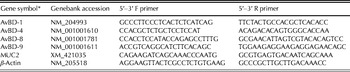
After slaughter on day 35, the mucosal contents of the jejunum (the region anterior to Meckel's diverticulum) from 12 chicks/treatment (four chicks per group) were scraped with a metal spatula. Mucosal contents of 1 ml were immediately mixed with 5 ml of RNAlater solution (Ambion, Austin, TX, USA) and left overnight, then stored at −80 °C until isolation of total RNA. The total RNA of jejunal mucosa samples were extracted by RNeasy® Mini Kit (QIAGEN, Valencia, CA, USA) according to the manufacturer's recommendations. RNA integrity and quantity were analysed using agarose gel electrophoresis and NanoDrop® ND-1000 Spectrophotometers (Thermo Fisher Scientific, Waltham, MA, USA). Reverse transcription was conducted on the cleaned RNA samples using High Capacity cDNA Reverse Transcription kit (Roche, Mannheim, Germany) according to the manufacturer's recommendations. The abundance of avian β-defensin (AvBD) 1, 4, 8 and 9, and mucin 2 (MUC2) were determined using quantitative reverse transcription-PCR. Primers for the target genes were selected from the NCBI database (Table 2).
Sequences of primers for avian MUC2, AvBD-1, AvBD-4, AvBD-8, AvBD-9 and β-actin in real-time PCR

* AvBD, avian beta-defensin; MUC2, mucin 2.
For each reverse-transcription product, 1 μg/μl cDNA was serial-diluted to 1000, 100, 10 and 0·1 ng/μl, respectively, as a template for real-time PCR. The PCR reaction for each sample was run in triplicate using SYBR Green I kit (Roche, Mannheim, Germany) and the LightCycler Real-Time PCR System (Roche, Mannheim, Germany) according to the manufacturer's instructions. A total of 41 cycles of reactions were run: 1 cycle at 95 °C for 15 min; and 40 cycles at 95 °C for 15 s, at 60 °C for 30 s and at 72 °C for 30 s. Melting curve analysis was determined after reaction at 60 °C for 15 min, followed by cooling to 40 °C for fluorescence signal detection.
For all specific mRNAs amplified, linear inverse correlations were observed between the amount of RNA and C T values (number of cycles at threshold lines). Gene expression was normalized to β-actin expression and was expressed relative to the same value for the control group (Lehmann & Kreipe Reference Lehmann and Kreipe2004).
Statistical analysis
The experimental data (expected feeding efficiency) were analysed by the SAS (PROC GLM; SAS Institute 1997) with a two-stage nested design, with chick group nested within treatment. The factor in the first stage was treatment and the nested factor in the second stage was chick group within treatment (the effect of treatment and chick group were fixed). Multiple comparisons were performed on least squares means, the probability of committing type I errors was fixed at P<0·05. The model for statistical analysis was:
 $$Y_{ijk} = \mu + \alpha _i + \beta _{\,j(i)} + \varepsilon _{ijk} $$
for testing the effect of treatment; the null hypothesis pertains to the equality of population treatment effect: H 0: α 1=α 2=α 3=0, where Y ijk is the observed dependent variable; μ is the overall mean; α i is the ith treatment effect; β j(i) is the jth effect of the chick group nested within the ith treatment; ε ijk is the random error of the kth observation from the jth chick group within the ith treatment. Means were compared pairwise (pdiff) and results are given as least squares means and pooled standard error (s.e.) in tables. In the figures, the results are presented as means and standard deviations (s.d.). Since feed intake data were collected from the total feed consumption of each chick group, the feeding efficiency (feed per gain) data were analysed appropriately for the complete block design using the one-way analysis of variance of GLM procedures of SAS.
RESULTS
Growth performance
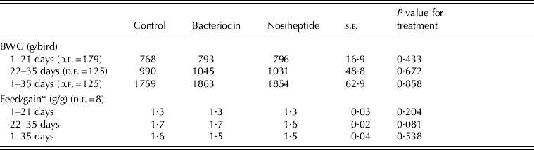
The data for body weight gain (BWG) and feed conversion ratio (FCR) are presented in Table 3. Dietary supplementation by bacteriocin or NHT had no significant effect on BWG during the starter (days 1–21) or grower (days 22–35) periods. During the grower period, NHT supplementation appeared to result in better FCR performance, but the differences were not significant either between the treatment or control groups.
Effect of bacteriocin and NHT on the growth performance of broiler chicks
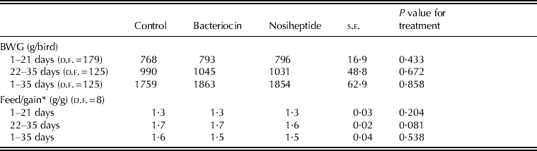
* Feed efficiency was calculated by (total feed consumption/total body weight gain) of all animals in each chicks group.
Caecal microbial population
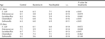
The results of the caecal bacterial population analysis are shown in Table 4. Viable counts of E. coli, Salmonella and Clostridium were high in 21-day-old broiler chicks fed with NHT supplementation, but decreased as the birds grew older. The NHT treatment group had lower Lactobacillus counts than either the control or bacteriocin treatment groups at two sampling times (days 21 and 35). The groups receiving either bacteriocin or NHT supplementation had lower E. coli counts than the control group at day 35, but only bacteriocin supplementation also significantly decreased the counts of Enterococcus and Salmonella. Bacteriocin supplementation also significantly affected Lactobacillus growth in the chicks’ caeca during the experimental period. A strong relationship was evident between the manipulation of caecal flora by bacteriocin treatment and the presence of Lactobacillus.
The effect of bacteriocin and NHT supplementation on caecal bacterial populations (log cfu*/g) of broiler chicks at 21 and 35 days (d.f.=53)

* cfu, colony-forming unit.
Caecal SCFA composition
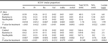
The results of SCFA, ammonia and lactate analysis of the caecal samples are shown in Table 5. The proportion of acetic acid in the NHT treatment caecal sample was significantly lower than that of the other groups at days 21 and 35 (P<0·05). However, the concentration of acetic acid did not differ significantly between the treatment or control groups. NHT treatment significantly increased the propionic acid concentration at day 35. Total SCFA concentration as well as the proportions of iso-butyric acid and iso-valeric acid increased as the broiler chicks aged. Chicks fed with bacteriocin had higher lactate concentration in the caecal samples at day 21 (P<0·05), but lactate concentration of all treatment groups dropped rapidly at day 35 (c. 4–10 times lower than at day 21). The NHT treatment resulted in higher ammonia concentrations at day 21 relative to the other two groups, but at day 35 there was no significant difference between any of the groups.
The effect of bacteriocin and NHT supplementation on SCFA, ammonia (NH3) and lactate concentration in caeca of broiler chicks (d.f.=53)

* Ac, acetic acid; Pr, propionic acid; Bu, butyric acid; Val, valeric acid; isoBu, iso-butyric acid; isoVal, iso-valeric acid.
Immunity function analysis
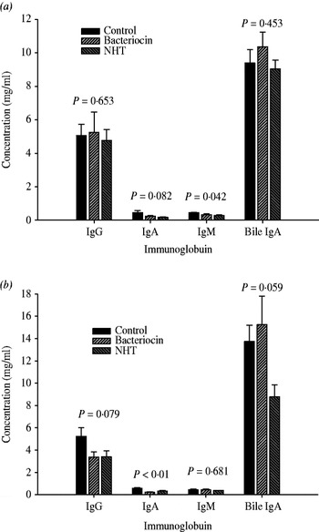
The effect of bacteriocin and NHT supplementation on serum immunoglobulins (Ig) and bile IgA is shown in Fig. 1. The NHT treatment appeared to decrease serum IgA and IgM concentrations at day 21, but showed no significant effect on IgG concentration. On day 35, both bacteriocin and NHT groups displayed lower IgA concentration (P<0·05) and a trend of decreased IgG concentration in the serum, compared with the control group. No differences among the three groups were observed for bile IgA at day 21. However, the bacteriocin treatment group tended to have increased bile IgA, and showed significantly higher bile IgA than the NHT treatment group at day 35.
Effect of dietary bacteriocin and NHT supplementation on serum Ig G, A and M content and bile IgA content in gallbladder in broiler chicks at 21 (a) and 35 days old (b). Bars equal means±s.d. (d.f.=53).

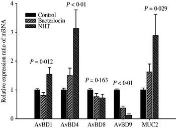
Greater AvBD1, AvBD4 and MUC2 mRNA expression were observed in the NHT treatment group relative to the other groups (P<0·05), but no significant differences emerged between the control and bacteriocin treatment groups (Fig. 2). However, both bacteriocin and NHT supplementation significantly decreased the AvBD9 expression level (P<0·05), especially in the NHT treatment group (which was three times lower than the control group). The AvBD8 expression was not affected by either bacteriocin or NHT supplementation.
Effect of dietary bacteriocin and NHT supplementation on AvBD (1, 4, 8 and 9) and MUC2 genes mRNA expression in broiler chicks at 35 days old. Bars equal means±s.d. (d.f.=35).

DISCUSSION
The results of BWG analysis indicated no significant differences among the three groups during the experimental period. An earlier extensive study of broiler chicks indicated that NHT was an effective growth promoter at low (0·35 mg/kg) and high levels (221 mg/kg), but without a predictable dose response (McGinnis et al. Reference McGinnis, Johnson and Fox1978). The results of pathogenic bacteria counts in caeca, enteric bacterial translocation to internal organs and the drug-resistance determinant in a previous study of broiler chicks (Hsieh et al. Reference Hsieh, Wang, Chen and Chen2010) indicated that the applicable NHT supplementation level in broiler diet was 2·5 mg/kg.
The preliminary study (40 chicks in each treatment group) compared the growth performance of broiler chicks fed with yeast with bacteriocin or yeast control (2·5 g/kg of S. cerevisiae DBY747). The results indicated that broilers fed yeast with bacteriocin had significantly higher BWG (38±5·4 g/day for days 1–21; 68±7·3 g/day for days 22–35) than the yeast control group (34±2·2 g/day for days 1–21; 57±5·1 g/day for days 22–35). These findings suggest that the better growth performance of bacteriocin treatment groups might be because of the antibiotic substitute function of bacteriocin and to the growth-promoting effect of yeast. The FCR data in Table 3 also suggested that bacteriocin improved nutrient utilization as effectively as did NHT.
Previous studies indicated that yeast cell walls containing mannan oligosaccharide (MOS) could reduce the intestinal Salmonella count by 26%, and significantly reduced E. coli concentration in the caeca of broiler chicks compared with a control group (Spring et al. Reference Spring, Wenk, Dawson and Newman2000; Baurhoo et al. Reference Baurhoo, Ferket and Zhao2009). However, lactobacilli concentrations did not differ when broiler chicks were fed different levels of MOS or a control diet (Baurhoo et al. Reference Baurhoo, Ferket and Zhao2009). Fernandez et al. (Reference Fernandez, Hinton and Van Gils2002) also reported that MOS supplementation could increase the caecal Enterococcus and Bifidobacteria concentration in hens, but had no effect on the lactobacilli concentration. This finding suggested that the higher lactobacilli concentration in the caeca of the bacteriocin supplementation group could result from the effect of bacteriocin expression in yeast cells.
The analysis of flora composition indicated that the caecal bacterial population of the bacteriocin supplementation group was probably influenced by the synthetic effects of bacteriocin and yeast. NHT is produced by S. actuosus and is active against Gram-positive organisms (Cromwell et al. Reference Cromwell, Stahly, Speer and O'Kelly1984). A recent study of antibiotic growth promoters in broiler chicks indicated that NHT supplementation decreased c. 1 log (i.e. to about a tenth of that previously) of the numbers of Lactobacillus, but did not affect the count of Coliform or Enterococci (Lee et al. Reference Lee, Lyu, Wang, Weng and Chen2011). The current results shown in Table 4 also indicated that NHT supplementation led to a significant decrease in the caecal Lactobacillus count, but that Salmonella concentration was not inhibited by NHT.
It was found that broiler chicks fed with an NHT supplementation diet showed significantly lower proportions of acetic acid compared with other treatment or control groups. However, the higher total SCFA concentration resulted in a similar acetic acid concentration among three treatment groups. Higher concentrations of branch chain SCFA and NH3 were evident in the NHT treatment group on day 21, suggesting that proportionately more amino acids were being formed in the chick caeca. Previous research reported that NH3 can disturb the development of the mucosa of the intestine (Visek Reference Visek1984), and NH3 was found to correlate negatively with a reduction in villus height (Nousiainen Reference Nousiainen1991).
However, despite the known properties of NHT against Gram-positive bacteria, lower E. coli and Salmonella concentrations were found in the current study in the NHT treatment group at day 35 relative to the control group. This finding might be explained by the higher propionic acid and butyric acid concentration in caecal samples of the NHT treatment group. Previous studies indicated that increasing propionic and butyric acid concentration led to decreased numbers of E. coli in all sections of the intestinal tract (Gálfi & Neogrády Reference Gálfi, Neogrády and Piva1996; Williams et al. Reference Williams, Verstegen and Tamminga2001).
The current study observed that caecal lactate concentration dropped rapidly after day 21 (Table 5), but that lactic acid bacterial concentration was still higher at day 35 than at day 21. This result might be explained by the increase in caecal content and lactate consumed by propionate bacteria (Chassard & Bernalier-Donadille Reference Chassard and Bernalier-Donadille2006). Van Der Wielen et al. (Reference Van Der Wielen, Biesterveld, Notermans, Hofstra, Urlings and Van Knapen2000) found no significant correlations between numbers of lactobacilli and volatile fatty acids, or among the different bacterial groups in the caeca. The bacteriostatic action of SCFA is exerted when the acid is in the dissociated lipophilic state, and increases as the pH of the intestine decreases (Corrier et al. Reference Corrier, Hinton, Ziprin and Deloach1990). Because the current study did not determine the caecal content pH or undissociated SCFA concentration, the particular effect of bacteriocin and NHT on SCFA-related antibacterial activity should be verified by future studies.
Antibody titer responses have been used as a measure of the humoral immune status of birds (Sklan et al. Reference Sklan, Melamed and Friedman1994). Mucosal immunity is an important part of humoral immunity and secretory IgA (sIgA) is the effector of mucosal immunity. This is the most prominent antibody present at mucosal surfaces, and it provides passive immunoprotection against invading pathogens in the gastrointestinal tract. Bile IgA is the major source of intestinal IgA (Savage et al. Reference Savage, Cotter and Zakrzewska1996).
The current study found a higher bile IgA concentration in the bacteriocin treatment group relative to the other groups on day 35. This finding may have resulted from the higher Lactobacillus concentration in the intestinal tract and the effect of cell wall components from yeast with bacteriocin. Lactobacilli have long been recognized for their ability to stimulate the sIgA immune system, increasing resistance to disease (Fuller Reference Fuller1977). However, species of Lactobacillus vary in their immunoregulatory properties (Mountzouris et al. Reference Mountzouris, Tsitrsikos, Palamidi, Arvaniti, Mohnl, Schatzmayr and Fegeros2010). The MOS from the yeast cell wall has also been shown to enhance IgA titres in the plasma of poultry (Savage et al. Reference Savage, Cotter and Zakrzewska1996).
To date, however, limited studies have been conducted to determine the effects of NHT on serum Ig concentrations. A study by Li & Zou (Reference Li and Zou2005) showed that NHT had an inhibitory effect on broiler chicks’ immune organs and lymphocyte proliferation of peripheral blood and milt at day 42. Supplementation with NHT also significantly decreased the IgA-positive cell numbers and sIgA concentration in the ileum (Lee et al. Reference Lee, Lyu, Wang, Weng and Chen2011). One potential mechanism by which growth-promoting antibiotics may exert their effect is to decrease immunogenic bacteria inhabiting the small intestine. A study by Roura et al. (Reference Roura, Homedes and Klasing1992) suggested that antibiotic nutritional supplements might enhance chicks’ growth by preventing the immunogenic stress and associated metabolic changes caused by cytokines. By limiting the growth of intestinal bacteria, antibiotics may decrease the energetic costs associated with constitutive, low-level inflammation in the gut of animals.
Avian species express only β-defensin, and avian heterophils or epithelial cells have been reported to express these molecules (Akbari et al. Reference Akbari, Haghighi, Chambers, Brisbin, Read and Sharif2008). The current study found no significant differences in the gene expression ratio among all tested β-defensin between the control and bacteriocin supplementation groups. Bacteriocin supplementation significantly enhanced the lactobacilli growth in broiler chicks’ gut, but related studies have suggested that intestinal colonization by lactic acid bacteria, or when treated with probiotic only, did not enhance the expression levels of β-defensin genes (Menendez & Finlay Reference Menendez and Finlay2007; Akbari et al. Reference Akbari, Haghighi, Chambers, Brisbin, Read and Sharif2008). With the exception of AvBD11, mRNA expression of chicken β-defensin was found throughout the digestive tract (van Dijk et al. Reference Van Dijk, Veldhuizen and Haagsman2008). Enhanced expression of defensin genes is usually associated with pathogenic infections and inflammations caused by these infections (Menendez & Finlay Reference Menendez and Finlay2007). In the current study, the expression of AvBD1 and AvBD4 mRNA were higher in the NHT treatment group, but AvBD9 expression was significantly lower in the jejunal mucosal contents of the NHT group. However, all bacterial populations were relatively low in the NHT treatment group, which implied that NHT might regulate defensin expression by other mechanisms.
The current study found that MUC2 gene expression was significantly higher in the NHT treatment group compared with the controls. Previous research indicated an effect of antibiotics on the thickness of the intestinal wall. Nutritional supplementation with an antibiotic that acted against Gram-positive bacteria affected mucin biosynthesis by enhancing mucin mRNA expression in the jejunum and ileum (Smirnov et al. Reference Smirnov, Perez, Amit-Romach, Sklan and Uni2005). After mucus has been secreted by goblet cells throughout the gastrointestinal tract, it forms a gel adherent to the mucosal surface as a barrier layer in the intestine. The mucus adherent layer was of similar thickness throughout the chickens’ small intestines, but variations in the thickness of the mucus adherent layer would be expected to influence the nutrient digestion processes (Smirnov et al. Reference Smirnov, Sklan and Uni2004). Antibiotic supplementation may result in a thinner intestinal wall. However, in the current study, lower bacterial counts also showed in the NHT treatment group. This finding suggested that an increase in mucin synthesis was possibly because of the balance of the intestinal mucus layer.
The aspect of interest in bacteria–mucin interactions is the participation of the intestinal microbiota in mucin turnover (1) by stimulating mucin gene expression, and (2) by production of mucin-degrading enzymes (Mattar et al. Reference Mattar, Teitelbaum, Drongowski, Yongyi, Harmon and Coran2002; Smirnov et al. Reference Smirnov, Perez, Amit-Romach, Sklan and Uni2005). Previous studies (Mack et al. Reference Mack, Ahrne, Hyde, Wei and Hollingsworth2003; Smirnov et al. Reference Smirnov, Perez, Amit-Romach, Sklan and Uni2005) have reported that a probiotic supplement consisting mainly of Lactobacillus and Bifidobacterium species resulted in increased mucin synthesis and secretion. Derensy-Dron et al. (Reference Derensy-Dron, Krzewinski, Brassart and Bouquelet1999) also indicated that Bifidobacterium species possess high mucin degradation activity. Mack et al. (Reference Mack, Ahrne, Hyde, Wei and Hollingsworth2003) reported that Lactobacillus plantarum inhibited the adherence of pathogens to intestinal epithelial cells by increasing the mRNA expression of mucin genes. The current study found that MUC2 gene expression tended to be higher in the bacteriocin treatment group compared with the other groups. Because bacteriocin supplementation significantly enhanced the lactic acid bacterial population in chicks’ intestines, the mucin degradation ability of lactic acid bacteria might affect MUC2 gene expression. The results of gene expression analysis in the current study were consistent with those of previous studies. This suggests that both bacteriocin and NHT supplementation alter the intestinal flora and affect the expression of AvBD and mucin dynamics.
In conclusion, it has been demonstrated that bacteriocin and NHT supplementation significantly affect the bacterial population and SCFA composition of broiler chicks’ caeca. Furthermore, the effect of bacteriocin supplementation may result from enhancing the lactic acid bacterial growth and physiological characterization of yeast cell wall composition. Improvement in gut flora and the immunoregulatory properties associated with bacteriocin supplementation indicated that bacteriocin might be a viable alternative to antibiotics in broiler chicken feed. However, further knowledge on the mechanisms behind the relationship between bacteriocin and immune activity is required to clarify the role of bacteriocin in broiler chicks’ gut physiology.