Introduction
In human-assisted reproduction, in vitro fertilization (IVF) success rates are strongly dependent on embryo implantation, which occurs in only 20–30% of cases after fresh embryo transfer. Moreover, about 10% of patients have unexplained repeated implantation failures (RIF) after several IVF treatments. Understanding and mastering the molecular mechanisms that govern this key phase of human reproduction would therefore improve significantly the IVF success rate and provide a therapeutic response to numerous RIF cases.
At present, many hypotheses have been put forward to explain RIF, one of these is the exploration of immuno-physiological aspects of implantation and pregnancy for these kinds of patients and also to elucidate maternal–fetal immuno-tolerance mechanisms, based on the fact that the embryo is a semi-allogenic conceptus to which the maternal immune system should become adapted (Hill et al., Reference Hill, Polgar and Anderson1995). Therefore, it appears likely that any dysfunction that affects the maternal immune response could explain the RIF for the same patient. Nevertheless, the maternal immune system should modulate implantation and pregnancy more by local mechanisms gathered at the maternal–fetal interface rather than by systemic mechanisms across the organism.
Indeed, it is accepted today that implantation much depends on endocrine mechanisms widely described as immune local mechanisms involving a broad spectrum of cytokines. A real cross-talk, particularly rich and complex and not yet elucidated, is established between endometrium and embryo even before and during implantation through a myriad of molecules such as cytokines, chemokines, growth factors, adhesion molecules etc. (Johnson et al., Reference Johnson, Christmas and Vince1999). This accurate and organized cross-talk involves maternal immune cells, particularly lymphocytes.
In fact, early before implantation, semen presence in the uterus causes an influx of T lymphocytes and macrophages that initiate uterine inflammatory conditions suitable for implantation. Later, natural killers (NK) cells begin to accumulate in the endometrium during the latter middle period of the menstrual cycle (Bulmer et al., Reference Bulmer, Longfellow and Ritson1991; King, Reference King2000). These large granular lymphocytes called uterine NKs (uNKs) are phenotypically different from peripheral NKs (pNKs) as they are characterized by the expression of CD56, but do not express CD16 and CD3 (Bulmer & Lash, Reference Bulmer and Lash2005). At the time of implantation these cells are the most represented immune cells at the maternal–fetal interface and are 50–90% of all decidual leukocytes, while pNKs represent only 5–15% of circulating leukocytes (Parham, Reference Parham2004). Infiltration of uNK cells is clearly influenced by sex hormones, particularly progesterone. Nevertheless, it has been reported that endometrium-derived IL15 and IL11 have also been implicated in proliferation and differentiation of these cells (Ashkar et al., Reference Ashkar, Black, Wei, He, Liang, Head and Croy2003; Becknell & Caligiuri, Reference Becknell and Caligiuri2005). This increase in uNK cell levels in the uterus suggests that they play an important role in the implantation mechanism (Saito et al., Reference Saito, Shima, Nakashima, Shiozaki, Ito and Sasaki2007). The endometrium uNK rate seems to be correlated with IVF success rate (Johnson et al., Reference Johnson, Christmas and Vince1999) and therefore should be a good indicator of predictive IVF success.
Many studies have investigated the uNK contribution and action mechanism in implantation. It has been shown that uNKs control endometrium decidualization and vascularization through vascular endothelial growth factor (VEGF), placental growth factor (PLGF), leukaemia inhibitory factor (LIF) and interferon (IFN) secretion (Kimber, Reference Kimber2005; Croy et al., Reference Croy, Esadeg, Chantakru, van den Heuvel, Paffaro, He, Black, Ashkar, Kiso and Zhang2003). They participate in embryo–maternal interactions during implantation and fetal development by decidual spiral artery remodelling to provide the embryo with the necessary nutrients (Ashkar et al. Reference Ashkar, Black, Wei, He, Liang, Head and Croy2003; Hanna et al. Reference Hanna, Goldman-Wohl, Hamani, Avraham, Greenfield, Natanson-Yaron, Prus, Cohen-Daniel, Arnon, Manaster, Gazit, Yutkin, Benharroch, Porgador, Keshet, Yagel and Mandelboim2006; Fettback et al. Reference Fettback, Domingues, Hassun Filho, Motta, Serafini and Baracat2009; Lash & Bulmer, Reference Lash and Bulmer2011).
It has been reported that the presence of lymphocytes during implantation contributes, in concert with the endometrium and blastocyst, to cytokine production, giving a profile that is dominated by Th1 proinflammatory cytokines: tumour necrosis factor (TNF), IFNγ, LIF, IL1, IL2, IL6, IL12 and IL15. The presence of these factors as inflammatory response actors seems to be essential for early stages of implantation (Kachkache et al., Reference Kachkache, Acker, Chaouat, Noun and Garabedian1991; McMaster et al., Reference McMaster, Newton, Dey and Andrews1992; Sanford & Wood, Reference Sanford and Wood1992; Stewart et al., Reference Stewart, Kaspar, Brunet, Bhatt, Gadi, Köntgen and Abbondanzo1992). However, the initial inflammatory response must be selectively attenuated thereafter for maintenance of pregnancy (Chaouat et al., Reference Chaouat, Dubanchet and Ledee2007), that is why too early an inflammatory deficit would lead rather to embryonic implantation failure, while inflammatory excess leads to acute (spontaneous miscarriage) or chronic (pre-eclampsia, delayed intrauterine growth) rejection.
Conversely, the maintenance of pregnancy would necessitate a specific cytokine profile, characterized by the predominance of Th2 cytokines: IL3, IL4, IL10, IL5, IL13 and granulocyte–macrophage colony-stimulating factor (GM-CSF) that cause an anti-inflammatory state that is adequate for uterine receptivity and, therefore, for pregnancy (Miyazaki et al., Reference Miyazaki, Tsuda, Sakai, Hori, Sasaki, Futatani, Miyawaki and Saito2003; Challis et al., Reference Challis, Lockwood, Myatt, Norman, Strauss and Petraglia2009; David Dong et al., Reference David Dong, Aplin and Nicosia2009; Nagamatsu & Schust, Reference Nagamatsu and Schust2010; Mor et al., Reference Mor, Cardenas, Abrahams and Guller2011).
Thus, the maternal immune system seems to be involved in the establishment and maintenance of pregnancy through the Th1/Th2 profile balance. An imbalance between these two systems could explain implantation failures in some patients (Wegmann et al., Reference Wegmann, Lin, Guilbert and Mosmann1993; Darmochwal et al., Reference Darmochwal, Leszczynska, Rolinski and Oleszczuk1999; Raghupathy et al., Reference Raghupathy, Makhseed, Azizieh, Hassan, Al-Azemi and Al-Shamali1999). In the mouse model, it has been demonstrated that embryo rejection is controlled by the Th1 system while the Th2 system plays a protective role (Vassiliadis et al., Reference Vassiliadis, Ranella, Papadimitriou, Makrygiannakis and Athanassakis1998), knowing that autoimmune factors are the cause of 30% of human miscarriages (Pandey et al., Reference Pandey, Rani and Agrawal2005). Many studies have explored the ability of T lymphocytes and uNKs to control the Th1/Th2 balance and it seems increasingly clear that uNK is much more relevant than other cells in the establishment of the Th1 cytokine profile.
Implantation failure is a main limiting factor in assisted reproductive technologies (ART) and many authors have shown clearly the contribution of cytokines and other factors to endometrium immuno-modulation to improve ongoing pregnancy (Makrigiannakis et al., Reference Makrigiannakis, Minas, Kalantaridou, Nikas and Chrousos2006, Reference Makrigiannakis, Karamouti, Drakakis, Loutradis and Antsaklis2008, Reference Makrigiannakis, Petsas, Toth, Relakis and Jeschke2011; Toth et al., Reference Toth, Würfel, Germeyer, Hirv, Makrigiannakis and Strowitzki2011; Hutter et al., Reference Hutter, Heublein, Knabl, Andergassen, Vrekoussis, Makrigiannakis, Friese, Mayr and Jeschke2013). It has been estimated that inadequate endometrial receptivity is related in up to two-thirds of cases (Simon et al., Reference Simon, Moreno, Remohi and Pellicer1998). As immune cells are known to modulate endometrial receptivity through a large panel of cytokines, it seemed important in patients with RIF to investigate the benefit on embryo implantation rate of intrauterine insemination (IUI) of their own lymphocytes in utero at the preimplantation stage. In vitro experiments in mice have demonstrated an increased embryo implantation rate with human peripheral blood mononuclear cells (PBMC) (Nakayama et al., Reference Nakayama, Fujiwara, Maeda, Inoue, Yoshioka, Mori and Fujii2002; Yu et al., Reference Yu, Yang, Guo, Fang, Yin, Luo, Li, Li, Zhao, Zou and Xu2014). Many recent studies have investigated this innovative immune approach to the implantation mechanism based on the hypothesis that, in RIF patients, lymphocyte recruitment is not able to induce an uterine inflammatory condition suitable for implantation (Nakayama et al., Reference Nakayama, Fujiwara, Maeda, Inoue, Yoshioka, Mori and Fujii2002; Yoshioka et al., Reference Yoshioka, Fujiwara, Nakayama, Kosaka, Mori and Fujii2006; Zhylkova et al., Reference Zhylkova, Feskov, Feskova, Somova and Chumakova2010; Okitsu et al., Reference Okitsu, Kiyokawa, Oda, Miyake, Sato and Fujiwara2011; Yu et al., Reference Yu, Yang, Guo, Fang, Yin, Luo, Li, Li, Zhao, Zou and Xu2014).
The practice of immuno-modulation techniques to improve implantation and pregnancy could be an interesting prospective therapeutic method for ART as reported by several studies (Yoshioka et al., Reference Yoshioka, Fujiwara, Nakayama, Kosaka, Mori and Fujii2006; Zhylkova et al., Reference Zhylkova, Feskov, Feskova, Somova and Chumakova2010; Okitsu et al., Reference Okitsu, Kiyokawa, Oda, Miyake, Sato and Fujiwara2011) and should even revolutionize the conventional IVF protocols actually practiced.
The aim of this work is to understand how PBMC culture and intrauterine transfer on the day of ovulation induction can modulate implantation rate in patients with RIF in order to improve pregnancy. This preliminary study was clearly beneficial to our patients and needs to be continued and developed for more improvement.
Materials and methods
Patients’ selection
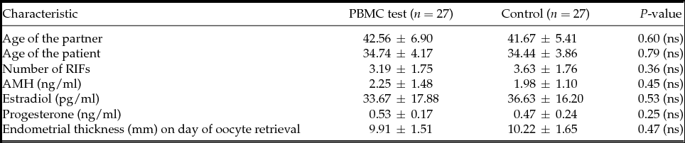
This is a prospective randomized study design of parallel group trials with the same size using block randomization. It includes a sample of 54 patients selected within the Anfa Fertility Center and spread over 1 year. Selected women were assigned randomly to receive autologous PBMCs cultures by an intrauterine non-invasive insemination 2 days before embryo transfer representing the PBMC test or no treatment group without receiving any cell transfer prior to embryo transfer to serve as control. The effect of immunotherapy PBMCs was evaluated by the implantation rate and clinical pregnancy. Each patient contributed a single cycle to this study, so there was no crossover between groups. As anticipated, PBMC test and control groups were comparable (Table 1). After the study recieved ethical approval, patients provided written informed consent.
Comparison of patient characteristics included in the study with at least 2 RIFs

All patients without exception were selected on the following inclusion–exclusion criteria:
-
• Inclusion criteria: at least two previous failures of implantation after IVF/intra-cytoplasmic spermatozoa injection (ICSI) (mean = 3), primary infertility, endometrial thickness < 6 mm in ovulation induction, age < 40 years (mean 35 years) (Table 1), regular menstrual cycles, BMI < 30, absence of uterine pathology and infectious negative balance.
-
• Exclusion criteria: polycystic ovary syndrome and uterine pathology.
Ethical standards
The authors assert that all procedures contributing to this work comply with the ethical standards of the relevant national and institutional committees on human experimentation and with the Helsinki Declaration of 1975, as revised in 2008. All patients who participated in this study signed an informed consent after being informed about the terms and issues of study.
Stimulation protocol
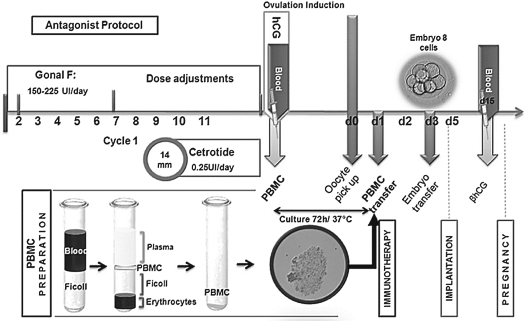
In order to eliminate the effect of protocol and better align the sample and follicular cohort, it was preferable to use the antagonist protocol using r-FSH (Cetrotide 0.25 and Gonal-F) (Fig. 1). Further r-FSH administration (Gonal-F; Serono Laboratories, Saint Cloud, France) was started by daily subcutaneously injections (150–225 IU/day), which were maintained constantly for 5 days and were adjusted according to usual parameters of follicle growth determined by serum estradiol concentrations and ultrasound monitoring. Ultrasound scans were performed to evaluate the number and sizes of early antral follicles and to calculate mean ovarian volume. When at least one follicle exceeded 14 mm in diameter, a potent, third-generation GnRH antagonist, cetrorelix acetate (Cetrotide, 3 mg; Serono Laboratories), was administered subcutaneously (0.25 IU/day). An intramuscular injection of 10,000 IU of human chorionic gonadotrophin (HCG; Gonadotrophines Chorioniques Endo®, Organon) was performed after obtaining follicles ≥ 17 mm. Luteal phase was supported by vaginal administration of micronized progesterone 400 mg/day (Utrogestan®, Besins International, Montrouge, France) from the day of oocyte pick-up to the day of the pregnancy test. If a pregnancy occurred, progesterone administration was extended until evidence of fetal heart activity at ultrasound was found.
Immunotherapeutic strategy by transferring peripheral blood mononuclear cells (PBMCs) in patients with repeated implantation failures (RIF). After antagonist ovarian stimulation protocol, a blood sample is scheduled on the day of ovulation induction in order to isolate PBMCs using a separation protocol based on Ficoll (PREPARATION PBMC). PBMC are well prepared after a culture for 72 h and then transferred to the patient in utero at D1 (immunotherapy).

Collection, culture and transfer of PBMCs
Each sample of blood was taken from RIF patients undergoing ART cycles on the day of ovulation induction and it was collected in citrated tubes to perform a separation based on Ficoll-Hypaque solution with a volume of 3 ml (Histopaque®-1077; Sigma). After centrifugation at 18–20°C for 30–40 min at 400 g, we obtained a distinct layer of PBMC under the median plasma (Fig. 1). The PBMC layer was transferred to another tube and three volumes of phosphate-buffered saline (PBS) was added and then centrifuged at 18–20°C and 60–100 g for 10 min to obtain a PBMC pellet. The lymphocyte pellet was washed twice with PBS and then resuspended at 37°C in complete culture medium ready for use (supplied by ATL R & D laboratory. 78 320 La Verrière France) with 75 IU of HMG-Menoupur®. After 72 h of incubation, 1 × 10⁶ cells in 0.4 ml were transferred into the endometrium cavity 2 days prior to embryo transfer.
During the 3 days of PBMC incubation, special attention was paid to monitoring the culture including the convergence of lymphocyte cells PBMC forming clusters of cytoplasmic processes (Fig. 2), this criteria is crucial for transfer PBMC efficiency. Therefore, two patients were excluded from the study due to unsatisfactory PBMC culture.
Photomicrograph (original magnification ×40; scale bar, 100 μm) of peripheral blood mononuclear cells (PBMCs) cultures isolated from blood samples of patients. After a separation phase using protocol based on Ficoll, PBMCs are in isolated mononuclear cells and inactive form (0 h). Throughout incubation (72 h, 0 h), cells are activated and thus have a more granular appearance to bind them and increase convergence more condensed form clusters.

IVF/intra-cytoplasmic spermatozoa injection (ICSI) protocol
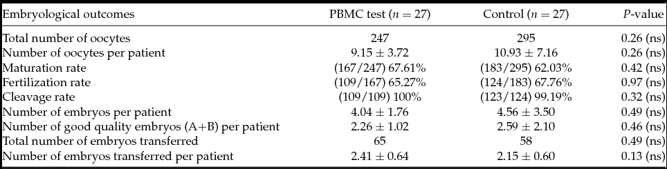
Follicles were collected 35–36 h post HCG injection. The oocytes obtained were denuded by hyaluronidase (hyaluronidase, LifeGlobal). After washing in buffered medium (Ferticult, JCD, France), oocytes were placed in a Petri dish that contained culture medium (Global®, LifeGlobal) and subsequently transferred to a box containing sperm microinjection immobilized in polyvinylpyrrolidone (PVP, LifeGlobal). Under a microscope, ICSI sperm is injected by pipette into the oocyte injection held in place with a drop of culture medium (Global®, LifeGlobal) through a contention pipette. The microinjected oocytes are cultured in microdroplets of 50 μl IVF30 medium (VitroLife) in each box in a 60 mm Nunc dish, covered with NidOil (Nidacon), then incubated in an oven at 37°C and in 5% CO2 in air. Thereafter, fertilization was confirmed by observation of the second polar body expelled and two pronuclei (2PN) in the oocyte cytoplasm. Embryos were kept in culture until the day 3 and their quality was evaluated in order to transfer the two best day 3 embryos into the uterus of the patient-treated PBMC or not. Adequate embryo quality was defined as embryos that had uniformly sized and shaped blastomeres, embryos with ooplasm that had no granularity and a maximum fragmentation of 10% were considered good quality embryos (A + B). Depending on the patient, one, two or three embryos were transferred in utero using a Frydman catheter (CCD Laboratories, Paris, France) (Table 2).
Comparison of patient embryological outcomes included in the study with at least 2 RIFs

Management of clinical pregnancy
The success of implantation is estimated by ultrasound via the observation of the embryo sac. The occurrence of clinical pregnancy is controlled by observing by ultrasound the development of embryo after 6–8 weeks.
Statistical analysis
Data were presented as mean ± standard deviation (SD) or standard number representing the total. Thus, these data were analyzed by Student's t-test for comparison of mean values or chi-squared test for comparison of percentages using the Statistical Package, Statistica (version 6.0) to compare significantly different populations: a P-value < 0.05 indicates significant difference. Then, mean values of clinical outcomes were evaluated to calculate study power by post-hoc test using G*Power software (version 3.0.10).
Results and Discussion
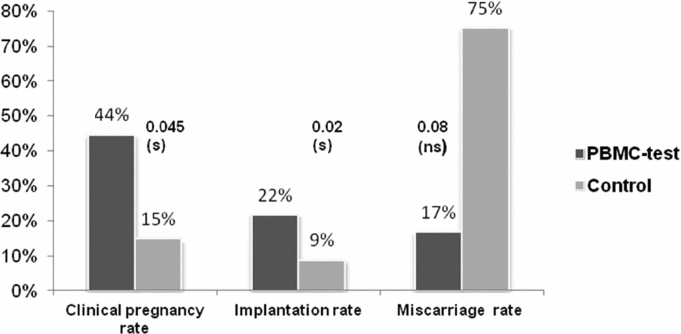
This study shows that in utero administration of autologous PBMC in patients with at least two RIF at improves implantation rate significantly (21.54 vs. 8.62%) compared with untreated patients and also the clinical pregnancy rate (44.44 vs. 14.81%) (Fig. 3). Such immunotherapy had no significative effect on the miscarriage rate of these patients when implantation has occurred, which could be a consequence of chromosomal disorders.
PBMC immunotherapy effect on clinical outcomes of patient with at least two RIFs. Note: Results are expressed as n, n(%). A statistically significant difference is considered when P < 0.05 (s). P > 0.05 is not significant (ns). Implantation rate is expressed by calculating the total number of embryonic sacs by total number of transferred embryos.

Our results are in agreement with the immunotropic theory, which stipulates that maternal immune cells positively influence implantation, placental and fetal growth. However, the precise mechanism of PBMC action is still unclear.
One of the hypotheses we can postulate for these patients with RIF is a poor recruitment of their lymphocytes at the endometrial level, which could be due to inadequate or insufficient endometrial signals. Therefore, in those patients, the inflammatory endometrial state usually required for implantation success should be affected. Then, uterine PBMCs supplementation 2 days before embryo transfer should compensate the endometrial initial inflammatory deficit.
Many studies complement our hypothesis, showing that uNK and T lymphocyte rates are decreased in patients with unknown infertility reporting a dysregulation of cytokines production (Klentzeris et al., Reference Klentzeris, Li, Dockery and Cooke1992; Fukui et al., Reference Fukui, Ntrivalas, Gilman-Sachs, Kwak-Kim, Lee, Levine and Beaman2006; Karami et al., Reference Karami, Boroujerdnia, Nikbakht and Khodadadi2012; Chernyshov et al., Reference Chernyshov, Sudoma, Dons’koi, Kostyuchyk and Masliy2010; Sacks et al., Reference Sacks, Yang, Gowen, Smith, Fay and Chapman2012; Zhou et al., Reference Zhou, Wang, Zhao, Wang, Sun and Hu2012; Dons’koi et al., Reference Dons’koi, Chernyshov, Sudoma, Honcharova and Osypchuk2013a,Reference Dons’koi, Chernyshov, Sirenko, Strelko and Osypchukb). Furthermore, an interesting and recent study demonstrated that PBMCs intrauterine administration had no effect on implantation rate in patients with only one IVF failure (34 vs. 33%) but significantly increased in patients with at least three IVF failures (42 vs. 25%) (Okitsu et al., Reference Okitsu, Kiyokawa, Oda, Miyake, Sato and Fujiwara2011).
Clinical best practice guidelines stipulate to look into possible underlying causes that may necessitate therapeutic investigation only after three implantation failures have taken place, and only if four good quality embryos of fresh or frozen cycles have been transferred minimally in women under the age of 40 (Coughlan et al., Reference Coughlan, Ledger, Wang, Demirol, Gurgan, Cutting, Ong, Sallam and Li2014). However, RIF as a clinical entity is yet to have a universally accepted definition but it refers to repeated failure in achieving clinical pregnancy. We, therefore, carried out a comparative analysis of the PBMC-treatment effectiveness as a function of the number of RIFs (e.g., two RIFs vs. three RIFs), as depicted in Table 3.
Comparison of patients’ characteristics in the two RIFs subgroup and the three RIFs at least subgroup

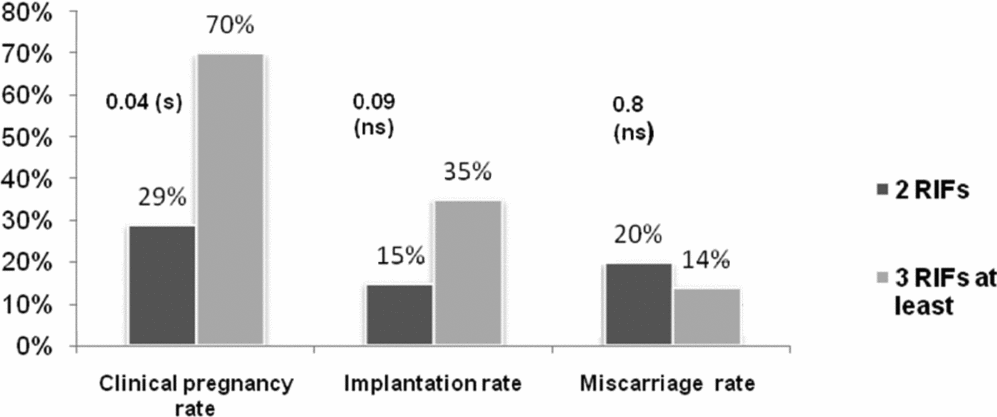
Clinical pregnancy rates strongly increase in patients with at least three RIFs as compared with control patients (70 vs. 29%; Fig. 4), in contrast with a lack of a significant difference between implantation and miscarriage rates in the same patients.
Comparison of PBMC immunotherapy effect on clinical outcomes between two RIFs patients subgroup and at least three RIFs patients subgroup. Note: Results are expressed as n, n(%). A statistic significant difference is considered when P < 0.05 (s). P > 0.05 is not significant (ns). Implantation rate is expressed by calculating the total number of embryonic sacs by total number of transferred embryos.

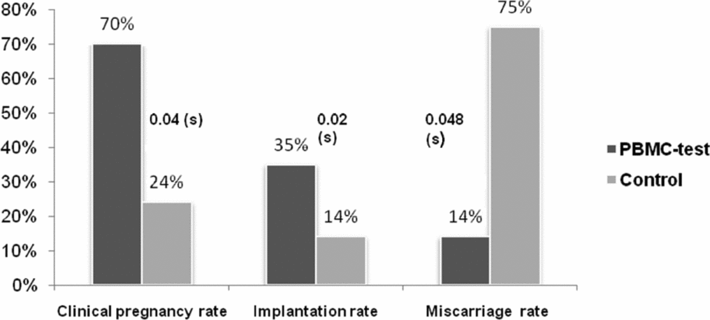
In addition, our study showed that when analyses were restricted to patients with at least three RIF (n = 27), which included 10 patients from the PBMC test versus 17 patient controls (Table 4), clinical pregnancy and implantation rates were approximately tripled in these treated patients compared with the controls (70, 35 vs. 24, 14% respectively) (Fig. 5), agreeing with Okitsu et al.'s (Reference Okitsu, Kiyokawa, Oda, Miyake, Sato and Fujiwara2011) findings. In addition, a significant five-fold decrease in miscarriage rate could be noted (14.29% in PBMC test vs. 75% in control (P = 0.048) and power 1 – β = 84%) compared with patients with at least two RIFs. Generally, other studies also support our observations (Nakayama et al., Reference Nakayama, Fujiwara, Maeda, Inoue, Yoshioka, Mori and Fujii2002; Zhylkova et al., Reference Zhylkova, Feskov, Feskova, Somova and Chumakova2010), confirming the effectiveness of our immunotherapy treatment in the patients who suffered from multiple location failures probably caused by insufficient presence of Th1 cytokines or from high miscarriages rate induced by Th2/Th1 deficit.
Characteristics of three RIFs at least patients subgroup

PBMC immunotherapy effect on clinical outcomes in the at least three RIFs patients subgroup. Note: Results are expressed as n, n(%). A statistically significant difference is considered when P < 0.05 (s). P > 0.05 is not significant (ns). Implantation rate is expressed by calculating the total number of embryonic sacs by total number of transferred embryos.

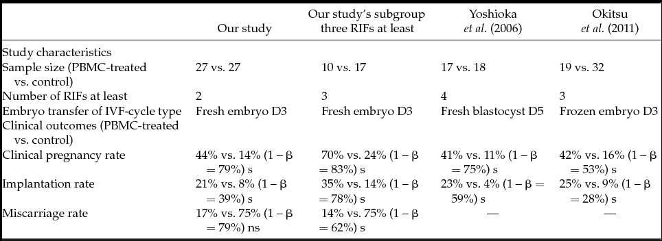
Those observations were supported by the statistical evaluation of our study power, which was about 66% for all patients and 74% in the three RIFs subgroup. Thus, our implantation and clinical pregnancy rates showed great improvements over previously published studies and were 59% for two RIFs and 81% for three RIFs (67% for Yoshioka et al., Reference Yoshioka, Fujiwara, Nakayama, Kosaka, Mori and Fujii2006 and 40% for Okitsu et al., Reference Okitsu, Kiyokawa, Oda, Miyake, Sato and Fujiwara2011) (Table 5).
Statistic power evaluation of different PBMC immunotherapy studies on clinical outcomes

Although the observed effects are in all likelihood due to the transferred PBMC, we could not totally exclude the possibility that the increased implantation rates in treated patients could be due to possible endometrial inflammation caused by the intrauterine devices used to deliver the PBMCs. While this can be controlled in women that undergo the same procedure minus the PBMCs, such a practice is ethically controversial and was not proposed in similar studies (Yoshioka et al., Reference Yoshioka, Fujiwara, Nakayama, Kosaka, Mori and Fujii2006; Okitsu et al., Reference Okitsu, Kiyokawa, Oda, Miyake, Sato and Fujiwara2011). Furthermore, others have shown that uterine mechanical stimulation prevents implantation in women (Rivera et al., Reference Rivera, Yacobson and Grimes1999), and intrauterine devices in rats evoked a minor inflammatory reaction that did not induce implantation (Schulten et al., Reference Schulten, Lobl and Ward1975). Together, it seems improbable that the increased implantation rates observed in our study would be attributable to uterine mechanical stimulation.
In the context of our study, it was interesting to investigate how administrated PBMCs could be activated to produce cytokines. It is well known that leukocyte activation begins during diapedesis when leukocytes move through blood vessels to colonize tissues. Furthermore, leukocytes are exposed in uteri to many signals that emanate from the endometrium and blastocyst. In our IVF protocol, it seems evident that many of these activating signals are short-circuited as in vivo embryo migration in maternal genital tracts does not occur. However, it is well established that PBMC, especially NK and monocytes, have the property to be activated by contact with their in vitro culture support. Therefore, we can easily suppose that, in our study, administered PBMCs were activated during the 3 days of in vitro culture knowing that after 24 h of PBMCs culture, IL1α, IL1β and IL8 transcripts are abundant (Ideta et al., Reference Ideta, Sakai, Nakamura, Urakawa, Hayama, Tsuchiya, Fujiwara and Aoyagi2010). In addition, PBMCs in our protocol stay in uteri for 2 days before embryos transfer and then can be submitted to various activating endometrial signals to produce cytokines.
When transferred in utero, PBMCs can modulate implantation quality using direct or indirect pathways by cytokine and growth factor secretion or by acting on adhesion proteins. It has been shown that uNKs but not pNKs produce at implantation specific cytokines such as GM-CSF, CSF1, IL2, IFNγ and LIF (Jokhi et al., Reference Jokhi, King, Sharkey, Smith and Loke1994; King et al., Reference King, Jokhi, Smith, Sharkey and Loke1995; Ideta et al., Reference Ideta, Sakai, Nakamura, Urakawa, Hayama, Tsuchiya, Fujiwara and Aoyagi2010; Yu et al., Reference Yu, Yang, Guo, Fang, Yin, Luo, Li, Li, Zhao, Zou and Xu2014). This observation suggests an important local action for these maternal immune cells to promote implantation. It is now well established that uNKs induce endometrial decidualization phenomena in order to prepare implantation through LIF secretion (Kimber, Reference Kimber2005; Yu et al., Reference Yu, Yang, Guo, Fang, Yin, Luo, Li, Li, Zhao, Zou and Xu2014) and stimulate angiogenesis in the decidua (Croy et al., Reference Croy, Chantakru, Esadeg, Ashkar and Wei2002; Hanna et al., Reference Hanna, Goldman-Wohl, Hamani, Avraham, Greenfield, Natanson-Yaron, Prus, Cohen-Daniel, Arnon, Manaster, Gazit, Yutkin, Benharroch, Porgador, Keshet, Yagel and Mandelboim2006; Oh & Croy, Reference Oh and Croy2008) remodeling endometrial spiral arteries by VEGF, PLGF and angiogenic substances (Segerer et al., Reference Segerer, Kammerer, Kapp, Dietl and Rieger2009; Lash & Bulmer, Reference Lash and Bulmer2011; Yu et al., Reference Yu, Yang, Guo, Fang, Yin, Luo, Li, Li, Zhao, Zou and Xu2014). Furthermore, VEGF concentration peaked significantly in first-trimester decidua and seemed indispensable for implantation success and the maintenance of pregnancy (Salamonsen, Reference Salamonsen2003; Segerer et al., Reference Segerer, Kammerer, Kapp, Dietl and Rieger2009).
Aberrant maternal immune responses and an unbalanced cytokine profiles specially the ones secretes by uNKs are influencing feto-maternal immune tolerance during implantation and could induce RIF or even recurrent pregnancy losses (Makrigiannakis et al., Reference Makrigiannakis, Minas, Kalantaridou, Nikas and Chrousos2006, Reference Makrigiannakis, Petsas, Toth, Relakis and Jeschke2011; Toth et al., Reference Toth, Würfel, Germeyer, Hirv, Makrigiannakis and Strowitzki2011). Patients with miscarriages present over-expression of IL15 which be related to vascularization with consecutive placental and fetal rejection (Toth et al., Reference Toth, Haufe, Scholz, Kuhn, Friese, Karamouti, Makrigiannakis and Jeschke2010).
uNKs also promote an increase in decidual monocyte-attracting chemokines secretion, especially GM-CSF, which decreased level has been clearly correlated with implantation failure rate in RIF patients (Perricone et al., Reference Perricone, De Carolis, Giacomelli, Guarino, Sanctis and Fontana2003). In such patients, embryos cultures supplementation with GM-CSF demonstrated an increase in implantation rate or even included in the basic treatment for RIF patients for immune modulation (Ziebe et al., Reference Ziebe, Loft, Povlsen, Erb, Agerholm, Aasted, Gabrielsen, Hnida, Zobel, Munding, Bendz and Robertson2013; Toth et al., Reference Toth, Würfel, Germeyer, Hirv, Makrigiannakis and Strowitzki2011).
The function of uNK cells includes more than cytokine production; they contribute to regulate trophoblastic invasion through cytotoxicity, which is controlled by activating signals and inhibitory receptors. Excessive cytotoxicity is known to provoke implantation failures. However, in this study, PBMC supplementation has a positive effect on implantation rate so it is not very probable that their action includes, at least in this protocol, an increase in cytotoxicity. An interesting study has shown that most uNK cells are considered to be cytokine-producing cells rather than cytotoxic cells (Tabiasco et al., Reference Tabiasco, Rabot, Aguerre-Girr, El Costa, Berrebi, Parant, Laskarin, Juretic, Bensussan, Rukavina and Le Bouteiller2006). It is interesting to note that once implantation has occurred in patients with two RIFs, we found no significant effect of immunotherapy on miscarriage rate. This result suggests that PBMC transfer has an effect only when the implantation process fails. Thus immunotherapy appears to be ineffective in patients with RIF caused by an early dysregulation in the maintenance of pregnancy. Patient with unexplained recurrent spontaneous abortions (URSA) probably present with over-activation of cytotoxic uNKs characterized by up-regulation of CD69stim and CD158a as reported elsewhere (Chernyshov et al., Reference Chernyshov, Sudoma, Dons’koi, Kostyuchyk and Masliy2010; Sacks et al., Reference Sacks, Yang, Gowen, Smith, Fay and Chapman2012; Dons’koi et al., Reference Dons’koi, Chernyshov, Sudoma, Honcharova and Osypchuk2013a,Reference Dons’koi, Chernyshov, Sirenko, Strelko and Osypchukb; Wu et al., Reference Wu, Luo, Zhang, Li, Xu, Zhou, Luan and Liu2014). In these patients, another therapeutic strategy needs to be developed.
In this study, we have not considered the PBMCs-T lymphocytes fraction role, as it is known that there is no adhesion of these cells in in vitro culture support in contrast with NKs and monocytes. This reason is why T lymphocytes are probably eliminated during culture cell washing.
Another action of PBMCs on implantation and clinical pregnancy rates in our patients may be mediated by macrophages. Maternal macrophages represented 30–35% of all decidual leukocytes in first-trimester deciduas (Bulmer et al., Reference Bulmer, Lunny and Hagin1988) and should be implicated through their cytokine secretion in the control of trophoblastic invasion (Abrahams et al., Reference Abrahams, Straszewski-Chavez, Guller and Mor2004). Furthermore, uNK activation requires IL2 produced by macrophages and macrophage interactions through the NKG2D signalling pathway (Basu et al., Reference Basu, Eriksson, Pioli, Conejo-Garcia, Mselle, Yamamoto, Wira and Sentman2009). Indeed, decidual macrophages are considered as local immuno-modulators during implantation and eventually essential for normal pregnancy with abundance of M2 phenotype. Conversely, hypothetically, the over-expression of Fas ligand (FasL) as proapoptotic cytokine by M1 phenotype induced trophoblast apoptosis in interaction with decidual corticotropin-releasing hormone (CRH) (Guenther et al., Reference Guenther, Vrekoussis, Heublein, Bayer, Anz, Knabl, Navrozoglou, Dian, Friese, Makrigiannakis and Jeschke2012; Kalantaridou et al., Reference Kalantaridou, Zoumakis, Weil, Lavasidis, Chrousos and Makrigiannakis2007). Fas/FasL and petformin expressed by trophoblasts play a crucial role in feto-maternal immune tolerance during implantation (Makrigiannakis et al., Reference Makrigiannakis, Karamouti, Drakakis, Loutradis and Antsaklis2008). For implantation, CRH is initially expressed in the endometrium to finely control stromal cell decidualization; for maintenance of pregnancy, CRH is produced more by the placenta following urocortin action (Zoumakis et al., Reference Zoumakis, Kalantaridou and Makrigiannakis2009). However, the increase in the decidual macrophages population is present in patient with spontaneous miscarriages and other gynaecological disorders as pre-eclampsia and endometriosis with over-expression of G-protein-coupled oestrogen receptor (GPER) (Guenther et al., Reference Guenther, Vrekoussis, Heublein, Bayer, Anz, Knabl, Navrozoglou, Dian, Friese, Makrigiannakis and Jeschke2012; Heublein et al., Reference Heublein, Lenhard, Vrekoussis, Schoepfer, Kuhn, Friese, Makrigiannakis, Mayr and Jeschke2012; Hutter et al., Reference Hutter, Heublein, Knabl, Andergassen, Vrekoussis, Makrigiannakis, Friese, Mayr and Jeschke2013).
Our results support the hypothesis that in RIF patients lymphocytes recruitment is not able to induce uterine inflammatory condition suitable for implantation. As this uterine infiltration of uNK cells is clearly influenced by endometrial cytokines secretion we can suppose that RIF patient endometrium has inadequate cytokine secretion, especially IL15 and IL11, which are known to be implicated in proliferation and differentiation of uNKs (Ashkar et al., Reference Ashkar, Black, Wei, He, Liang, Head and Croy2003; Becknell & Caligiuri, Reference Becknell and Caligiuri2005).
Thus, in utero administration of autologous PBMCs increases implantation rate in patients with RIF should be an effective therapeutic response to implantation repeated failures in order to improve classical IVF protocols success rate knowing that implantation is the most determinant mechanism in ART.