INTRODUCTION
Investigations of the functional neuromuscular biology of parasitic platyhelminths historically focused on small molecule, classical neurotransmitters such as acetylcholine, serotonin and catecholamines. The excitatory action of serotonin and the inhibitory action of acetylcholine (ACh) on fluke muscle preparations was described over 50 years ago (Chance & Mansour, 1953). These systems have provided targets for anthelmintic drugs; for example, cholinergic agonists have been used to treat tapeworm infections.
In comparison, it was only recently that the importance of neuropeptides in platyhelminths has been recognised by parasitologists. The previous chapter of this supplement (Ribiero, El-Shehabi & Patocka, in this supplement) exposes some of the impediments to the study of flatworm neuromuscular biology. Add to these the relatively recent awareness of the biological importance of neuropeptides in the phylum, and the understanding of the neuromuscular role of flatworm neuropeptides is truly in its nascency. However, even at this early stage, there are reasons to believe that neuropeptidergic systems could yield novel drug targets for a whole new generation of anthelmintic drugs.
There is clear evidence that neuropeptides are central to the biology of flatworms and, as illustration, neuropeptide immunostaining outstrips that reported for classical transmitter molecules in flatworms (Halton & Maule, 2004). Two distinct classes of neuropeptides have been identified within the phylum. Firstly, there are FMRFamide-like peptides (FLPs), which are relatively short peptides (typically <20 amino acids) ending in a carboxy-terminal RFamide motif. Secondly, there are neuropeptide Fs (NPFs), which are larger peptides (36–40 amino acids) that are related to the vertebrate neuropeptide Y (NPY) family of peptides.
In addition to playing a central role in flatworms, the importance of neuropeptides in nematodes has also become apparent. One inaccurate concept is that the two phyla of worms are very similar, such that things learned from the nematodes can be easily extrapolated to the platyhleminths. The other extreme would also be inaccurate – that is, to think that the worms are completely dissimilar such that there will be no commonalities between the two phyla. This principle can be illustrated in the context of a quick overview of platyhemlinth neuropeptides in comparison to nematode neuropeptides.
The most easily noted disparity between the two phyla is the dramatic disproportion in the breadth of neuropeptides expressed by worms from the two groups. Each nematode species has a staggering complement of apparent neuropeptides, with at least 236 distinct peptides encoded in the genome of C. elegans and 67 of these being FLPs (Li, Kim & Nelson, 1999; Pierce et al. 2001; Kim & Li, 2004; McVeigh et al. 2005). This wide neuropeptide complement is apparent in parasitic nematodes as well, where biochemical and genetic information has confirmed at least 27 distinct FLPs in Ascaris suum (Davis & Stretton, 1996; Yew et al. 2005). Compared to this staggering complement of FLPs present in typical free-living and parasitic species of nematodes, only one or two distinct FLPs have been found in each platyhelminth species thus far examined. While it is possible that some of these species may contain as yet undetected FLPs, it does not seem possible that there is anywhere close to the complement of FLPs in flatworms that are found in nematodes. Another difference between the phyla relates to the neuropeptide F (NPF) family of peptides. The NPFs are abundant in every platyhelminth thus far examined. However, the presence of the NPF family of peptides has not yet been established in the nematodes, and it seems likely that they may be absent from the phylum altogether.
Pointing out these differences might lead to the impression that there is little commonality in the neuropeptidergic components of the two phyla of worms, but that is not the case. Although the breadth of FLPs is different in the two phyla, some of the FLPs share a great deal of similarity. As a testimony, FLPs from each phylum have activity in the other; for example, a number of nematode FLPs are myoexcitatory in flatworm preparations (Mousley et al. 2004). Also, there are components of the neuropeptidergic systems that appear to be common to both phyla, such as the neuropeptide receptors, the proteins involved in signal transduction, the processing enzymes involved in the generation of active neuropeptides and, possibly, the proteases involved in terminating neuropeptide signalling.
There is reason to believe that these neuropeptidergic systems could yield novel drug targets. Firstly, the neuropeptide signalling systems are widespread and important in parasitic flatworms (Halton & Gustafsson, 1996). Although we have much to learn, there is little doubt of the central role of neuropeptides in flatworm biology; one clear role for neuropeptides is in the control of motor function (Day & Maule, 1999). In contrast, at least some of the neuropeptides present in parasitic flatworms have restricted roles in their mammalian hosts. Also, the endogenous neuropeptides are remarkably potent in parasitic worms; in schistosomes, some peptides have threshold activities in the low nanomolar or high picomolar range (Day et al. 1997; Humphries et al. 2004).
This paper will provide an overview of what is known about the role of neuropeptides in platyhelminths, keeping an eye on the consideration that molecules involved in the neuropeptidergic signalling could be attractive targets for novel drugs to treat infections with platyhelminths. Elsewhere in this supplement, the possibility of these targets crossing phyla lines is considered in great detail (Mousley, Maule & Marks, in this supplement).
FMRF AMIDE-LIKE PEPTIDES (FLPS)
Structure of flatworm FLPs
FLPs (also referred to as FMRFamide-related peptides, or FaRPs) are, as their name suggests, somewhat like the seminal neuropeptide sequence of FMRFamide, isolated from the Venus clam Macrocallista nimbosa by virtue of its cardioexcitatory activity (Price & Greenberg, 1977). This group now represents the largest family of neuropeptides known in the invertebrates. FLPs are abundant and widespread within flatworms, but the structural diversity characteristic of FLPs in other invertebrate phyla is lacking. All currently identified flatworm FLPs conform to the formal FLP carboxy terminal motif of (F/Y)XRFamide, where X denotes a variable hydrophobic residue (Greenberg et al. 1988; Shaw, Maule & Halton, 1996; Espinoza et al. 2000).
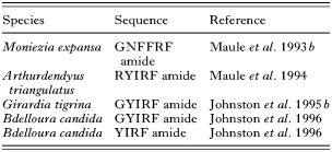
In contrast to the structural complexity of the platyhelminth nervous system, with its characteristic orthogonal arrangement (Halton & Gustafsson, 1996), flatworms appear to be rather straightforward in terms of neuropeptide diversity. There was a flurry of reports identifying four flatworm FLPs around a decade ago (Maule et al. 1993b, 1994; Johnston et al. 1995b, 1996) (see Table 1), but no novel structural or molecular characterizations have since been reported, and no FLPs have yet been isolated from the trematodes.
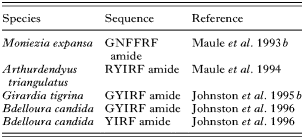
The current lack of structural peptide data from these important parasites can be attributed to their generally small size and scarcity, which practically preclude peptide discovery through the tissue-hungry techniques of acid-ethanol extraction, HPLC and peptide sequencing. As an example, isolation and structural analysis of the cestode FLP, GNFFRFamide, required an initial amount of 1 kg Moniezia expansa tissue (Maule et al. 1993b; Shaw et al. 1996). Such relatively massive amounts of tissue are viable when dealing with large, easily obtained species such as tapeworms, but are problematic when dealing with the small and relatively rare trematodes. Greater success has been achieved with isolations from turbellarian tissue. These free-living platyhelminths express higher levels of neuropeptides in their nervous systems, a feature which could be at least partly attributable to the greater mobility demanded by free-living worms as compared to some of their parasitic cousins who have a lower overall requirement for neuromuscular dexterity when residing within the host environment (Johnston et al. 1995a). This results in a higher ratio of peptide to wet weight of tissue in turbellarians and, in addition to the readily available supply of many of these species, accounts for the greater success of neuropeptide extractions from these worms.
The first identified flatworm FLP, GNFFRFamide from the sheep tapeworm M. expansa (Maule et al. 1993b), has since been resolved as an atypical member of the small family of platyhelminth FLPs. The FLPs characterized from turbellarians are tetra/pentameric and share a characteristic YIRFamide carboxy terminus. Despite its double aromatic motif, GNFFRFamide is still consistent with the generalised FLP structure, since phenylalanine possesses both aromatic and hydrophobic characteristics. The distinct structure of this cestode FLP may reflect the relative evolutionary relationships between the platyhelminth classes; cestodes are considered the most derived class of flatworms. With further regard to evolutionary lineages, it has been postulated that the turbellarian FLPs – not FMRFamide – represent the ancestral FLP structure in invertebrates, since bilaterian flatworms could represent the evolutionary progenitors of all invertebrate phyla (Shaw, Maule & Halton, 1996).
No molecular data have yet been published regarding FLP gene structure in platyhelminths. Although molecular techniques eliminate many of the inherent difficulties associated with lack of source tissue, effectively allowing amplification of cDNA from milligram amounts of tissue (Matz, 2002, 2003), neuropeptide genes are nevertheless expressed at low levels, compounding the difficulties associated with the oft-attempted but rarely successful approach of PCR amplification using degenerate oligonucleotides. Further, flatworm biology has not, until very recently, benefited from the establishment of genome sequencing programmes and associated expressed sequence tag (EST) generation. In contrast, parasitic nematodes have enjoyed such attention, resulting in the successful identification of numerous flp gene candidates from a range of species (McVeigh et al. 2005). The ongoing surge of interest in bioinformatics has now led to the initiation of genome projects for Schistosoma (El-Sayed et al. 2004; LoVerde et al. 2004) and the turbellarian Schmidtea mediterranea (Sanchez Alvarado et al. 2002; Reddien et al. 2005), in addition to the EST programmes in S. mansoni and S. japonicum and other parasitic species including Echinococcus granulosus, E. multilocularis, Mesocestoides corti, Echinostoma paraensi, Fasciola hepatica and Clonorchis sinensis as well as the important free-living models Dugesia ryukyuensis and D. japonicum (http://www.ncbi.nlm.nih.gov/dbEST dbEST release 051305). Even cursory searches of these databases reveal many sequences encoding putative FLPs, but as yet no flp genes have been characterized. The impending completion of the S. mansoni genome project (LoVerde et al. 2004) should provide resources for the molecular characterization of transcripts encoding neuropeptides and their receptors in a manner similar to those of the Caenorhabditis elegans and Drosophila melanogaster projects. S. mansoni genome data should also provide a springboard for the similarity-based identification of homologous genes in other flatworms.
Localisation of flatworm FLPs
FLP localisation studies have traditionally employed immunocytochemical (ICC) techniques coupled to confocal scanning laser microscopy (CSLM) (Halton & Maule, 2004). Such methods have been useful because they typically require only a short peptide sequence to enable antigen preparation and subsequent generation of specific antisera. Consequently ICC has been applied to the localisation of endogenous FLPs isolated from flatworms in the absence of the molecular sequence information which would allow employment of the much more specific technique of in situ hybridisation (ISH). However, the current and continuing increase in genetic sequence data from platyhelminths will allow design of DNA/RNA probes for use in ISH techniques in the near future, enabling useful and interesting comparisons to be made regarding the specificity of existing FLP immunolocalisation.
FLP distribution has been extensively reviewed over the years (Shaw et al. 1996; Day & Maule, 1999; Maule et al. 2002; Halton & Maule, 2004) and will not be repeated in detail here. In short, FLP immunoreactivity (FLP-IR) has been reported in a wide variety of species, including representatives of all major platyhelminth groups. The typical flatworm nervous system consists of a central nervous system (CNS), comprised of a bi-lobed brain and paired longitudinal nerve cords, and a peripheral nervous system, comprised of a number of nerve nets somewhat reminiscent of the plexuses seen in cnidaria. Generally FLP-IR is widespread within flatworms and has been described in the innervation of locomotory muscle, attachment structures, the alimentary system and reproductive organs. Distribution includes both these peripheral neuronal elements and neurones of the central nervous system. FLP distribution patterns mirror those of the classical transmitter ACh in monogeneans (Maule et al. 1990b; Cable et al. 1996; Zurawski et al. 2001) and trematodes (Stewart, Marks & Halton, 2003). In neuromuscular innervation at least, the apparently complementary distribution of an inhibitory transmitter (ACh) with the wholly excitatory FLPs seems rational in terms of these two neurotransmitters performing a co-modulatory role in neuromuscular co-ordination. The existence of separate neuronal pathways for FLPs and serotonin have been demonstrated in the majority of species examined (Maule et al. 1990a; Biserova et al. 2000; Zurawski et al. 2001; Kotikova et al. 2002; Stewart, Marks & Halton, 2003; Stewart et al. 2003a,b) with the exception of ootype innervation in the trematode Echinostoma caproni, which includes cells displaying the apparent co-localisation of serotonin and FLP immunoreactivities (šebelová et al. 2004). Since there is evidence that serotonin acts to maintain the contractile activity of flatworm muscle through stimulation of cAMP levels (Day, Bennett & Pax, 1994), it has been suggested that serotonin might maintain muscular energy levels through elevation of cAMP in ootype cells, and that the FLP(s) trigger the onset of egg production (šebelová et al. 2004). However, no data are available from flatworms regarding interactions between FLP signalling and cAMP levels.
Such observations as those of šebelová et al. (2004) regarding FLP involvement in flatworm reproductive function have long been a focus for parasite neurobiologists, because pharmacological interference with reproductive function could have significant therapeutic value. Perhaps the strongest evidence implicating FLPs in reproductive function of flatworms is that regarding the FLPergic system of Polystoma nearcticum, a monogenean parasite of the grey treefrog, Hyla versicolor. This parasite lives within the host urinary bladder, in reproductive synchrony with its anuran host. An ICC study has shown that while serotonin-IR of the parasite's reproductive apparatus remains constant, FLP-IR can only be demonstrated during host spawning (Armstrong et al. 1997), suggesting an intrinsic and regulated role for FLPs in the modulation of flatworm reproduction. The involvement of FLPs in reproductive function is further supported by a recent developmental study showing that FLP innervation of the reproductive system of two strigeid trematodes could only be demonstrated following the onset of egg production (Stewart, Marks & Halton, 2003). The collective evidence strongly supports a role for FLPs in flatworm reproductive function.
Physiology of flatworm FLP signalling
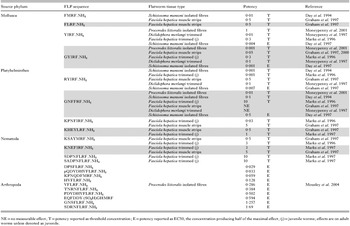
The earliest studies aiming to characterize the effects of putative transmitters on flatworm muscle utilised muscle strip or whole worm assays (Chance & Mansour, 1949, 1953; Paasonen & Vartiainen, 1958; Terada et al. 1982). These approaches have been successful in characterizing the excitatory action of FLPs on flatworm somatic muscle, including Diclidophora merlangi (Maule et al. 1989b; Moneypenny et al. 1997), E. caproni (Humphries et al. 2000) and F. hepatica (Marks et al. 1996; Graham, Fairweather & McGeown, 1997). These investigations provide evidence of the myoexcitatory actions of platyhelminth FLPs, but the inherent drawback of such studies is that they cannot discriminate between neuropeptide effects on muscle and those on nerve or other tissues. For example, it is difficult to determine if the excitatory effects on a whole worm are due to direct excitation of the muscle, an effect on motor neurones or a combination of both. The acoelomate anatomy of flatworms renders the denervation of muscle tissue by dissection practically impossible. An alternative method of separating muscle from nerve tissue was developed using schistosomes (Blair et al. 1991) in which enzymatic digestion produced dispersed, individual muscle fibres which retained their contractile qualities. This preparation not only allows direct study of neurotransmitter effects on muscle fibres free from interference caused by extraneous tissues (which in standard tissue preparations can hinder access of polar compounds to muscle-based receptors), but with the removal of these extraneous tissues the site of excitation can be more definitively localised to the muscle. The excitatory effects of flatworm FLPs on isolated muscle fibres have been reported in S. mansoni (Day et al. 1994), Bdelloura candida (Johnston et al. 1996) and Procerodes littoralis (Moneypenny et al. 2001), suggesting that an FLP receptor resides on flatworm muscle fibres. The presence of an FLP-specific receptor on the somatic musculature is further substantiated by the specificity of FLP myoexcitation. For example, single amino acid substitutions were sufficient to cause flatworm FLPs to lose their activity on schistosome muscle preparations (Day et al. 1997).
Further confirmation of the existence of a single muscle-based FLP receptor is provided by studies utilising peptide analogues incorporating the d isomer of phenylalanine (dF). These inactive forms competitively inhibit the excitatory effects of flatworm FLPs. A GYIRFamide analogue (GYIRdFamide) blocks the contractile effects of GYIRFamide, YIRFamide and GNFFRFamide on isolated turbellarian muscle cells (Moneypenny et al. 2001). Similarly, FMRdFamide blocked the contractile effects of both FMRFamide and GNFFRFamide on S. mansoni muscle fibres (Day et al. 1994) (Table 2).

FLP signalling
Graham, Fairweather & McGeown (2000) published the only report on FLP signalling, investigating GYIRFamide-induced excitation in muscle strips of the trematode F. hepatica. This study used a physiological approach to monitor the activity of GYIRFamide in the presence of pharmacological modulators of intracellular signalling pathways. Results suggest that GYIRFamide signals through a G protein-coupled receptor (GPCR) pathway involving activation of phospholipase C (PLC) and protein kinase C (PKC). These are both constituents of the phosphatidylinositol pathway, which involves G-protein activation of PLC, an enzyme which processes phosphatidylinositol (4,5) bisphosphate (PIP2) into inositol (1,4,5) triphosphate (IP3) and diacylglycerol (DAG). DAG stimulates PKC to activate effector proteins such as enzymes, contractile proteins and ion channels. The myomodulatory activity of PKC has also been demonstrated in S. mansoni, where phorbol esters, well known activators of PKC, produced spastic paralysis of schistosome musculature (Blair, Bennett & Pax, 1988), and in Bdelloura candida where these compounds caused partial tonic paralysis of whole worms (Blair & Anderson, 1994). IP3 acts to release sequestered Ca2+ from the sarcoplasmic reticulum (SR) through IP3 receptor channels. Although other studies have shown that PLC stimulation leads to IP3 production in the cestode Hymenolepis diminuta (Samii & Webb, 1996) and S. mansoni (Wiest et al. 1992), these have not yet been directly linked to the FLP signalling system. These pathways indirectly implicate the presence of a functional SR within flatworm muscle cells. In fact, SR components have been demonstrated in flatworm tissue, including the presence of ryanodine receptor (RyR) channels capable of supporting contraction of schistosome muscle (Day et al. 2000). The S. mansoni SR is also imbued with thapsigargin-sensitive Ca2+ pumps, which allow sequestration of cytosolic Ca2+ (Noel et al. 2001).
The involvement of extracellular Ca2+ influx through voltage-gated channels in contractile activity has been demonstrated in the turbellarian Dugesia tigrina (Cobbett & Day, 2003), and the trematodes F. hepatica (Graham, McGeown & Fairweather, 1999) and S. mansoni (Day et al. 1994). Although there are some data implicating extracellular Ca2+ in the FLP-induced contractions (Day et al. 1994), the conduit for that influx and how it is coupled to PKC is unclear.
In summary, although FLPs are abundant and important in flatworms their biological functions and the associated signalling pathways remain unclear. There are reasons to believe that some of these issues can be illuminated in the next few years. Firstly, the study of dispersed muscle fibres is expanding and more data should accumulate regarding the events associated with FLP-induced myoexcitation. For example, the protocol has been successfully applied to F. hepatica (Kumar et al. 2003, 2004). Secondly, more techniques are now becoming available to investigate the functional biology of flatworms, such as RNA interference (Boyle et al. 2003; Skelly, Da'dara & Harn, 2003) and genetic transformation (Wippersteg et al. 2002, 2003, 2005; Gonzalez-Estevez et al. 2003; Heyers et al. 2003). These should lead to the application of a range of molecular manipulations such as gene silencing or overexpression, as well as fluorescent protein-based expression analyses.
NEUROPEPTIDE FS (NPFS)
Structure of flatworm NPFs
The earliest suggestions that platyhelminths possessed neuropeptides similar to the vertebrate neuropeptide Y (NPY) family came from immunocytochemistry. In the 1980s and into the early 1990s, there were many reports of NPY family-IR in flatworms, and the most intense staining was usually noted with antisera targeting the carboxy terminus of pancreatic polypeptide (PP), a member of the NPY family (Larhammar, 1996; Balasubramaniam, 1997, 2003). PP-IR has been recorded throughout the nervous systems of every species examined; as well as the extensive staining throughout the CNS PP-immunopositive nerve plexuses serve the reproductive structures, the sub-tegumental muscle layers, the holdfast organs and the alimentary systems of flatworms. For example, in S. mansoni, there is extensive PP-IR in the nerve fibres surrounding the muscular egg chamber and at the entrances of the oviducts (Skuce et al. 1990; Marks et al. 1995). There is also extensive staining in the nerve plexuses serving the subtegumental, somatic muscle layers (Skuce et al. 1990), and the suckers that the male worms use for attachment within the vasculature (Marks et al. 1995). PP-IR is also abundant in nerves approaching the oesophagus of schistosomes that leads from the mouth to the blind-ending gut.
The neuronal location of PP-IR in flatworms suggested that the parasite antigens responsible were akin to vertebrate NPY. Furthermore, there was chromatographic evidence that the parasite antigens were structurally similar to the 36 amino acid peptide common to NPY family members. Chromatographic analysis of peptide extracts from D. merlangi (Maule et al. 1989a), M. expansa (Maule et al. 1991) and S. mansoni (Humphries et al. 2004) found a single peak of PP immunoreactivity which co-eluted with other NPY family members from columns which separate on the basis of size and hydrophobicity. This type of evidence led to the hypothesis that there was an NPY-like neuropeptide present in flatworms even before the identification of a specific peptide from the phylum. There was also evidence for similar peptides in other closely-related invertebrates (Maule, Halton & Shaw, 1995), and they also appeared to be abundant and widespread in the nervous system.
The first identification of an NPY-like neuropeptide in a flatworm was via biochemical purification of the PP immunoreactive peak from the tapeworm M. expansa (Maule et al. 1991, 1992a). The tapeworm neuropeptide is 39 amino acids in length and has striking similarity to peptides of the NPY family, which are typically 36 amino acids in length. The peptide was named neuropeptide F (NPF) because it features a carboxy terminal Phe (F) residue, in contrast to NPY's carboxy terminal Tyr (Y). The discovery of M. expansa NPF (mxNPF) was the first NPF identified in any animal, and it was also the first neuropeptide identified in a platyhelminth. Subsequent to the identification of mxNPF, similar peptides have been identified in other animals, including other platyhelminths (Table 3).

In addition to the cestode Moniezia, NPFs have been identified in the turbellarian Arthurdendyus triangulatus (Curry et al. 1992; Dougan et al. 2002) and the trematodes S. mansoni and S. japonicum (Humphries et al. 2004). All of these NPFs feature a carboxy terminal GRPRFamide motif, are 36 amino acids in length (except mxNPF which is 39 amino acids long), and have conserved Y residues at positions 10 and 17 relative to the carboxy terminus, the hallmark feature of NPY family peptides.
Immunolocalisation of flatworm NPFs
NPF-immunoreactivity (NPF-IR) is abundant and widespread throughout the nervous system of every flatworm species examined, which includes over 30 species. Previous reviews have provided extensive reports of these staining patterns (Day & Maule, 1999; Maule et al. 2002; Halton & Maule, 2004), and only a summary will be provided here.
Firstly, NPF-IR occurs exclusively in neural elements in flatworms. NPF is abundantly expressed in the CNS of flatworms, including in the bi-lobed brain, the main nerve cords and the commissures that connect the two main nerve cords as they progress away from the brain. NPF-IR is also present in much of the peripheral nervous system. The nerve fibres serving most of the distinct muscle systems of flatworms are replete with NPF-IR, including the somatic muscle layers, the holdfast organs, the reproductive structures and the alimentary system. Though the specific orientation and grouping of somatic musculature in the different classes within the platyhelminths is variable, there is consistently NPF-IR throughout the nerves associated with these muscle layers. Schistosomes are an apt example. Schistsomes have a rather complex organization of body muscle which lies in three distinct layers – circular, longitudinal and diagonal – directly underneath the worm's outer tegument (Mair et al. 2000b). These muscle layers are invested with NPF-immunoreactive neural elements (Skuce et al. 1990), and this appears typical as it is repeated in all other flatworms examined, which includes examples from each class (Maule et al. 1993a; Reuter et al. 1995a,b). The parasitic platyhelminths also feature distinct muscle groupings in their holdfast organs, which are critical for the maintenance of proper position within or on the host and which are also supplied with NPF-immunopositive innervation (Maule et al. 1990a, 1992a,b; Marks et al. 1995).
NPF is also present in the peripheral nerve plexuses associated with the reproductive structures of flatworms. For example, NPR-IR is rich in nerves surrounding the muscular egg chambers of the important trematode parasites S. mansoni (Skuce et al. 1990; Marks et al. 1995) and F. hepatica (Magee et al. 1991; Marks et al. 1995). Much of the pathology associated with these parasites is directly attributable to the eggs, yet a great deal remains unknown regarding the processes regulating egg production. There are also special muscle groups associated with the alimentary systems that include innervation that displays NPF-IR. For example, the muscular pharynx of the turbellarians P. littoralis (Reuter et al. 1995b) and Archiloa unipunctata (Reuter et al. 1995a) are heavily innervated, as is the oral sucker of schistosomes (Skuce et al. 1990; Marks et al. 1995; Mair et al. 1998).
Physiology/biochemistry of flatworm NPFs
Even though the first NPFs were identified over a decade ago, and even though NPFs appear widely distributed throughout the nervous systems of flatworms and other invertebrates, there is still very little known about the biological function of these peptides in flatworms or other invertebrates. The localisation of NPFs could provide clues, suggesting a role for the peptide in the modulation of somatic muscle, reproduction and feeding. However, the distribution of NPF is so broad that it eliminates few possibilities.
There are data emerging that link NPF signalling to a specific biochemical pathway, the inhibition of cyclic AMP (cAMP) accumulation. Structural data suggest a relationship between invertebrate NPFs and vertebrate NPYs, and new functional data also connect the peptides. NPY family peptides act on a family of G protein-coupled receptors (GPCRs) known collectively as Y receptors. All five Y receptor subtypes have been linked to the inhibition of adenylyl cyclase, and two of the subtypes are also associated with increases in cytoplasmic Ca2+ (Motulsky & Michel, 1988; Mihara, Shigeri & Fujimoto, 1989; Aakerlund et al. 1990). The inhibition of cAMP accumulation is so regularly associated with Y receptors that it has been called the universal signalling mechanism for Y receptors (Michel, 1991; Michel et al. 1998).
Two invertebrate GPCRs have been cloned which, when in heterologous expression systems, respond to NPFs; one is from Lymnaea stagnalis (Tensen et al. 1998) and one from Drosophila melanogaster (Garczynski et al. 2002). Like receptors for vertebrate NPYs, the invertebrate NPF receptors produce an inhibition of cAMP accumulation, suggesting that this signalling pathway could be conserved between NPYs and NPFs. A more compelling case for linking invertebrate NPF receptors to the inhibition of cAMP comes from schistosomes. Schistosome NPF inhibits the accumulation of cAMP in schistosome homogenates at a threshold of 10−11 M (Humphries et al. 2004). The inhibition is quite specific, with the most potent action reserved only for the endogenous schistosome peptide, but could also be reproduced less potently by mxNPF and porcine NPY. This potent and specific endogenous inhibition of cAMP by NPF in an invertebrate preparation is a more substantial link between NPF-receptor signalling pathways in invertebrates and those of vertebrate NPY family peptides. This conservation of downstream signalling pathways lends support to an evolutionary relationship between these peptides.
There are also data implicating NPF in flatworm muscle function. The earliest data suggesting that NPF might act on muscle came from the myoexcitation observed when a truncated version of mxNPF was applied to F. hepatica muscle strips (Marks et al. 1996). Subsequently, mxNPF was found to be excitatory on intact larvae of the cestode Mesocestoides vogae at concentrations greater than 100 μM (Hrčkova et al. 2004). Since M. vogae larval musculature is also stimulated by the cestode FLP GNFFRFamide, it could be hypothesised that the mxNPF myoexcitation is simply due to a rather inefficient interaction with the excitatory muscle-based FLP receptor. However, the FLP myoexcitation is blocked by GNFFRdFamide and the mxNPF myoexcitation is not, suggesting that they are interacting with different receptors. Although the high concentration required could question the physiological relevance of the findings, the large and polar neuropeptide would not penetrate the whole animal very efficiently providing a plausible reason for the observed low potency.
The mxNPF-induced myoexcitation observed in whole animals could be attributable to a muscle-based receptor, but it could also be due to NPF effects on neuronal or other tissue. In fact, a number of diverse inhibitors blunted or blocked NPF's myoexcitation, including voltage-gated calcium channel blockers, sarcoplasmic reticular calcium channel blockers, calcium ATPase inhibitors, PKC inhibitors and even cAMP inhibitors. This suggests most parsimoniously that NPF's action on muscle is indirect and depends on complex signalling interactions. The studies did reveal that the NPF effect involves a GPCR and, contrary to expectations based on other reports of NPF and NPY function, could include an increase in cAMP levels (Hrčková et al. 2004). Again, this could be because an NPF receptor really is directly coupled to a stimulation of cAMP, or it could be that the cAMP occurs in the muscle as an indirect result of NPFs action on another cell type or its non-specific interaction with a Gs coupled receptor.
NPF summary
The structural similarities of flatworm and molluscan NPFs leave little doubt of their relationship to each other. However, the relationship between invertebrate NPFs and vertebrate NPYs is still unclear. The case for an evolutionary relationship between invertebrate NPFs and vertebrate NPYs is based on the following: (1.) The conservation of the biologically-critical carboxy terminal, RXR(Y/F)amide. (2.) The conservation of tyrosine residues at positions 10 and 17 relative to the carboxy terminus. (3.) The conservation of size of the mature peptides, most frequently 36 amino acids, always from 36–40 amino acids. (4.) The presence of a phase 2 intron in the codon for the penultimate arginine in M. expansa NPF (Mair et al. 2000a). Phase 2 introns are in themselves less common, and one can be found in the codon for the penultimate arginine in vertebrate NPY family peptides. (5.) The conservation of downstream signalling pathways coupled to inhibition of cAMP.
Although these data are somewhat compelling, they are not conclusive. There does not appear to be a phase 2 intron present in the genes for A. triangulatus or schistosome NPFs (Dougan et al. 2002; Humphries et al. 2004). The invertebrate peptides also lack the conservation of prolines in a PXXPXXP motif which is present in almost all vertebrate NPY family peptides (although some of the NPFs feature less organized prolines in the N-terminal half of the peptide; see Table 3).
Despite some knowledge of the biochemical pathways involved in NPF signalling and a potential role in muscle modulation, the physiological function of NPF in flatworms remains unresolved. One of the major impediments to determining NPF function has been a general lack of tissue or organ-based bioassays. The active neuropeptide is quite polar and, therefore, does not cross the surfaces of whole animals very well at all, while most platyhelminth bioassays utilize the whole animal. The elucidation of NPF function in platyhelminths will almost certainly require the development of new bioassays.
ELEMENTS COMMON TO FLP AND NPF SIGNALLING SYSTEMS
Although neuropeptide receptors seem to be alluring targets for new anthelmintic drugs, they are not the only component of the neuropeptidergic systems with such appeal. Neuropeptidergic transmission requires more than simply the interaction of a peptide signal with a receptor; both the generation of the appropriate neuropeptides and the termination of the neuropeptide signal present processes that are potentially vulnerable to pharmacological interference.
Generation of neuropeptides
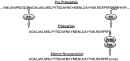
Rather than being enzymatically produced from chemical precursors, neuropeptides are derived from genetically encoded propeptides. The processing of the original transcripts into mature signalling molecules requires a number of enzymatic steps (Fig. 1). For example, a typical neuropeptide originates as a pre-propeptide from which propeptides are cleaved by prohormone convertases, and these propeptides, which are glycine-extended intermediates, are enzymatically converted to amidated mature peptides. Each of these two steps (1) the liberation of the propeptide from the original transcript and (2) the amidation of the propeptide to form active mature neuropeptides, each represents potential targets for disruption of the neuropeptidergic signalling systems that are vital to flatworm biology. The specific appeal of these targets is that they could represent a site of vulnerability for all of the neuropeptide signalling systems in the worm. While interfering with a single amidated neuropeptide by antagoinsing the neuropeptide receptor could have important consequences in the worm, blocking all amidated neuropeptide signalling by blocking the generation of active peptides would likely be even more calamitous to the parasite. However, a major hurdle for the exploitation of these potential targets would be selectivity, since the enzymes responsible for neuropeptide processing in the parasites could be very similar to those performing the same function in the host.

Fig. 1. Schematic of the enzymatic processing involved in the production of mature, bioactive amidated neuropeptides in flatworms. The schematic uses the Schistosoma mansoni NPF sequence as an example (Humphries et al. 2004). Prohormone convertases (PC) clip the pre-propeptide at mono- and di-basic sites to form the glycine-extended propeptide. Although the N-terminus of neuropeptides can be produced by cleavage of the signal peptide, three of the four flatworm peptides in which the pre-propeptide structure is known appear to have the N-terminus formed by PC cleavage (see Table 3). The only case in which PC cleavage does not produce the carboxy terminus of the glycine-extended propeptide is M. expansa NPF (Mair et al. 2000a) where a stop codon follows the codon for glycine, such that the original translated product requires no cleavage. The carboxy-terminal glycine is then converted to a more stable α-amide group by the sequential action of peptidyl α-hydroxylating monooxygenase (PHM) and peptidyl α-hydroxyglycine α-amidating lyase (PAL). This is the only known mechanism for producing α-amidation, and neurally-distributed PHMs have been identified in schistosomes (Mair et al. 2004) and planaria (Asada et al. 2005).
Prohormone convertases
The prohormone convertase (PC) gene family is widespread throughout the phyla, from prokaryotes to eukaryotes and, because the catalytic domain is highly conserved, all PCs are thought to have originated from a common gene (Seidah et al. 1998). The only direct evidence for a PC in platyhelminths is from the free-living Dugesia japonica where a cDNA encoding a PC2 homologue has been characterized which is 67% similar to the Aplysia PC2 (Agata et al. 1998). Although there have been no functional studies on the D. japonica PC2, in situ hybridization shows it to be distributed throughout the CNS – in fact, it is used as a CNS-specific marker to track CNS regeneration (Agata et al. 1998).
There is a great deal of indirect evidence for functional PCs in platyhelminths. Most convincingly, the parent pre-propeptides of some platyhelminth neuropeptides are known and the resultant mature peptides demonstrate cleavage at the same mono- and di-basic sites typically targeted by PCs. For example, the sequences of M. expansa (Mair et al. 2000a), Arthurdendyus triangulatus (Dougan et al. 2002) and S. mansoni (Humphries et al. 2004) pre-proNPFs are known, and production of the known proNPFs requires cleavage at signature basic sites. Further, a search of platyhelminth sequences reveals a number of other potential convertase sequences within the phylum, but there are as yet no functional data. However, experimental evidence from the model platyhelminth Schmidtea mediterranea supports the importance of PCs in flatworms. These planarians feature a cDNA encoding a protein most similar to PCs in the molluscs L. stagnalis (Smit, Spijker & Geraerts, 1992) and Aplysia californica (Ouimet et al. 1994). RNAi-mediated silencing of that S. mediterranea gene produces worms with motility defects, including paralysis (Reddien et al. 2005). Since neuropeptides putatively processed by PCs are known to be myoexcitatory in a number of flatworms, deletion of PCs might be expected to produce motor defects.
In summary, although there is compelling evidence for the presence and biological importance of PCs in flatworms, at this time very little is known about the specific nature of these enzymes within the phylum. Certainly, not enough is known to speculate as to whether pharmacological differences between host and parasite PCs could be sufficient for pharmacological exploitation.
Amidating enzymes
Most mammalian and insect neuropeptides and hormones feature an alpha amide group at the carboxy terminus. This alpha amide group is generated by the post-translational modification of a C-terminal glycine-extended propeptide, and the process includes two sequential reactions; the first is catalysed by peptidyl α-hydroxylating monooxygenase (PHM) and the second is catalysed by peptidyl α-hydroxyglycine α-amidating lyase (PAL). PHM and PAL are two separate catalytic moieties but, in many eukaryotes, both exist on a single bifunctional protein called peptidylglycine α-amidating monoxygenase (PAM) (Eipper, Stoffers & Mains, 1992; Eipper et al. 1993). In some invertebrates such as Drosophila, Calliactis and Hydra, PHM and PAL are expressed as independent proteins (Hauser, Williamson & Grimmelikhuijzen, 1997; Kolhekar et al. 1997; Williamson, Hauser & Grimmelikhuijzen, 2000). The end result of the two reactions is the conversion of the ionisable free carboxylic acid on the carboxy terminus of the propeptide to a non-ionisable alpha amide group. The carboxy-terminal amidation is usually essential for the bioactivity of these peptides (Prigge et al. 2000).
The presence of these amidating enzymes in flatworms is testified to by the number of amidated neuropeptides that have already been structurally characterized from the phylum. As described earlier, the first such flatworm neuropeptide was M. expansa NPF, which ends with the signature motif GRPRFamide (the amide group can be represented biochemically as –NH2, in this review the ‘amide’ notation is used) (Maule et al. 1991). Later, A. triangulatus NPF, GNFFRFamide, RYIRFamide, YIRFamide and GYIRFamide were structurally characterized from platyhelminths. In all of these cases, the mature peptides were not predicted from molecular data, rather the neuropeptides were purified and structurally characterized. The presence of these amidated carboxy termini on endogenous peptides is indication of the presence of amidating enzymes in the worms.
The importance of the amidation reaction to the function of these neuropeptides in flatworms is evident from physiological data; the absence of the amide group renders both the FLPs and the NPFs inactive. The potent myoactivity of FLPs on schistosome muscle is abolished in the absence of the carboxy-terminal amide (Day et al. 1997). For example, YIRFamide has a half-effective concentration of 4 nM, while the YIRF lacking the amidation is completely without effect. Likewise, while porcine NPY can mimic schistosome NPF's inhibition of cAMP in worm homogenates, a non-amidated porcine NPY is without effect (Humphries et al. 2004).
There is now direct evidence for the presence of amidating enzymes in flatworms; the enzyme which catalyses the first step has been identified in S. mansoni (Mair et al. 2004) and named smPHM. The enzyme is widely distributed throughout the schistosome nervous system, including both the central and peripheral nervous system. In this regard, the smPHM immunoreactivity looks much like the staining observed with antisera targeting FLPs and NPFs. In fact, immunoreactivity for the amidating enzyme appeared even more widespread than that observed for FLPs and NPF – suggesting that some neurones negative for FLPs and NPFs may express PHM and, therefore, other as yet-unidentified amidated neuropeptides. Another PHM has recently been identified in the free-living platyhelminth Dugesia japonicum (Asada et al. 2005); in this planarian, PHM is also localised to the nervous system.
Heterologously expressed schistosome PHM has functional PHM activity, but it also has some characteristics different from typical PHMs. For example, one requirement for PHM activity is Cu2+ and both schistosome and rat PHM lose activity when the available Cu2+ is chelated by EDTA. However, while other PHMs are dramatically inhibited by the absence of exogenous (0·5–1·0 μM) Cu2+, smPHM is not significantly inhibited, suggesting that the schistosome enzyme more avidly binds Cu2+. Also unlike other previously described PHMs, smPHM has little activity at neutral pH, with optimal activity at pH 3·5. Lastly, the schistosome enzyme showed a 10-fold Km greater compared to rat PHM for the peptidylglycine substrate. Although the functional significance of these differences between schistosome PHM and its mammalian homologues are unclear, the differences do suggest that there could be exploitable pharmacological differences between the host and parasite enzymes.
CONCLUSIONS
There is a great deal of recent evidence for the centrality of neuropeptides in the biology of parasitic flatworms. In common with each other, both NPFs and FLPs are very broadly expressed and conserved across flatworms suggesting fundamental roles in platyhelminth biology. Accepting this fundamental similarity, the contrast in comparative relationships between the two known neuropeptide messengers in flatworms and those in vertebrates could not be more stark. The FLPs form a signalling system that appears to be pivotal to co-ordinated motor function in flatworms and yet in vertebrates these peptides are very sparsely expressed and appear to be of limited functional significance with no known role in motor co-ordination. In complete contrast, the NPFs appear to be homologous to the most abundant neuropeptide in vertebrate brains, namely NPY, and to employ the same second messenger pathway to convey their cytosolic signals. Notwithstanding this contrast, they both are structurally quite distinct from known vertebrate neuropeptides – elevating the attraction of their receptors as novel targets for parasite control.
Although it is highly likely that other neuropeptide families occur within flatworms, until their discovery, the focus must remain on FLPs and NPFs. Indeed, the signalling systems associated with FLPs and NPFs in parasitic flatworms each have appeal as potential reservoirs of chemotherapeutic targets. In both cases, it seems likely that the receptors for the neuropeptides are the foremost candidates as novel anthelmintic targets, but there is presently very little known about these receptors. There is now a convergence of factors that should, in the near future, precipitate a harvest of knowledge regarding these receptors and other components of neuropeptide signalling. Firstly, the rapidly emerging genomic and EST data from flatworms will be of immense value in uncovering novel proteins and peptides.
Coincidentally, the recent discovery of a number of neuropeptide receptors from other invertebrates will inform homology searches of the new genetic data from platyhelminths (see Greenwood, Williams and Geary in this supplement). Lastly, assays to characterize, functionally express and screen newly discovered receptors are rapidly increasing in both ease and sophistication. Although the neuropeptide receptors may be the most obvious target, others hold promise as well. If neuropeptide processing enzymes such as prohormone convertases and amidating enzymes prove to have sufficient pharmacological distinctions from their mammalian homologues, these could be prime targets for drugs that would compromise the biology of parasitic platyhelminths.
ACKNOWLEDGEMENTS
The authors are supported by NIH Grant AI49162.