INTRODUCTION
Cystic echinococcosis (CE), caused by Echinococcus granulosus, is one of the most geographically widespread zoonoses in the world. In China, human CE has been recorded in 22 Provinces and Administrative Regions, with a particularly high endemic level over large areas of north-western Provinces and especially in the Xinjiang Uygur Autonomous Region (XUAR) (Craig, 2004). This 1·7 million km2 region is the largest provincial administrative region of China (1/6 of the total area). Topographically, Xinjiang is characterized by a sharp demarcation of mountains and basins. The Tianshan Mountains divide the region into two halves forming two basins (in the north, the Jungar basin and in the south, the Tarim basin).
According to the year 2000 census Xinjiang had a population of 19 million, divided in 13 nationalities mainly comprising Uygur (45%), Han (40%) and Hui (4·5%) ethnic groups, who are engaged mainly in industrial and agricultural production. Also the Kazakh (6·7%) and Mongolian (1%) groups, who are involved primarily with nomadic or semi-nomadic pastoralist life-styles (Craig et al. 1991). The latter normally engage in seasonal migration from winter to summer pastures to provide optimal grazing for livestock (sheep, goats, cattle, horses and camels) (Wang et al. 2001).
Since the 1950s, at least 21560 CE patients have been treated in 58 hospitals in Xinjiang (Wen and Yang, 1997; Craig, 2004) that represents more than half of the CE cases reported in China. Data from surgical records in XUAR indicated that the highest incidence (>80 per 100000 per annum) was observed in Han Chinese, especially from settled rural agricultural communities (Chi et al. 1990). However, active mass ultrasound screening surveys, performed in traditional semi-nomadic pastoral communities of Kazakh or Mongolian ethnicity, revealed a higher risk of CE infection (Chai, 1993, Wang et al. 2001, 2005).
The main definitive host of E. granulosus in XAUR is the domestic dog. The ratio of dogs to humans in the early 1990s was 1[ratio ]10–15 in agricultural areas and 1[ratio ]6 in pastoral areas. Canine prevalence rates (ranging from 6·9 to 70%) confirm the endemic status of the region (Liu et al. 1993).
Livestock husbandry is composed mainly of sheep (more than 23 million heads). Guard or shepherd dogs are kept in large numbers, usually with dogs and sheep in the same sheepfold. Thus sheep are in frequent contact with dog's faeces. More than 5 million sheep are slaughtered annually in households and thus represents a major problem for slaughter hygiene and management and an important risk factor in the widespread transmission of CE. However, other intermediate hosts, such as cattle, goat, pig, horse, yak and camel, can act in the parasite life-cycle (Liu et al. 1993). Due to the diversity of E. granulosus hosts, but also to the contrasted geographical, climatic and socio-ethnic conditions described above, the question of the strains or genotypes of E. granulosus that are involved in potential human infection is important. Previous genotyping studies of E. granulosus performed by McManus et al. (1994) and Zhang et al. (1998) in Xinjiang, analysing 87 intermediate hosts (sheep, cattle and camel), found a large predominance of the sheep strain or G1 genotype (84/87), but the camel strain or G6 genotype was also detected in 2 cattle and 1 camel hydatid cysts in northern Xinjiang. In these two surveys 11 human hydatid cysts were analysed, and identified as the G1 genotype (McManus et al. 1994; Zhang et al. 1998).
In order to further investigate the genotype(s) of E. granulosus involved in zoonotic transmission in this area, a panel (n=47) of surgically treated CE patients was investigated, from the Region's hospitals. We also compared the genotypes identified from these CE patients to E. granulosus adult worm genotypes collected from 13 dogs from 2 different endemic areas, in the south or north of the Tian Shan Mountains in XUAR.
MATERIALS AND METHODS
Human hydatid samples
From February to July 2005, hydatid cysts were surgically removed or drained from CE patients, resident in Xinjiang. The surgical interventions were performed at hospitals in the capital city, Urumqi, and in the northern city of Hobukesar. For multi-cyst cases, cysts were sampled independently. All hydatid cysts were checked under a microscope for the presence of protoscoleces, which were collected sterilely and preserved in 95% ethanol. For each patient, data were collected about ethnicity, past and current domicile, as well as dog and livestock contact history.
Hydatid cysts of E. granulosus were ultrasonography classified (CE1 to CE5) (WHO/OIE, 2001) for 44 CE patients exhibiting abdominal cysts.
Adult E. granulosus worms
From October 2004 to May 2005, stray dogs were collected in Bayanbuluk (Hejing County) in north Tarim Basin, and in Hobukesar County (northwest Jungger Basin). All dogs were humanly killed (using ketamin glycol). At necropsy, the pyloric and ileocecal ends of the small intestine were ligated. The small intestine was excised, opened length-wise and carefully examined. Any E. granulosus worms were picked up one by one, rinsed in water and stored individually in 95% ethanol for molecular examination.
DNA extraction
Total genomic DNA was isolated from protoscoleces aspirated from human hydatid cysts, using the High Pure PCR Template Preparation kit (Roche Diagnostics, Mannheim, Germany) based on Proteinase K digestion. For non-fertile cysts (i.e. cysts without protoscoleces), endocyst membranes were minced and broken up in 3 consecutive liquid nitrogen baths. The same process was applied for adult worms in order to destroy the eggs (Cabrera et al. 2002). Each sample was incubated in lysozyme at 37 °C for 1 h. Then, SDS was added to proteinase K and lysis buffer and incubated overnight at 55 °C. Manufacturer's recommendations were carefully followed for the last part of the extraction process.
Molecular analysis
A part of the mitochondrial cox1 gene of E. granulosus was amplified with the EgCOI 1/2 primers (Bart et al. 2006). For each template, 2 μl of genomic DNA were mixed with 2·5 μl of 10×Diamond™ Taq PCR buffer (BBI), 1·5 mM MgCl2, 20 μM dNTP (Sangon, Shanghai, China), 1 μM of each primer (Sangon) and 1 U Diamond™ Taq DNA Polymerase (BBI) in a 25 μl final volume. The PCR programs contained 35 cycles with, for each cycle, a denaturation step (30 s at 94 °C), a hybridization step (30 s interval at 56 °C and an elongation step (30 s at 72 °C). After confirmation of the amplification in a 1·5% agarose gel, each amplified fragment was purified and then sequenced (Takara Biotechnology, Dalian, China). The sequences were manually checked, aligned, using the BioEdit software (Hall, 1999) then compared to those of the GenBank data base (http://www.ncbi.nlm.nih.gov).
Serology
ELISA using recombinant Antigen B prepared from E. multilocularis (EmAgB8/1) (Mamuti et al. 2004) was applied for 31 serum samples available from surgically treated CE cases. In order to determine if the IgG antibody response could be linked with clinical (number, type, location and fertility of the cysts) or genetic features of the disease, Student's t-tests were calculated.
RESULTS
Epidemiological and clinical characteristic of CE patients
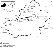
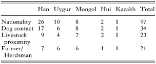
The 47 CE patients included in this study comprised Han (n=26), Uygur (n=10), Mongol (n=8), Hui (n=2) and Kazakh (n=1) ethnicities (Table 1). Patient ages ranged from 5 to 71 years with an average at 32 years, and they originated from 13 prefectures in Xinjiang (Fig. 1). Thirty-six CE patients had a history of dog or/and livestock ownership. Of 45 abdominal CE patients, 30 presented with only 1 cyst, mainly in the liver (27/30), and 15 patients exhibited 2 to 7 cysts, which were located in the liver, spleen, abdomen or pelvic cavity. Two CE patients had lung cysts.

Fig. 1. Location of the humans and dogs collected in XUAR.

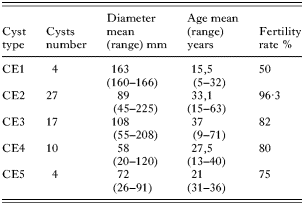
Analysis of cyst pathology showed that 44 out of 62 abdominal cysts (71%) were classed as CE2 or CE3 stages (i.e. with daughter cysts or evidence of detached endocyst); 4 cysts (6%) were of CE1 type (univesicular) and 14 (24%) were classed as in inactive stages CE4 (solid mass) and CE5 (calcifying). Cyst size tended to decrease from an average diameter of 163 mm in CE1 type cysts to 72 mm in CE5 cysts.
The fertility rate was high in all cysts types and surprisingly, 11/14 CE4 and CE5 type cysts contained viable protoscoleces (Table 2).

Genotyping of human CE
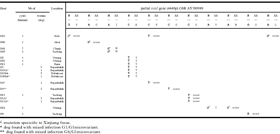
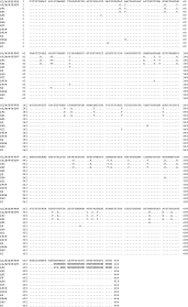
A total of 67 hydatid cysts were genotypically characterized. Among the 47 CE patients who were studied, 45 exhibited hydatid cysts of the sheep strain (G1 genotype). Two CE patients, with liver cysts, were infected by the camel strain (G6 genotype) (Fig. 2). Thirty-six patients (harbouring a total of 49 cysts) exhibited a 100% homology with the cox1 sequence taken as reference for the G1 genotype (AY389989). Table 3 shows 10 CE patients, with a total of 17 cysts, presenting 7 different haplotypes, all presenting transition mutations (i.e. T/C and A/G substitutions), with 1–3 nucleotides that differ from the G1 genotype. For one of the haplotypes found for 3 samples (H2, H21 and H43), the result of the 92C>T mutation is a substitution of alanine by a valine in position 31 according to the echinoderm mitochondria genetic code (Rice et al. 2000). A BLAST search found the 92C>T mutation reported in intermediate host in Romania, Turkey and Austria under the respective Accession numbers AJ686565, AJ508012 and AJ508019 and also in Tunisia (M'rad et al. 2005).

For another haplotype (GbR DQ356876) detected in 2 cases with multiple cyst presentations (i.e. H44 with 7 cysts, and H45 with 2 cysts), the 51A>G mutation causes a substitution of an isoleucine by a methionine in position 17. This mutation was not found elsewhere according to the BLAST search. The 4 remaining haplotypes detected did not cause any change in the protein sequence. Two of these (H40 and H51), registered under the GenBank Accession numbers DQ356875 and DQ356874, presented as original sequences, with 1 and 3 mutations respectively, compared to the reference G1 sequence. Finally, 2 G1 haplotypes were detected for 2 CE patients from north Xinjiang. The CE patient H11 presented 1 210C>T mutation, which was previously described in Argentina (AF458874); the CE patient H9 presented one 348G>A, previously recorded in Tunisia (AY679145). Alignment of the E. granulosus gene sequences, from patients H41 (originally resident in Inner Mongolia) and H53 (originated from Altai Prefecture, north Xinjiang), showed 100% homology with the camel strain (G6 genotype) (AF408687).
Genotyping of E. granulosus in dogs
Forty-five gravid E. granulosus adult tapeworms, collected from 13 dogs, were genetically identified. Twenty-three of them, recovered from 9 dogs (6 in Bayanbuluk and 3 in Hobukasar), exhibited 100% homology with the cox1 sequence taken as reference (G1 genotype, AY389989). As in the human CE hydatid cyst samples, 4 haplotypes, presenting transition mutations compared to G1, were observed in 19 E. granulosus tapeworms (Fig. 2). The sequence presenting the 92C>T mutation (found in CE patients H2, H21 and H43) was also detected in 11 worms recovered from 4 dogs (2 from Bayanbuluk and 2 from Hobukasar). In 1 worm from dog 4 (D4iv), a silent mutation (102C>T) was found (GbR DQ356881). This mutation, also found in the CE patient H51, was already described in other E. granulosus samples from China and India (AY386207 and DQ109036 respectively). In 2 other dogs (D1A and D13), 4 worms presented 100% homology with the H11 sequence, exhibiting a silent mutation (210C>T) described above. Finally, 2 adult worms, from 1 dog (D9) from Bayanbuluk, possessed a silent mutation (147C>T), that has been identified in Argentina (AF458875) and Australia (AJ508030). In this same dog, 3 other individual worms were identified as belonging to the G6 camel strain and, with 100% homology, with the G6 sequence detected in the H41 and H53 CE patients.
In summary, among the 10 dogs, for which more than 1 E. granulosus tapeworm had been characterized, 5 were infected by 2 different E. granulosus genotypes (4 dogs harbouring the common G1 genotype mixed with a G1 haplotype and 1 dog harbouring 1 G1 haplotype and the G6 genotype). Of these 5 dogs, 4 originated from Bayanbuluk (west Xinjiang) and 1 from Hobukasar (north), and both these areas are Mongol Autonomous Counties/Prefectures.
Serology
The recombinant AgB-ELISA was performed on 31 CE patients, of which 28 were seropositive. The 3 seronegative patients all exhibited a single cyst (CE2, CE4 or CE5) and underwent surgery for the first time.
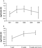
Due to the small number of patient's sera analysed, no significant differences (P>0·05) were found when we compared the anti-AgB response with the type of the CE cyst or the number of cysts per patient. Nevertheless, the trends in seroreactivity do indicate that the CE2 and CE3 cysts were more antigenic than the other stages, and that the more a patient harbours a higher number of cysts, the higher the serology is (Fig. 3A,B) (Daeki et al. 2000). No relationship could be drawn between the detected haplotypes (either within G1 haplotypes or between G1 and G6 genotypes) and the anti-AgB response.

Fig. 2. Partial cox1 gene nucleotide alignment. In total, 10 Echinococcus granulosus G1 haplotypes have been identified. H, Human isolate; D, Dog isolate.

Fig. 3. Relation between the AgB-ELISA serology and the clinical features of the CE for 31 patients. The tendency seems to indicate that the Type 2 and Type 3 cysts correspond to the highest immune response (A) and that the more a patient harbours a high number of cysts, the higher the serology is (B). Nevertheless, no significant differences were found after performing the Student's t-test.
DISCUSSION
The Xinjiang Uygur Autonomous Region (XUAR) includes a vast pastoral area of north-western China, and also is one of the most important foci of cystic echinococcosis (CE) in the world (Craig, 2004). The traditional life-cycle of E. granulosus in XUAR appears to predominantly involve the domestic dog and sheep, but other intermediate hosts such as cattle, yak, goats or camels have been reported (Chai, 1995). Previous studies, to investigate E. granulosus strains or genotypes in Xinjiang, have identified the common sheep strain (G1), as the major source of infection for human CE (McManus et al. 1994). Morphological studies have indicated the occurrence of at least one other form of adult worm or hydatid cyst/protoscoleces from dogs and cattle in northern Xinjiang (Craig et al. 1991; Chai 1995). The camel strain (G6 genotype) of E. granulosus was identified in camels and cattle using RFLP and PCR-RFLP methods in camel and in cattle (Li et al. 1992; Zhang et al. 1998).
In the current survey, our aim was to further genetically characterize E. granulosus infecting humans in Xinjiang. The genotyping results of 67 hydatid cysts, resected from 47 CE patients, who came from different areas of XUAR, confirmed that the majority of cysts (97%) were predominantly of the sheep strain (G1 genotype) as previously reported by McManus et al. (1994). Whatever the patient nationality, or geographical location, 45 CE patients (96%) were infected with hydatid cysts of the common sheep strain. Two CE patients (4%) harboured the camel strain (G6 genotype).
Contrary to the findings of Zhang et al. (1998), but in accordance with studies performed in other foci of E. granulosus in South America, North Africa or Romania (Kamenetzki et al. 2002; Haag et al. 2004; Bart et al. 2004, 2006), 7 G1 haplotypes, differing by 1–3 nucleotides compared to the reference G1 sequence, have been found in CE patients from Xinjiang. Nevertheless, no relationship has been drawn between these G1 haplotypes (found for 16 hydatid cysts) and the geographical location of the patients, their nationality, the location, the type, cyst fertility or size, or their AgB antigenicity. From these haplotypes, we can track the origin of the multiple cysts for one CE case. Among 13 patients (a total of 32 hydatid cysts ranging from 2 to 7 cysts per patient), all the cysts belonging to the same individual CE patient exhibited the same genotype. Clear evidence is described for one CE patient (H44), who presented with a total of 7 hydatid cysts. All of the cysts from patient H44 possessed a mutation (51A>G) which indicates most likely a single source of the infection from a definitive host in space and certainly in time. This hypothesis is confirmed by the fact that this patient (H44) had already undergone surgery twice before, stressing the risk of contamination by protoscoleces during the operation.
In contrast to Africa, South America and the Middle East, no previous report existed concerning the involvement of the camel strain (G6) in human CE infection in China (Zhang et al. 2000; Harandi et al. 2002; Bardonnet et al. 2002; Guarnera et al. 2004; Dinkel et al. 2004). We report here the first 2 cases in China of patients harbouring a G6 genotype hydatid cyst. One of these 2 patients, a Uygur farmer with a history of dog ownership, came from Buerjin County (Altai prefecture) where the G6 genotype was previously identified in 1 CE infected camel (McManus et al. 1994). The likely location of infection for the second patient, a 22-year-old Mongol man, is not clear. He spent his youth (from birth until 17 years old) in Inner Mongolia (an endemic province that borders Mongolia), then came to Xinjiang (Kashgar then Urumqi). Assuming that the infection in an endemic area can occur in 3-year-old children (Guarnera et al. 2004), and that the cyst (5 cm) was classified as a type CE3 (with daughter cysts present), there is a high probability that this patient became infected in Inner Mongolia rather than in Xinjiang.
The majority (96%) of the human CE cases analysed in the current study were characterized as the sheep strain (G1 genotype), and it is therefore interesting to speculate why the occurrence of the camel strain (G6 genotype) was apparently so low in humans in Xinjiang. Actually, 2 hypotheses are under discussion: (i) humans are refractory or poorly susceptible to G6 infection (Zhang et al. 1998) and/or (ii) the level of contamination of the environment with ‘G6’ eggs from dog faeces is not very high (Guarnera et al. 2004). In our study, among the 45 E. granulosus adult worms analysed from dogs, 3 exhibited the G6 genotype. The dog infected by these G6 worms originated in the southern Tianshan Mountains, suggesting that the camel strain does not occur only in the north of the region. The apparent low occurrence of the G6 strain in dogs (3 worms/45), in accordance with our human data, suggests that in Xinjiang, as in other countries, the rate of human G6 infection is closely linked with the parasitic biomass available in the environment. Guarnera et al. (2004) showed that for the Argentinian CE cases it was not possible to discern any differences in hydatid cyst pathogenicity or cyst type between patients infected by the G1 versus the G6 genotype. Similarly, the 2 ‘G6 CE patients’ in the current study did not present different clinical pathology compared to the other 45 CE patients examined in Xinjiang.
The sympatric occurrence of 2 or more E. granulosus strains has been reported elsewhere (Wachira et al. 1993; Kamenetzky et al. 2002; Bardonnet et al. 2003; Bart et al. 2006). In these studies, animal intermediate hosts naturally infected with E. granulosus were examined and each host possessed only 1 strain. A definitive host, fed by the offal infected by G1 hydatids then subsequently by the offal infected by G6 hydatids could, however, theoretically develop both genotypes of adults. We have now reported, based on molecular analysis, the occurrence of a mixed infection of G1 and G6 genotypes of E. granulosus in a dog in a sympatric focus. The co-existence of these two strains/genotypes of E. granulosus raises the interesting question of the possibility of cross-fertilization events in the dog host and thus, the emergence of a hybrid genotype. Microsatellite analysis will be performed on the E. granulosus worms collected from this dog to check for the possibility of genetic features that indicate recombination of G1 and G6 genes.
We are very grateful to Professor Sheyhidin for supplying the two lung hydatid cysts. We thank Wang Gang and all the Xinjiang Hydatid Clinical Research Institute members for their technical help. This work was supported in part by a grant from the Ecology of Infectious Diseases Program, USA (NSF/NIH, EID #1565).