INTRODUCTION
Neospora caninum is an obligate intracellular parasite that invades many cell types of its main hosts, cattle and dogs. Neosporosis can cause abortion in adult cattle and severe neurological disorders in cases of congenital infection in cattle and dogs (Dubey et al. 2002). Recent work has been invested in identifying and characterizing N. caninum stage-specific antigens, which could be useful markers for improved immunodiagnosis as well as candidate immunogens in vaccine development. To date, however, only 1 bradyzoite stage protein has been described (Fernandez-Garcia et al. 2006), and there have been several reports on the characterization of proteins from the tachyzoite stage (reviewed by Hemphill et al. 1999). DNA cloning showed that surface protein NcSRS2 is shared by both the tachyzoite and bradyzoite stages whereas the surface protein NcSAG1 is expressed only in tachyzoites (Hemphill and Gottstein, 1996; Hemphill et al. 1997; Howe et al. 1998). With regard to microneme secretory organelle proteins NcMIC1, NcMIC2, NcMIC3 and NcSUB1 (Louie and Conrad, 1999; Lovett et al. 2000; Sonda et al. 2000; Keller et al. 2002) have been cloned together with several dense granule proteins NcGRA6, NcGRA7 and NcNTPaseI (Jenkins et al. 1997; Lally et al. 1997; Liddell et al. 1998; Asai et al. 1998; Jenkins et al. 2005). Stage conversion studies and the use of tissues from infected animals provided evidence for the expression of some of them in both the tachyzoite and bradyzoite stages. Some, in particular NcGRA6 and NcGRA7 (Lally et al. 1996), NcSAG1 (Howe et al. 2002; Chahan et al. 2003), and NcSRS2 (Nishikawa et al. 2001a; Schares et al. 2000, 2005; Gaturaga et al. 2005; Liao et al. 2005) have proved of diagnostic value in infected bovines, whereas NcSRS2, NcMIC3 and NcGRA7 have shown efficacy against neosporosis as immunization antigens (Nishikawa et al. 2001b; Cannas et al. 2003; Liddell et al. 2003; Jenkins et al. 2004; Alaeddine et al. 2005; Staska et al. 2005). One of the antigens, NcGRA7, is a tachyzoite-dense granule-associated 33 kDa protein, which has been recently associated to the cyst wall, together with the NcGRA1 and NcGRA2 proteins (Vonlaufen et al. 2004), and shares similarity with Toxoplasma gondii dense granule proteins (Lally et al. 1997). In addition, NcGRA7 induces a strong immune response in naturally and experimentally N. caninum-infected animals (Lally et al. 1996; Jenkins et al. 1997; Schares et al. 1998). When mice were immunized with plasmid DNA coding for NcGRA7, partial protection against congenital neosporosis was achieved (Liddell et al. 2003; Jenkins et al. 2004).
Infection by N. caninum has been shown to elicit a characteristic immune response that is mainly directed against several N. caninum tachyzoite antigens of low (17–18 kDa) and medium (60–62 kDa) molecular weight (Schares et al. 1998; Alvarez-Garcia et al. 2002; Okeoma et al. 2004). The Neospora 17–19 kDa fraction (p17) is a tachyzoite immunodominant antigen (IDA) recognized by sera and foetal fluids from naturally infected bovines. In this work, we have sought to characterize the genes coding for p17 antigens, through the construction and immunoscreening of a Neospora tachyzoite cDNA library, in combination with protein analysis by 2D-PAGE, Western blot, cloning of the recombinant NcGRA7 protein and mass spectrometry. We report here the identification and molecular characterization of the Neospora immunodominant p17 antigen.
MATERIALS AND METHODS
Neospora caninum cultivation
Neospora tachyzoites of the Nc-1 isolate were grown in vitro by continuous passage in Vero cells. Infected cultures were maintained in minimum essential medium (MEM) (Sigma Chemical Co., Madrid, Spain) supplemented with L-glutamine, penicillin, streptomycin (Gibco BRL Life Technologies Ltd, Paisley, Scotland) (Lindsay and Dubey, 1989) and 2% horse serum (Sigma). The cells were scraped off the culture vessel and tachyzoites were purified by filtration through 5 μm pore size filters to remove host cell debris. The tachyzoites were washed twice in cold sterile phosphate-buffered saline (PBS), pH 7·2 and pelleted by centrifugation at 600 g for 10 min prior to storage at –80 °C.
Polyclonal antibody production
Polyclonal rabbit anti-N. caninum antiserum was raised in female New Zealand White rabbits (Harlan Interfauna S.A., Barcelona, Spain). Samples of pre-immune serum were collected to confirm the absence of antibodies against N. caninum tachyzoites by IFAT. Rabbits were immunized intradermally with whole extract from 108 tachyzoites in a 50% emulsion of Freund's complete adjuvant and thereafter boosted twice at 4-week intervals by intramuscular injection of the same immunogen and adjuvant. Sera were collected 2 weeks after the third immunization and used for affinity purification of anti-p17 antibody (APA17).
APA17 was prepared as described by Hemphill et al (1997). Briefly, 100 μg of N. caninum proteins (Alvarez-Garcia et al. 2002) was fractionated by SDS-PAGE and transferred onto a nitrocellulose membrane. A nitrocellulose strip with the immunoreactive antigens was excised by first immunostaining the edges of the membrane to locate the position of the proteins of interest. The strip was incubated overnight at 4 °C with 5 ml of the rabbit immune serum diluted 1[ratio ]10, and subsequently washed in Tris-buffered saline with 0·05% Tween-20 (TBS-T) 3 times for a total of 30 min. Bound antibodies were eluted in 50 mM Tris, 50 mM glycine, 0·05% Tween-20, pH 2·6 buffer for 5 min on ice, and the acidic buffer was then neutralized by the addition of 1M Tris base solution. Finally, the antibody solution was clarified by centrifugation (10000 g, 20 min, 4 °C) and BSA added to a final concentration of 0·3%. The specificity of the antibody preparation for the N. caninum p17 antigen was confirmed by Western blot.
Construction of a cDNA library
Total N. caninum RNA was isolated from 2×10⁹ tachyzoites with Tripure reagent according to the manufacturer's instructions (Roche Diagnostics S.L., Barcelona, Spain). PolyA+ RNA was isolated using oligo-dT cellulose (FastTrack system, Invitrogen S.A., Barcelona, Spain) and the integrity of total RNA and mRNA was assessed by standard agarose gel electrophoresis and ethidium bromide staining.
A Neospora cDNA expression library was prepared with reagents from Stratagene (Madrid, Spain). Briefly, first strand cDNA was synthesized from 5 μg of tachyzoite polyA+ RNA using a poly-dT primer with an internal XhoI restriction site. After the second strand synthesis, the cDNA was blunt-ended, ligated to EcoRI adaptators, phosphorylated with T4 polynucleotide kinase, and run on a Sepharose 400 spin column (Amersham Biosciences, Barcelona, Spain) to remove molecules under ~400 pb. The final cDNA product was then ligated into the EcoRI- and XhoI-digested arms of the UNI ZAP XR λ bacteriophage vector and packaged into viable virions using the Gigapack III Gold packaging extracts. The primary cDNA library contained 3×10⁵ phages, 97% of which were recombinant as determined by a β-galactosidase test. Subsequently, one round of library amplification was carried out in E. coli XL1-Blue MRF′.
Immunoscreening of the cDNA library
Expression and screening of the library with APA17 were performed by standard procedures. 2×10⁵ recombinant phages were plated on a lawn of E. coli XL1-Blue MRF′, incubated at 37 °C for 3·5 h and adsorbed to IPTG-impregnated nitrocellulose membranes. Each nitrocellulose membrane was quenched with TBS-T containing 3% BSA and then probed with APA17 diluted 1[ratio ]20 in TBS-T with 0·3% BSA (1 h, 37 °C). Next, the membranes were washed in TBS-T and incubated for 1 h with an anti-rabbit IgG peroxidase conjugated antibody (Sigma) diluted 1[ratio ]1000 in TBS-T prior to visualization with 4-chloro-1-naphthol (Bio-Rad Laboratories, Madrid, Spain). Positive plaques were re-screened twice until pure clones were obtained.
DNA sequencing
cDNA inserts from positive clones were amplified by PCR using universal forward and reverse T3 and T7 primers (Sigma). Phages from a positive plaque, 20 pmol of each primer, 0·2 mM dNTPs, PCR buffer and 1 U Taq polymerase were combined in a 25 μl reaction mix. Thermal cycling involved 1 cycle at 95 °C for 10 min, 40 cycles at 94 °C for 30 s, 56 °C for 30 s, and 72 °C for 1·3 min, followed by 1 cycle at 72 °C for 5 min and final cooling to 4 °C. The PCR products were sized on a 1% agarose gel, purified using the ExoSap-IT PCR Clean-Up kit (USB Corporation Cleveland, Ohio, USA), and analysed by cycle sequencing in their entirety. Matching open reading frames were identified through BLAST searches against the NCBI and TIGR sequence databases.
One-dimensional PAGE and Western blot
N. caninum soluble antigens were fractionated by 15% SDS-PAGE and transferred to nitrocellulose membranes for Western blot as described (Alvarez-Garcia et al. 2002), using sera from a Neospora-naturally infected cow at a dilution of 1[ratio ]20 and APA17 at a dilution of 1[ratio ]50. Coomassie staining of SDS-PAGE was performed following standard procedures.
Two-dimensional PAGE
Purified tachyzoites (2×10⁹) were suspended in 1 ml of 10 mM Tris-HCl buffer, pH 7·2, containing 2 mM phenylmethylsulfonyl fluoride (Sigma), disrupted by ultrasound treatment (Sonifier 450, Branson Ultrasonic Co., USA) in an ice-bath, and then centrifuged at 10000 g for 20 min at 4 °C. Protein concentration in the soluble fraction was determined using a Micro BCA protein assay kit (Pierce, Rockford, USA). Protein samples of 300 μg (analytical gels) or 5–10 mg (preparative gels) were solubilized in lysis buffer (7 M urea, 2 M thiourea, 2% CHAPS, 65 mM dithioerythritol, bromophenol blue traces) with 2% Pharmalyte pH 3–10 (Amersham Biosciences), and applied onto Immobiline pH 3–10 non-linear DryStrips (18 cm long, Amersham Biosciences). Isoelectric focusing was performed on a IPGphor system (Amersham Biosciences) at 15 °C using the following programme: (i) for analytical gels: passive rehydration for 16 h, 500 V for 1 h, 500–2000 V for 1 h, and 8000 V for 5·5 h, or alternatively (ii) for preparative gels: 30 V (active rehydration) for 13 h, 500 V for 1 h, 1000 V for 1 h, 2000 V for 1 h, 2000–5000 V for 3 h, and 8000 V for 11 h. Next, immobilized pH gradient strips were reduced with 2% dithioerythritol and then alkylated with 2·5% iodoacetamide in equilibration buffer (6 M urea, 50 mM Tris-HCl, pH 6·8, 30% glycerol, 2% SDS). The second dimension run was carried out on homogeneous 10% T, 1·6% C (piperazine diacrylamide) polyacrylamide gels, 1·5 mm thick, at 40 mA per gel for 6 h in a Protean II gel tank (Bio-Rad). Values for Mr and pI were estimated using internal two-dimensional SDS-PAGE and external SDS-PAGE standards (Bio-Rad).
Two-dimensional Western blot: following standard procedures
Following analytical two-dimensional PAGE, proteins were electroblotted onto nitrocellulose membranes in Towbin buffer at 50 mA overnight. Immunodetection was performed with APA17 (1[ratio ]200), and with positive serum (1[ratio ]1600 IFAT titre) from a breeding cow that had previously aborted due to natural infection with N. caninum (1[ratio ]100). Immunoreactive spots were detected using 1[ratio ]1000 horseradish peroxidase-labelled anti-rabbit (Sigma) or 1[ratio ]250 anti-bovine IgGs (Hipra Laboratories S.L., Amer, Spain), as necessary, and the ECL chemiluminescent detection system (Amersham Biosciences).
Expression of recombinant NcGRA7 (rNcGRA7)
The NcGRA7 protein was produced in the prokaryotic expression vector system pRSET (Invitrogen) as a recombinant protein. An NcGRA7 gene fragment corresponding to nucleotides 82-654 was amplified by PCR with oligonucleotides FNcGRA7 (TAATGGATCCGAGGAGACTTGGCAACC- GAA) and ReNcGRA7 (TTGGAATTCCCTAT-TCGGTGTCTACTTCC). PCR was performed on 0·1 μg of N. caninum genomic DNA, isolated from purified tachyzoites using the GenomicPrepTM Cells and Tissue DNA Isolation kit (Amersham Biosciences). Each PCR mixture consisted of 1·5 mM MgCl2, 200 μM of each dNTP, 0·2 μM of each oligonucleotide, 1X Expand High Fidelity PCR reaction buffer (Roche, Barcelona, Spain) and 2·6 U Expand High Fidelity enzyme mix (Roche), in a reaction volume of 50 μl. PCR was carried out according to manufacturer's instructions, at 56 °C annealing temperature. The amplified DNA fragment was purified, digested by Bam HI and Eco RI and cloned into the pRSET-C plasmid previously linearized with the same endonucleases.
For rNcGRA7 protein expression, Rosetta (DE3) pLysS Escherichia coli cells (Novagen, Madrid, Spain) transformed with the recombinant plasmid were cultured in selective medium (Luria Bertoni with 100 μg/ml ampicillin and 34 μg/ml chloramphenicol) until the OD600 reached 0·7. Recombinant protein synthesis was initiated by adding isopropyl-β-D-thiogalactopyranoside (IPTG; 1 mM final concentration) to the culture. After incubation (4 h, 37 °C), bacteria were harvested (3500 g, 15 min), suspended in 5V of the pellet weight of cooled lysis buffer (20 mM phosphate buffer, pH 7·4, 0·5 M ClNa, 5% glycerol, 0·5% Triton X-100, 40 mM imidazole and proteases inhibitors (1 ml/20 mg crude extract, Calbiochem, Madrid, Spain) and disrupted by ultrasound. Treatment consisted of 1 sec pulses on plus 1 sec off during 20 min (Sonifier 450, Branson Ultrasonic Co., USA), in an ice-bath, followed by centrifugation at 16000 g for 20 min at 4 °C. The rNcGRA7 was produced as a soluble protein and purified by IMAC using HisTrap HP columns (Amersham Biosciences) in native conditions (20 mM phosphate buffer, pH 7·4, 0·5 M ClNa and 500 mM imidazole) using an ÄKTA prime (Amersham Biosciences). Protein concentration was measured using the Micro BCA Protein Assay Reagent kit (Pierce, Rockford, USA).
Mass spectrometric (MS) analysis: MALDI-TOF MS and MALDI-TOF/TOF MS
Spots of interest were manually excised from preparative Coomassie-stained two-dimensional gels. Stained gel plugs were destained with acetonitrile and washed twice with 50% acetonitrile in 25 mM ammonium bicarbonate (NH4HCO3), and the remaining solvent was removed by drying in a SpeedVac. Protein spots were reduced with 10 mM dithioerythritol in 25 mM NH4HCO3 for 30 min at 56 °C and subsequently alkylated with 55 mM iodoacetamide in 25 mM NH4HCO3 for 20 min in the dark. Gel plugs were successively washed with 25 mM NH4HCO3 and acetonitrile, and vacuum-dried prior to overnight digestion at 37 °C with 12·5 ng/μl sequencing grade trypsin (Roche) in 25 mM NH4HCO3. Following crude extract separation, peptides were extracted from the plugs first into 50% acetonitrile, 1% trifluoroacetic acid and then into 100% acetonitrile. The peptide fractions were pooled, and the volume was reduced using a SpeedVac.
One μl of each sample (crude extracts and extracted peptides) and then 0·4 μl of 3 mg/ml alpha-cyano-4-hydroxycinnamic acid matrix (Sigma) in 50% acetonitrile/0·01% trifluoroacetic acid (TFA) were spotted onto a MALDI target. Samples for MS/MS sequencing were mixed 1[ratio ]1 with alpha-cyano matrix (5 mg/ml in 50% acetonitrile/0·3% TFA) and spotted on the stainless steel plate. MALDI-TOF MS analyses were performed on a Voyager-DE STR instrument (PerSeptive Biosystems, Framingham, MA, USA). Peptides were selected in the mass range of 700–4500 Da. All mass spectra were externally calibrated with the Sequazyme peptide mass standards kit (PerSeptive Biosystems) and internally with trypsin autolysis peaks. MS/MS sequencing analyses were carried out using the MALDI-tandem time-of-flight mass spectrometer 4700 Proteomics Analyzer (Applied Biosystems, Framingham, MA).
Database search
The peptide mass fingerprinting and peptide fragmentation data obtained from MALDI-TOF MS and MS/MS analyses, respectively, were used to search for protein candidates on the non-redundant GenBank and Swiss-PROT/TrEMBL databases using the Mascot algorithm (http://www.matrix-science.com). Initial search parameters were as follows: Cys as S-carbamidomethyl derivate and Met in oxidized form, 1 missed cleavage site, peptide mass tolerance of 50 ppm, and MS/MS tolerance of ±0·5 Da. The sequences determined from MS/MS data were used to search for homologous proteins using the NCBI and TIGR basic local alignment search tool (BLAST).
RESULTS
Immunoscreening of a N. caninum tachyzoite cDNA library
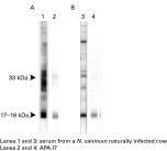
Testing of the affinity-purified antibody, APA17, against a N. caninum whole soluble extract in one-dimensional Western blotting showed the antibody to react mainly with a 17–19 kDa fraction in native and reducing conditions although a weak reaction was also detected at 33 kDa under native conditions (Fig. 1). Once specificity of the selected antibody was checked by western blotting, clone selection by APA17 showed that most of the APA17-positive library clones had a 1210-pb cDNA insert whose sequence was identical to that of the previously described NcGRA7 gene. The open reading frame of 654 base pairs (bp) predicted a protein of 217 amino acids with an amino-terminal signal peptide spanning residues 1–27, the processing of which would produce a mature protein of 19795·6 Da. This is in good agreement with the apparent molecular mass of p17. The NetGlyc 1.0 and TMpred proteomics tools at ExPASy respectively predicted 1 N-glycosylation site and 2 transmembrane domains apart from the signal peptide, as previously described by Lally et al. (1997).

Fig. 1. Recognition of tachyzoite Neospora caninum antigens by a serum from a N. caninum naturally infected cow and affinity-purified antibodies developed against p17 (APA17). (A) Non-reducing conditions (without 2β-mercapthoethanol. (B) Reducing conditions.
Characterization of p17 by 2D-PAGE and Western blot
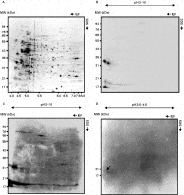
The p17 protein fraction was analysed by 2D-PAGE (Fig. 2A) combined with Western blotting and mass spectrometry. In Western blots of the 2D gels, APA17 stained 2 different spots at 33 kDa and 4 spots at 17 kDa in the low pH range (Fig. 2B). Interestingly, the isoelectric point for the predicted mature GRA7 protein is 4·43, which made this protein a good component candidate of the p17 fraction.

Fig. 2. Characterization of p17 by 2D-PAGE and Western blotting. (A) 2D proteome map of Neospora caninum tachyzoites. Migration of molecular weight and pI standards was as indicated. The p17 and p33 fractions are recognized by APA17 (B) and by a serum from a naturally infected cow (C) following isoelectric focusing of the Neospora soluble extract on an immobilized pH 3–10 non-linear gradient and 2D-Western blot. (D) p17 immunostaining (arrow) by a serum from a naturally infected cow following isoelectric focusing of Neospora soluble extract on an immobilized pH 3·5–4·5 non-linear gradient and 2D-western blot.
In a similar way to APA17 Neospora-positive serum from a naturally infected breeding cow intensely immunostained 2 spots at 33 kDa and 4 spots at 17 kDa in 2D Western blots (Fig. 2C). While densitometric analysis of ~700 silver-stained spots showed p17 and p33 to represent a minor percentage of N. caninum total protein, these 2 protein fractions are thus highly immunogenic, consistent with previous observations made on one-dimensional Western blots (Alvarez-Garcia et al. 2002).
EST database search with p17 MALDI-TOF peptide mass fingerprinting data
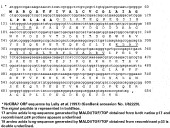
Since p17 was faintly visualized in a silver-stained gel and clearly detected in the acidic region of the gel by Western blotting, a high-resolution preparative 2D-PAGE fractionation in the low pH range was used to prepare for p17 sequencing. The spot in the 17 kDa size range detected by Coomassie staining and by immunostaining in Western blots (Fig. 2D) was subjected to tryptic peptide mass fingerprinting. The resulting sequence data were used in database searches for matching individual or consensus expressed sequence tags (ESTs). The experimental masses obtained by fingerprinting did not match any theoretical tryptic digests in the databases and further analysis of the sample by peptide fragmentation using tandem mass spectrometry was required to provide unambiguous identification. Peptide fragmentation data obtained from MALDI/TOF/TOF generated an unambiguous sequence of 17 amino acids (Fig. 3) with perfect identity to the predicted amino terminus of the mature N. caninum GRA7 dense granule protein, demonstrating that this is a component of the immunodominant p17 fraction.

Fig. 3. Gene peptide fragmentation data obtained from MALDI/TOF/TOF.
NcGRA7 recombinant protein (rNcGRA7) expression and EST database search with rNcGRA7 MALDI-TOF peptide mass fingerprinting data
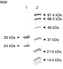
rNcGRA7 was expressed as a fusion soluble protein containing an N-terminal 6 kDa sequence from the plasmid which included a 6 histidine tag. The purified rNcGRA7 was run in a SDS-PAGE and 2 bands of 24 (p24) and 33 kDa (p33) were detected by Coomasssie staining (Fig. 4). The apparent molecular mass of p24, which is almost equivalent to the 17–19 kDa antigen plus 6 kDa sequence from the plasmid, matched perfectly with the predicted molecular weight of the mature NcGRA7 protein. When both bands, p24 and p33, were subjected to fingerprinting the experimental mass of p33 matched NcGRA7 theoretical tryptic digests. In order to corroborate our data, further MALDI/TOF/TOF analysis was accomplished and generated 2 unambiguous sequences of 17 and 19 amino acids corresponding to p24 and p33, respectively. The 17 amino acid-sequence was identical to that obtained for p17 described above, whereas the 19 amino acid-sequence was located between 96 and 116 residues in the NcGRA7 ORF (Fig. 3).

Fig. 4. Detection of the purified rNcGRA7 in a Coomassie-stained gel. rNcGRA7 antigen (lane 1) and the molecular weight standard (lane 2) were resolved in SDS-PAGE. Recombinant p33 and p24 NcGRA7 were submitted for mass spectrometry analysis.
DISCUSSION
In a continued effort to identify useful N. caninum antigens for the immunodiagnosis of neosporosis, we report here the molecular characterization of the 17–19 kDa antigenic protein fraction, p17, previously identified as the major IDA in one-dimensional PAGE by sera from bovines naturally infected with Neospora (Alvarez-Garcia et al. 2002; Alvarez-Garcia et al. 2006). Further characterization of p17 was accomplished by 2 complementary approaches. Firstly, a Neospora tachyzoite cDNA expression library was constructed and immunoscreened with a specific antibody developed against the p17 antigenic fraction (APA17), to identify cDNAs encoding some of its polypeptide components. Secondly, a proteomic approach consisting in 2D-PAGE and Western blot combined with mass spectrometric analysis was adopted to determine the amino acid sequence of any component of the immunodominant p17 fraction. Finally, cloning and expression of rNcGRA7 was done. The purified rNcGRA7 yielded 2 bands of 24 and 33 kDa in SDS-PAGE which were submitted for mass spectrometric analysis. The generated amino acid sequences matched perfectly with NcGRA7 proving that both p33 and p24 proteins correspond to NcGRA7.
Recombinant clones isolated by the immunoscreening of the cDNA library encompassed a cDNA sequence of 1210 bp that showed 100% identity with a tachyzoite cDNA encoding the dense granule protein GRA7. NcGRA7 was initially described by Lally et al. (1997), and its immunogenicity and diagnostic value was reported in other studies (Lally et al. 1996; Jenkins et al. 1997). The predicted size of the mature protein after removal of the putative signal peptide is approximately 19·8 kDa, in good agreement with the apparent molecular mass estimated for p17. Similar data have been reported for GRA7, with predicted molecular sizes for the mature protein ranging from 20·3 to 23 kDa (Lally et al. 1996, 1997). The difference between the apparent molecular weight of 17 kDa for p17 and the predicted molecular weight of 19·8 kDa for the encoded protein is within the resolution of one-dimensional SDS-PAGE, where p17 locates to the 17–19 kDa molecular weight range, a result confirmed by 2D-PAGE.
The above results are supported by those from our proteomic approach to determine whether the GRA7 protein is present in the p17 fraction. 2D-PAGE offers greater protein resolution potential than any other current separation technique and, when followed by Western blot, it allows minor but highly immunogenic proteins to be detected. In combination with mass spectrometry, it constitutes a powerful technique for antigen characterization. In the present study, the APA17 antibody simultaneously detected 2 antigenic fractions at 17–19 and 33 kDa, respectively. In this study, we observed 4 acidic isoforms in the p17 fraction. The 33 kDa fraction is composed of 2 acidic isoforms and exhibits a migration pattern similar to those of NcGRA7 (33 kDa and pI 4·3) (Lee et al. 2003) and Toxoplasma gondii GRA7 (Fischer et al. 1998). The similar detection pattern observed with APA17 and with bovine serum proves that the polypeptide components of p17 and p33 are immunodominant antigens which are of interest to the immunodiagnosis of bovine neosporosis. Although previous authors have reported that the native GRA7 protein may not be a major Neospora antigen in a Western blot assay (Lally et al. 1997), p17 has proved the most important IDA recognized by antibodies from naturally infected foetuses and breeding cattle under reducing conditions (Alvarez-Garcia et al. 2002). Dense granule proteins of T. gondii have been reported to be highly immunogenic in studies of human patients and in experimental toxoplasmosis, and have also been suggested to be involved in the development of concomitant immunity (Cesbron-Delauw, 1994; Vercammen et al. 2000).
Mass spectrometry on the tryptic digests of the17 kDa immunogen spot and both p24 and p33 obtained from the purified rNcGRA7 produced N-terminal sequences of 17 and 19 amino acids with a perfect match to NcGRA7. The analysis of the amino acid sequence of GRA7 reveals a clear partition of the hydrophobic and hydrophilic residues. The first hydrophobic region strongly suggests a signal peptide of 27 amino acids, which is consistent with GRA7 being a secreted protein. Excision of the signal peptide would produce a mature protein immunogen addressed to the secretory pathway, with an apparent molecular weight of 17–19 kDa. The absence of the signal peptide sequence in the mature protein is supported by the fact that when MALDI-TOF was carried out after tryptic digestion of the 17–19 kDa antigen, no matches were obtained when compared with ESTs available in the databases, whereas peptide fragmentation provided a 17 amino acidic sequence located immediately after the signal peptide. It must also be pointed out that the triptic peptide lacked the first 4 residues corresponding to the last residues of the signal peptide according to the GRA7 sequence. Our results can be readily explained by the fact that peptide mass fingerprinting data from MALDI-TOF are often unsuitable for EST database searches, since EST sequences are usually too short for reliable identification and there is uncertainty on the correct reading frame. Furthermore, ensuring confident matches requires a peptide mass fingerprint that must show identity to those available in the databases, whilst peptide fragmentation produces primary sequence information that can be used to search protein databases. This advantage points to peptide fragmentation data obtained either by post-source decay analysis or tandem mass spectrometry as a valuable approach for the study of Neospora spp., an organism still lacking genomic analysis.
It appears unlikely that p17 is the precursor form of NcGRA7 since the theoretical molecular weight of GRA7 is 19·8 kDa after removal of the signal peptide, and the predicted post-transductional modifications of the protein cannot explain these differences in molecular weights between the precursor and the final secretory product. Data from one-dimensional Western blots demonstrate that APA17 is highly specific, which is also strongly supported by mass spectrometry analysis of rNcGRA7, and support the hypothesis that NcGRA7 and p17 are encoded by the same open reading frame and that p17 is the monomer of NcGRA7. Dimer formation may involve hydrophobic interactions, while the absence of cysteine residues from the mature NcGRA7 rules out inter-chain disulfide linkage (Mercier et al. 2005). A similar processing of the MIC3 protein has been recently described in the closely related coccidian parasite, T. gondii, where TgMIC3 requires pro-peptide cleavage and dimerization to function as an adhesin (Cerede et al. 2002).
Our results provide evidence that p17 is encoded by the NcGRA7 gene. Further work will be required towards a more indepth molecular characterization of p17 and, importantly, the elucidation of GRA7 and p17 functions in host-parasite interactions.
We gratefully acknowledge the valuable support from the Proteomics Centre of Universidad Complutense de Madrid in the mass spectrometry analysis. We also thank Vanessa Navarro for her technical assistance. This work was supported by Project AGL2004-02529/GAN from Ministerio de Educación y Ciencia (MEC), and by the European Project QRLT-2001–2004 and COST Actions 854 and 857. Gema Álvarez García was funded by Consejería de Educación, Comunidad de Madrid.