Aortic coarctation is a congenital cardiac malformation that often requires surgical repair in the neonatal period. In general, low mortality rates for the repair of isolated aortic coarctation have been reported in recent years.Reference Ziemer, Jonas, Perry, Freed and Castaneda1 However, prematurity and very low birth weight are the risk factors associated with increased rates of morbidity and mortality.Reference Curzon, Milford-Beland and Li2 Prostaglandin E1 allows for temporary palliation of aortic coarctation, but is associated with side effects that can impair the overall outcome of an extremely low birth weight neonate.Reference Freed, Heymann, Lewis, Roehl and Kensey3
At present, there is insufficient evidence to favour either early surgical repair, interventional procedures such as balloon dilatation and stent implantation, or prolonged palliation with prostaglandin infusion in extremely low birth weight infants with critical congenital cardiac disease.Reference Ades, Johnson and Berger4–Reference Brodlie, Chaudhari and Hasan7
We report for the first time a staged surgical procedure for critical coarctation in an extremely low birth weight neonate with primary clipping of the duct and continued prostaglandin E1 infusion until delayed resection of the coarctation and end-to-end anastomosis.
Case report
The patient was a pre-term twin infant born at our institution at 25 weeks and 4 days of gestation after completing a course of betamethasone by Caesarean section because of feto–fetal transfusion syndrome and signs of severe centralisation. The birth weight was 740 grams; Apgar scores and umbilical arterial pH were normal. The patient was intubated for respiratory distress and a total of four doses – 400 milligrams per kilogram – of surfactant were administered.
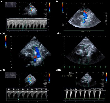
A routine echocardiography on the first day of life revealed a preductal coarctation with a hypoplastic distal aortic arch (Fig 1a).
(a) Continuous-wave Doppler echocardiogram on day 1 of life. There is a pronounced diastolic antegrade flow in the distal segment of the arch, which appeared markedly hypoplastic – less than 2 millimetres diameter. (b) Two-dimensional colour Doppler echocardiogram after clipping of the duct and continued prostaglandin E1 infusion on day 27. The distal arch is relatively wide – 2 millimetres diameter – and there is no significant flow acceleration or turbulence. (c) Two-dimensional colour Doppler (upper panel) and continuous-wave Doppler echocardiogram (lower panel) during continued prostaglandin E1 infusion with 30 nanograms per kilogram per minute on day 72. The upper panel shows constriction of the distal aortic arch, the lower panel shows flow acceleration compatible with a peak pressure gradient over the distal aortic arch of 48 millimetres of mercury. (d) Two-dimensional (upper panel) and continuous-wave Doppler echocardiogram (lower panel) showing a widely patent distal aortic arch with very mild flow acceleration at discharge on day 170.

During the initial days of life, the duct remained open without prostaglandin E1 administration and the ductal shunt was bidirectional, predominantly right to left. The medical team and the parents reached consensus that intensive medical therapy should be continued, but that the surgical repair of coarctation should be postponed as long as possible to enable growth before surgery.
Blood-stained tracheal aspirates and increasing fractions of left-to-right shunt through the duct on day 3 prompted permissive hypercapnia to increase pulmonary vascular resistance. Owing to arterial hypotension, negative-end diastolic flow in mesenteric and renal arteries and oligo and anuria, dopamine – 5 micrograms per kilogram per minute – was started on day 11. On day 15, prostaglandin E1 – 5 nanograms per kilogram per minute – was started because of constriction of the duct and isthmus. Systemic vasodilatation with nitroglycerine, at a dose of 3–10 micrograms per kilogram per minute, was started on day 18.
On day 26, – 1100 grams of body weight, including a significant oedema – an almost exclusive left-to-right shunt through the duct was observed, and the duct was clipped through a left posterolateral thoracotomy. Prostaglandin E1 was continued to prevent further constriction of the distal arch and isthmus (Fig 1b).
Presumably because of poor intestinal perfusion, the infant was unable to establish normal gastrointestinal motility and suffered from multiple intestinal perforations requiring repeated laparotomies.
An increase in pressure gradient across the isthmus to 35–40 millimetres of mercury despite prostaglandin E1 up to 30 nanograms per kilogram per minute finally triggered surgical resection of the coarctation with end-to-end anastomosis on day 75 through a left posterolateral thoracotomy at 1700 grams of weight, the clamp time being 15 minutes. The only complication was a transient right-sided phrenic nerve palsy.
The patient required laser therapy for rush-type retinopathy (grade 3 and above) and developed chronic lung disease of multifactorial origin – prematurity, inflammation, hyperperfusion and phrenic nerve palsy. Final extubation was on day 102, delayed by the phrenic nerve palsy. The patient was discharged with home oxygen requiring 0.03 litre per minute to achieve SpO2 values greater than 92%. Cranial ultrasound was normal at all times, and the neurological examination at discharge was also normal. Discharge echocardiography, 92 days after surgical repair, revealed an excellent result with widely open aortic anastomosis and normal flow across the distal aortic arch (Fig 1d). At 1-year postnatal age, flow velocity across the distal arch was 1.16 metre per second.
Discussion
Although there are reports of early repair of coarctation in infants with birth weights between 1000 and 2500 grams, prematurity is associated with increased mortality and increased rates of re-coarctation.Reference Curzon, Milford-Beland and Li2, Reference Sudarshan, Cochrane, Jun, Soto and Brizard8
In the past, surgical intervention for congenital cardiac disease was delayed in very premature infants, allowing for somatic growth and maturation, and prostaglandin E1 infusion is established for temporary palliation of aortic coarctation.Reference Freed, Heymann, Lewis, Roehl and Kensey3, Reference Brodlie, Chaudhari and Hasan7 In extremely low birth weight infants, however, management with prostaglandin E1 may result in complications associated with a large patent duct including intra- and periventricular haemorrhage, periventricular leucomalacia, pulmonary haemorrhage, bronchopulmonary dysplasia, and necrotising enterocolitis.
To avoid both morbidity and mortality associated with definite surgical interventions and prostaglandin E1-associated complications, both balloon dilatation and stenting of the coarctation have been reported.Reference Koch, Buheitel and Gerling5, Reference Radtke, Waller, Hebra and Bradley6 Balloon dilatation can be performed even in extremely low birth weight infants; however, severe problems associated with vascular access remain and re-coarctation rates are high.Reference Koch, Buheitel and Gerling5, Reference Fiore, Fischer and Schwartz9 Stent implantation would have required insertion of a 5 French sheath in the left carotid artery, and thus it was not considered to be an option in this extremely low birth weight infant.Reference Radtke, Waller, Hebra and Bradley6
Therefore, we chose for the first time an expectant staged surgical approach for the management of critical coarctation and hypoplastic distal aortic arch:
• Wait for signs of ductal or isthmic constriction.
• Use prostaglandin E1 to keep the duct open and the isthmus wide and apply adjuvant medical therapy to reduce pulmonary hyperperfusion.
• Clip the duct as soon as ductal steal is predominant and continue prostaglandin E1 infusion to inhibit constriction of ductal tissue located in the aortic isthmus.
• Perform definite surgical repair with resection and end-to-end anastomosis when the perfusion of the lower body can no longer be preserved by prostaglandin E1 infusion.
Strategies to minimise the side effects of the prostaglandin E1-mediated, persistent duct included permissive hypercapnia, high lung volume, dopamine as vasopressor, and nitroglycerine for systemic vasodilatation.
Despite these efforts, the clinical course was complicated by chronic lung disease, transient oligo and anuria, and intestinal dysmotility presumably because of impaired perfusion due to coarctation and left-to-right shunt across the patent duct.
In retrospect, earlier clipping of the duct might have reduced pulmonary hyperperfusion, lessened chronic lung disease, and preserved intestinal and renal perfusion. However, it was unknown for how long continued prostaglandin E1 could prevent constriction of the aortic isthmus and hence the need for ultimate surgical repair.
More than 20 months after extremely pre-term birth and 18 months after surgical repair of aortic coarctation, the patient revealed an excellent long-term surgical result with a widely open arch and normal flow velocity of vmax 1.1 metre per second in the descending aorta. Furthermore, medium-term neuro-developmental outcome was normal.
In summary, we report successful treatment of a 25-week, 740-gram infant with critical coarctation by a staged procedure. In retrospect, earlier clipping of the duct should have been considered. On the basis of this experience, it may appear advisable to close the duct as soon as ductal shunt is predominantly left to right and diastolic flow in mesenteric and renal arteries is impaired, while continuing prostaglandin E1 infusion to prevent further constriction of ductal tissue located in the aortic isthmus.
Acknowledgement
Anja Bialkowski, Winfried Baden contributed equally to this manuscript. The authors have no conflict of interest to declare.