Introduction
Canine visceral leishmaniosis (CVL) is a neglected tropical disease caused by protozoa of the genus Leishmania Ross, 1903, which affects dogs from all continents, except Oceania (Colwell et al., Reference Colwell, Dantas-Torres and Otranto2011; Dantas-Torres et al., Reference Dantas-Torres, Solano-Gallego, Baneth, Ribeiro, Paiva-Cavalcanti and Otranto2012; Otranto et al., Reference Otranto, Dantas-Torres and Breitschwerdt2019). More than 20 Leishmania species are known to exist, and in dogs, Leishmania infantum is the aetiological agent of CVL. Visceral leishmaniosis (VL) also affects humans in Europe, the Middle East, South Asia, Africa, and in Central and South America, where transmission may be anthroponotic, but where dogs with CVL can also act as reservoir hosts for human infection, transmitted by the bites of sand fly vectors (Chappuis et al., Reference Chappuis, Sundar, Hailu, Ghalib, Rijal, Peeling, Alvar and Boelaert2007; WHO, 2010). In the Americas, human VL is mainly if not exclusively zoonotic and found in at least 12 countries; however, 90% of the cases are reported in Brazil, causing approximately 4200–6300 infections per year, with a 7% mortality rate (Romero and Boelaert, Reference Romero and Boelaert2010; Alvar et al., Reference Alvar, Vélez, Bern, Herrero, Desjeux, Cano, Jannin and den Boer2012; WHO, 2017). The increasing spread of L. infantum into urban areas in the Americas is of great concern, and is associated with migration, population growth and poor living conditions (Werneck, Reference Werneck2008; Romero and Boelaert, Reference Romero and Boelaert2010; Harhay et al., Reference Harhay, Olliaro, Costa and Costa2011). CVL is a multisystemic disease with clinical signs that may be apparent, such as dermatitis, lymphadenomegaly, general muscular atrophy and renal disease (Baneth et al., Reference Baneth, Koutinas, Solano-Gallego, Bourdeau and Ferrer2008). However, control programmes for both the disease and infection are essential, because many dogs remain asymptomatic, which makes it difficult to diagnose infection and control transmission to humans (Gavgani et al., Reference Gavgani, Hodjati, Mohite and Davies2002; WHO, 2010). Current methods to prevent the spread of CVL include the use of topical insecticides on dogs and canine vaccination, which provides only a certain level of protection for dogs (Solano-Gallego et al., Reference Solano-Gallego, Koutinas, Miro, Cardoso, Pennisi, Ferrer, Bourdeau, Oliva and Baneth2009; Sousa-Paula et al., Reference Sousa-Paula, Silva, Sales and Dantas-Torres2019). In addition, many countries have also used euthanasia of seropositive dogs (Dantas-Torres et al., Reference Dantas-Torres, Solano-Gallego, Baneth, Ribeiro, Paiva-Cavalcanti and Otranto2012; BRASIL, 2014). However, scientific studies have not yet shown that this euthanasia strategy decreases the infection rate of VL (Romero and Boelaert, Reference Romero and Boelaert2010; Dantas-Torres et al., Reference Dantas-Torres, Miro´, Bowman, Gradoni and Otranto2019; Sousa-Paula et al., Reference Sousa-Paula, Silva, Sales and Dantas-Torres2019). Moreover, the method is not well-received by the population for ethical reasons (Costa, Reference Costa2011; Pereira-da-Silva et al., Reference Pereira-da-Silva, Marques, Lamounier, de Castro and Borja- Cabrera2017). The treatment of CVL-positive dogs is permitted in some countries; however, euthanasia may still be the only option for owners who cannot afford to have their dogs treated. Therefore, the development of more precise diagnostic methods for both rapid treatment of humans and control of CVL in dogs is urgently required (Foglia Manzillo, Reference Foglia Manzillo, Di Muccio, Cappiello, Scalone, Paparcone, Fiorentino, Gizzrelli, Gramiccia, Gradoni and Oliva2013; Fonseca et al., Reference Fonseca, Faria, Rodrigues, Nagem, Magalhães, Cunha, Bartholomeu and Andrade2014; Coelho et al., Reference Coelho, Costa, Lage, Martins, Garde and Jesus-Pereira2016).
For accurate diagnosis of CVL, several approaches are available. The parasitological method of biopsy followed by detecting the amastigote forms of L. infantum by microscopy is still considered the gold standard (BRASIL, 2014). However, it is an invasive method and there are complications with its use due to the need for a trained professional capable of recognising the parasites. Culture methods to supplement microscopy give improved sensitivity but are also highly susceptible to contamination (Boelaert et al., Reference Boelaert, Bhattacharya, Chappuis, El Safi, Hailu and Mondal2007; Goto et al., Reference Goto, Carter, Guderian, Inoue, Kawazu and Reed2010; de Vries et al., Reference De Vries, Reedijk and Schallig2015). Polymerase chain reaction (PCR) methods to detect Leishmania DNA show variable sensitivity, even though it is a species-specific method (Gomes et al., Reference Gomes, Cesetti, Paula, Vernal, Gupta, Sampaio and Roselino2017; Rampazzo et al., Reference Rampazzo, Solcà, Santos, Pereira, Guedes, Veras, Fraga, Kriegera and Costa2017). Various serological methods can be used including enzyme-linked immunosorbent assay (ELISA), indirect fluorescence antibody test (IFAT), direct agglutination test, Western-blot and immunochromatographic tests to detect specific antibodies present in serum samples; however, these can present problems in sensitivity and specificity (Georgiadou et al., Reference Georgiadou, Makaritsis and Dalekos2015; Magalhães et al., Reference Magalhães, Castro-Neto, Nascimento, Santos, Medeiros, Neto, Costa, Costa, dos Santos, de Carvalho, Oliveira and Melo Neto2017). Nonetheless, such serological techniques can be highly efficient in the detection of VL infection in humans and dogs. To standardize and optimize their use, many recombinant proteins have been studied in recent years with the aim of increasing the sensitivity and specificity of such tests (Celeste et al., Reference Celeste, Angel, Castro, Gidlund and Goto2004; Wolf et al., Reference Wolf, Failing, Taubert and Pantchev2014; Mniouil et al., Reference Mniouil, Fellah, Amarir, Sadak, Et-Touys, Bakri, Moustachi, Tassou, Hida, Lyagoubi, Adlaoui, Rhajaoui and Sebti2018).
In Brazil, the Ministry of Health has determined that the lateral flow strip test Dual-Path Platform assay (TR-DPP® LVC) produced by Bio-Manguinhos/Fiocruz, Brazil (BRASIL, 2014) should be used for serological screening of dogs, followed by an ELISA test (EIE-LVC) for confirmation of L. infantum infection (Grimaldi et al., Reference Grimaldi, Teva, Ferreira, dos Santos, Pinto, de-Azevedo and Falqueto2012; Faria et al., Reference Faria, de Castro Veloso, Coura-Vital, Reis and Damasceno2015). Some purified recombinant antigens, such as the rK39 antigen, of various Leishmania species have been produced and used in serological assays of VL (Badaró et al., Reference Badaró, Benson, Eulálio, Freire, Cunha, Netto, Pedral, Madureira, Burns, Houghton, David and Reed1996). Even though the TR-DPP® test uses a combination of recombinant proteins (rK26 and rK39) which should improve sensitivity (Bhatia et al., Reference Bhatia, Daiffalla, Jen, Badaró, Reed and Skeiky1999; Pattabhi et al., Reference Pattabhi, Whittle, Mohamath, El-Safi and Moulton2010), it can perform with low sensitivity, which compromises the efficacy of the diagnosis of infection in Brazil (Grimaldi et al., Reference Grimaldi, Teva, Ferreira, dos Santos, Pinto, de-Azevedo and Falqueto2012; De Santis et al., Reference De Santis, Santos, Souza and Chaves2013). Further, the crude parasite extract used in the EIE-LVC test has also shown false-positive results due to cross-reactivity with other pathogens, particularly Trypanosoma cruzi, in North, Central and South America (Ferreira et al., Reference Ferreira, de Lana, Carneiro, Reis, Paes, da Silva, Schallig and Gontijo2007; Solano-Gallego et al., Reference Solano-Gallego, Koutinas, Miro, Cardoso, Pennisi, Ferrer, Bourdeau, Oliva and Baneth2009). Cross-reactions are less likely to occur when using recombinant proteins in ELISA, such as rA2, rK9, rK26 and rK39 (Solano-Gallego et al., Reference Solano-Gallego, Koutinas, Miro, Cardoso, Pennisi, Ferrer, Bourdeau, Oliva and Baneth2009). Considering the need for new antigens for improved serodiagnosis of CVL that show both high specificity and sensibility, and no cross-reaction with other diseases, we undertook this study, in which we investigated the use of the recently described Lbk39 recombinant protein (De Souza et al., Reference De Souza, Carvalho, Bates, Petterle, Thomaz-Soccol and Bates2019) as an antigen in CVL diagnosis by the ELISA method.
Materials and methods
Production of Lbk39 recombinant protein
Lbk39 recombinant protein was produced essentially as previously described (De Souza et al., Reference De Souza, Carvalho, Bates, Petterle, Thomaz-Soccol and Bates2019). Briefly, the Lbk39 fragment of a kinesin-related gene of L. braziliensis is composed of 843 base pairs and encodes repetitive immunological amino acids. The protein is related to the kinesin-related gene of L. infantum (Burns et al., Reference Burns, Shreffler, Benson, Ghalib, Badaro and Reed1993), which has 6.5 repetitions of 39 amino acids. The synthetic gene Lbk39 was cloned with a 6xHis-tag in the recombinant vector pLEXSY-sat2 and inserted into the eukaryotic host L. tarentolae (De Souza et al., Reference De Souza, Carvalho, Bates, Petterle, Thomaz-Soccol and Bates2019).
An aliquot of transfected L. tarentolae was thawed and placed in 100 mL of culture medium. As soon as the cultures became slightly turbid after 24 h, the specific Streptothricin-class of aminoglycoside antibiotic Nourseothricin (LEXSY NTC, Jena, Germany, Jena Bioscience) for the pLEXSY-sat2 vector was added, and the culture was maintained under the same conditions by sub-passage every 4 days. Then the pre-inoculum was inoculated in BHI medium (2 L) supplemented with 2 mL of porcine hemin (Jena Bioscience), 5 mL of penicillin and streptomycin (Pen-Strep, Jena Bioscience) and 1 mL of NTC antibiotic (Jena Bioscience) and incubated at 26°C in the dark under aerated conditions. The culture was then centrifuged at 5000 g, 4°C, for 10 min, the supernatant medium removed and stored frozen at −80°C.
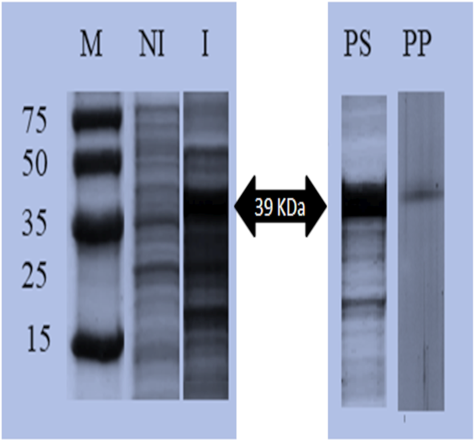
Lbk39 recombinant protein (250 mL) was purified from the supernatant medium using HisTrap HP column chromatography (GE HealthCare) by loading the thawed culture medium supernatant into the column and eluting the purified protein, according to the manufacturer's instructions. To remove salts and imidazole present in the purified protein, dialysis in PBS buffer 7.2 was performed at 4°C, once overnight, and twice for 2 h. The purified product (25 mL) was lyophilized to concentrate the Lbk39 recombinant protein. To determine the concentration of Lbk39 recombinant protein, the Micro BCA™ Protein Assay Kit (Thermo Fischer Scientific) was used, following the manufacturer's protocol. To verify that the protein was indeed purified, 15% SDS-PAGE SDS-polyacrylamide gel electrophoresis was performed on recombinant Lbk39 produced after the protein purification process, dialysis and lyophilization (for details, see De Souza et al., Reference De Souza, Carvalho, Bates, Petterle, Thomaz-Soccol and Bates2019). The gel was stained with nitrate silver (Fig. 1).
SDS-PAGE of induction and purification of LKb39.

Collection and sample processing
For this study, 152 canine serum samples were used to evaluate the diagnostic potential of Lbk39 in the main part of the study. Of these, 73 sera were from healthy animals from a non-endemic area of Brazil (Araucária, Parana), which can be assumed to be almost exclusively negative for CVL, with the possible exception of rare imported cases. For the positive sera, we used dog samples from the region of the outbreak Foz do Iguaçu (for more details, see Thomaz Soccol et al., Reference Thomaz Soccol, Pasquali, Pozzolo, Leandro, Chiyo and Baggio2017). To assess the clinical diagnosis of CLV, clinical signals were recorded in an epidemiological questionnaire. Owned dogs were examined by veterinarians for clinical signs of the disease and each dog was given an individual data file including identification, traits, behaviour, migration history and health issues. Signals investigated were weight loss, adenomegaly, alopecia, skin lesions, mucosal lesions, hyperkeratosis and muscle atrophy. Blood samples were collected by venipuncture of the jugular vein, transferred to 10 mL polypropylene tubes and processed 3–4 h after collection. In the laboratory, the blood was centrifuged at 1000 × g for 5–10 min and the sera were separated and stored at −20°C until analysed by serological methods, the TR-DPP (Bio-Manguinhos/Fiocruz) as recommended by the Brazilian Ministry of Health and used according to the manufacturer's instructions. The indirect ELISA using the total extract of L. infantum was performed as described by Maziero et al. (Reference Maziero, Thomaz-Soccol, Steindel, Link, Rossini, Alban and Nascimento2014). Each sample was tested in triplicate intra- and inter-plate. Three positive and negative dog sera were included in each plate as controls, when testing individual sera.
Dogs that tested seropositive were examined by puncture of popliteal lymph nodes. The collected material was used for culture in NNN media and PCR specific for Leishmania. DNA extraction was performed using the Wizard® Genomic DNA Purification Kit in accordance with the manufacturer's recommendations. Cytochrome B1/B2 was used as an internal control to verify DNA amplification (Oshaghi et al., Reference Oshaghi, Chavshin, Vatandoost, Yaaghoobi, Mohtarami and Noorjah2006). The PCR was performed using Internal Transcribed Spacer (ITS) primers (Schonian et al., Reference Schonian, Naserddin, Dinse, Scheweynoch, Schallig, Presber and Jaffe2003). The positive PCR products were sequenced. The sequencing was performed commercially by Macrogen Inc. (Seoul, South Korea). The 79 positive sera were from dogs in an area highly endemic for CVL (Foz do Iguaçu), and included 52 sera from clinical cases for CVL and 27 sera from no clinical dogs. In addition to these sera, to evaluate potential cross-reaction, 23 sera were used from dogs positive for Toxoplasma gondii infection.
Ethics statement
For collection of dog's samples, all procedures were carried out in strict compliance with the rules defined by the National Council for the Control of Animal Experiments (CONCEA), and every effort was made to minimize suffering. The work was approved by the Ethics Committee of the Federal University of Paraná, under protocol no. 044/2014. The owners have signed a consent form for the use of the samples.
Lbk39 antigen: ELISA standardization
The ELISA test was developed and standardized by analysing results using antigens at different concentrations (0.031, 0.062, 0.125, 0.250, 0.500 and 1000 μg well−1), two serum dilutions (1:100 and 1:200) and various dilutions of HRP-conjugated secondary antibodies (1:3500, 1:5000, 1:7500 and 1: 10 000). The Lbk39 antigen was diluted in carbonate-bicarbonate buffer, pH 9.6 and polystyrene microtiter plates (96-well EIA/RIA 12 × 8 well plates, Costar, Wilkes-Barre, Pennsylvania, USA) were coated with 100 μL well−1 of dilutions at all of the concentrations of antigens given above, and incubated overnight at 4°C. The next day, the plates were washed with 200 μL well−1 of washing solution (0.9% NaCl + 0.05% Tween 20), twice. Next, the wells were blocked with 120 μL of blocking solution composed of PBS + 0.1% casein at 37°C, for 1 h, and were then washed again, twice, with 200 μL well−1 of washing solution. After these steps, the serum samples were added and incubated at 37°C, for 1 h. The plates were then washed with 200 μL well−1 of washing solution four times. In total, 100 μL of various dilutions of a polyclonal goat anti-dog IgG HRP (2 mg mL−1) conjugate was plated at the concentrations given above at 37°C, for 1 h. Lastly, the reaction was developed by adding 100 μL well−1 of a solution prepared by combining 10.0 mL of citrate buffer (4.5% Na2PO4 + 3.25% citric acid + distilled water, pH 5.0), 2 mg of o-phenylenediamine dihydrochloride (2 mg tablet−1, Sigma, USA) and 2 μL of 30% H2O2, and incubating at room temperature for 30 min, avoiding light. The reaction was stopped by adding 20 μL well−1 of 1:20 H2SO4 solution. The plates were read in a PowerWave HT reader (BioTek) at 492 nm and the values were expressed in absorbance. The cut-off was calculated from the mean value of the negative controls.
Tests used for comparison with the Lbk 39 antigen
To confirm the CVL status of dogs within the study, the following three standard tests were used: parasitological diagnosis by culture of aspirates in Novy, Neal and Nicolle (NNN) medium; serological diagnosis using the TR-DPP test; and serological diagnosis using an ELISA test using total crude extract of L. infantum promastigotes (equivalent to the EIE-LVC test). These results were compared with those obtained using ELISA with Lbk39 recombinant protein.
Parasitological diagnosis by culture was done as recommended by Szargiki et al. (Reference Szargiki, Castro, Luz, Kowalthuk, Machado and Thomaz-Soccol2009). In brief, bone marrow aspirate, lymph node aspirate or the leucocyte layer of dogs were inoculated in NNN culture medium with 0.9% saline solution with weekly sub-passage, for 4 weeks at 24°C. The cultures were inspected every week for the presence of promastigotes. Positive cultures were identified as described by Thomaz Soccol et al. (Reference Thomaz Soccol, Pasquali, Pozzolo, Leandro, Chiyo and Baggio2017) and all found to be L. infantum.
Statistical analysis
The receiver operating characteristic (ROC) curve, sensitivity, specificity analysis and comparison between tests were analysed by MedCalc, version 18.11.6 (R Core Team, 2018; MedCalc Statistical Software, 2019); Graphpad prism 5.0 was used to plot the graphs and a significance level of P < 0.05 was adopted.
Results
Antigen production
A pre-inoculum culture was produced in 100 mL of medium, followed by the inoculation into 1 L of supplemented BHI medium for antigen production. After 4 days, the culture was centrifuged and separated into a pellet and supernatant medium. The total protein concentration in the supernatant medium was 8286.52 μg mL−1. The supernatant medium was processed in 250 mL aliquots, and each was used for protein purification using chromatography by loading the culture medium on to a 1 mL HisTrap HP column (GE HealthCare), according to the manufacturer's instructions. After purification by elution from the column, dialysis was performed to remove salts and imidazole from the eluate. Finally, the product was lyophilized and the Lkb39 protein was resuspended in ultrapure water and concentration was determined to be 337.68 μg mL−1 measured by the Micro BCA™ method.
Enzyme-linked immunosorbent assay
The ELISA test was evaluated for its ability to determine the level of anti-Leishmania antibodies in dog sera from L. infantum-infected and non-infected animals using as antigen the recombinant protein Lbk39. The ELISA was optimized by performing tests at six antigen concentrations and was able to differentiate sera positive for CVL from negative sera. The concentration chosen as standard was 125 μg protein per well, since this produced the best response for the most reasonable cost. In the assays performed, we observed a 34-fold increase in the absorbance values of positive controls compared with negative controls.
The cut-off value was calculated using ROC curve analysis. For serum dilution optimization, no significant differences were observed amongst the dilutions tested, thus the 200-fold dilution was chosen because it uses a small quantity of serum and the background was lower. The dilution of the goat anti-dog IgG HRP conjugate that gave the best results was 1:7500, so this was selected for use as standard. To determine the cut-off level of the reaction of positive and negative sera, the mean and standard deviation absorbances of the canine samples negative for CVL (73 sera samples) were determined. The absorbance cut-off was set at 0.156.
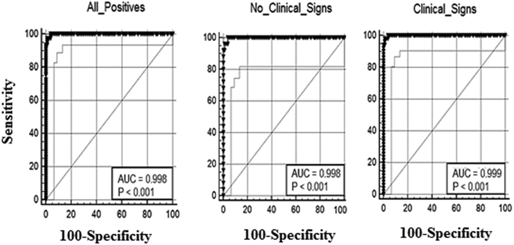
After standardization, we used the optimized ELISA with Lbk39 antigen to analyse all 152 canine sera in the study. From this analysis, of the 79 dog sera from the CVL endemic region of Foz do Iguaçu, 76 were positive and three were negative in the Lbk39 ELISA assay. Of these three negative animals, two were in the group with no clinical signs, one of these was negative in all other assays used and one was positive with the TR-DPP assay. The negative result from the dog with clinical signs was positive with the crude antigen ELISA and TR-DPP assays. Therefore, we conclude that two of these negative results from Foz do Iguaçu were false negatives and one was a true negative. All the dog sera from Araucária showed negative results with the Lbk39 assay. The ROC curve analysis of absorbance distribution against the Lbk39 antigen for various groups studied (all positives from any assay, dogs with clinical signs, dogs without clinical signs) is shown in Fig. 2. The difference in canine samples was significant between these groups (P < 0.001) and accurate. The positive group (78 samples) presented good accuracy (AUC, 0.998), similar to the no clinical signs group (AUC, 0.998), while the animals with clinical signs presented higher accuracy (AUC, 0.999) (Table 1).
Comparison of ELISA reactivity of canine sera against Lbk39 recombinant proteins. AUC, accuracy.

Characteristics and location of canine blood samples

a Clinical signs: the main clinical features observed for this classification were alopecia, peeling dry skin, brittle skin, nodules on the skin, ulcers, weakness, low weight and ocular lesions.
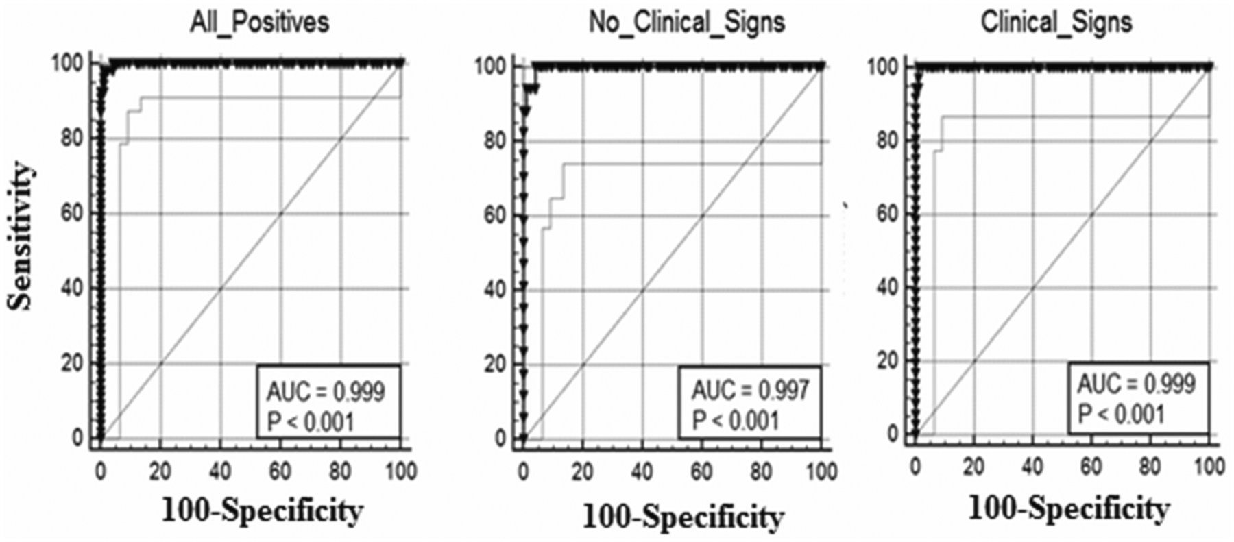
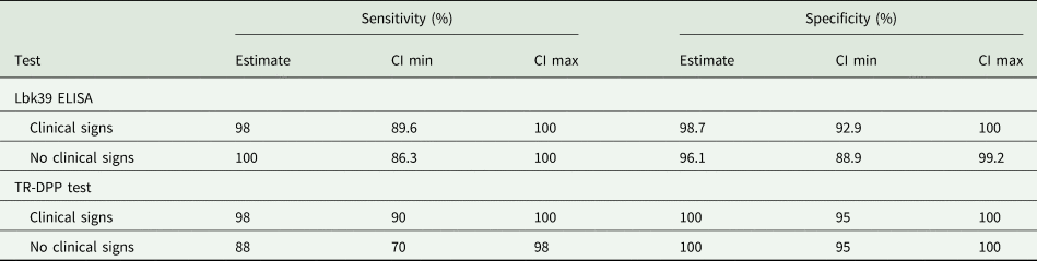
Culture is used as a standard test for CVL diagnosis, because although it may sometimes lack sensitivity, it has very high specificity, as when it is positive, it definitively means that the dog has the parasite. Therefore, we evaluated the subset of 56 dogs that presented with a positive culture. Table 2 shows the results for sensitivity and specificity of the Lbk39 assay with these sera, also subdivided into those with clinical signs or no clinical signs. As shown, the Lbk39 assay performed with very high sensitivity and specificity against this subset of dog sera. ROC curve analysis of the absorbance distribution for the Lbk39 ELISA against the positive culture subset confirmed that the test worked very well (P < 0.001) and was accurate (Fig. 3). The total positives group presented the same accuracy (AUC, 0.999) as the clinical signs subgroup (AUC, 0.999), while the no clinical signs subgroup presented with slightly lower accuracy (AUC, 0.997).
Comparison of ELISA reactivity of canine sera against Lbk39 recombinant proteins in true-positive cultures. AUC, accuracy.

Lbk39 test sensitivity and specificity for the sample of 56 positive canine sample cultures, according to clinical classification (with or without clinical signs)

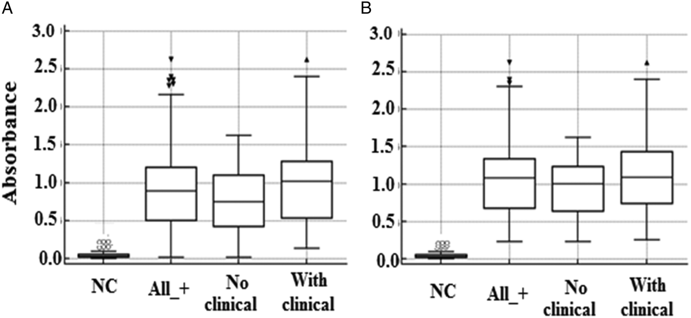
Analysis of the distribution of ELISA readings from the Lbk39 assay in various subgroups of dog sera is shown in Fig. 4. Comparisons are shown between negative controls, positives, those positives with clinical signs and those positives with no clinical signs, either for all sera (Fig. 4A) or for the subset of Lbk39 ELISA positives with positive culture (Fig. 4B). All groups and subgroups of positives were significantly different from the negative controls. When the analysis was confined to sera from dogs that also had a positive culture, as expected the discrimination was greater (Fig. 4B).
Box and whisker plot comparisons of absorbance distributions in different groups of dog sera. (A) Analysis of all samples. (B) Analysis of the subset of ELISA positives that also had a positive culture. The symbols represent outliers, where each corresponds to the result obtained with an individual serum.

Comparison of Lbk39 with other diagnostic tests
In addition to evaluating the performance of the Lbk39 ELISA against clinical and parasitological diagnostic tests, we also compared our standardized assay with other commonly used serological tests, ELISA with a crude antigen preparation and the TR-DPP assay. For these comparisons, we considered a true positive to be any animal that was positive in any of the assays used. This comparison showed that the Lbk39 assay outperformed all the other tests in terms of sensitivity (Table 3). The Lbk39 assay also performed well in terms of specificity (96.1%), with either TR-DPP being slightly better (97.2%). As mentioned previously, culture is highly specific, but this has a relative poor sensitivity (77%). Operationally, dogs with CVL are first identified by clinical signs and then the TR-DPP test is applied, sometimes this being the only testing performed. Therefore, we divided the positive group into cases with clinical signs and without clinical signs and evaluated the sensitivity and specificity of Lbk39 comparing with the TR-DPP assay (Table 4). When the individual groups were analysed, this showed that the group of sera from dogs with clinical signs were detected with similar and high sensitivity and specificity for both tests, the TR-DPP being slightly more specific. However, in dogs without clinical signs, the Lbk39 ELISA was significantly more sensitive, and slightly less specific than TR-DPP.
Comparison between Lbk39 antigen and other methods (TR-DPP®, iELISA and culture in artificial media) analysing 78 positive dogs and 75 negative controls

Comparison between Lbk39 antigen and TR-DPP for the sample of 78 positive controls, according to clinical classification (with or without clinical signs)

Cross-reaction with Lbk39
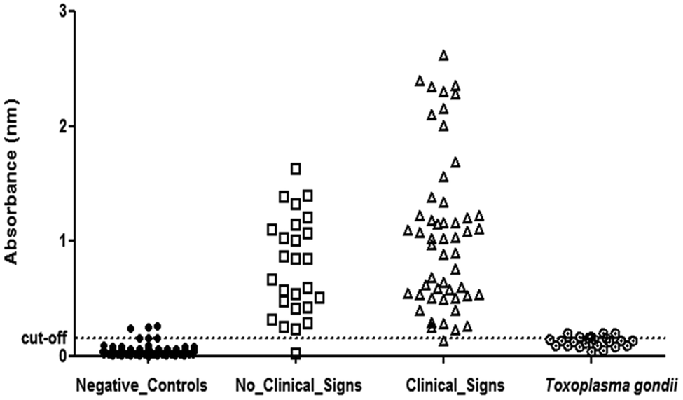
To check for potential cross-reactivity of the Lbk39 antigen with another intracellular parasite, we performed ELISA with Lbk39 against sera from toxoplasmosis-positive canine samples. No cross-reaction was observed (Fig. 5).
Analyses of cross-reaction with Lbk39 against Toxoplasma gondii compared with negative controls and dogs with and without clinical signs.

Discussion
Leishmania braziliensis is endemic in the study area, and L. infantum has been recognized since 2012 (Dias et al., Reference Dias, Thomaz-Soccol, Bisetto, Pozzolo, Chiyo and Freire2013; Thomaz Soccol et al., Reference Thomaz Soccol, Pasquali, Pozzolo, Leandro, Chiyo and Baggio2017); therefore, to work with specific species of CVL, dogs were subjected to clinical evaluation and identification of the parasite present in the popliteal ganglion. In endemic areas of L. braziliensis, diseased dogs generally have typical well-limited ulcerated lesions with raised edges. The main locations of ulcers are in the scrotal sac and on top of the ears (Dantas-Torres et al., Reference Dantas-Torres, de Paiva-Cavalcanti, Figueredo, Melo, da Silva, da Silva, Almeida and Brandão-Filho2010). Dogs with CVL generally exhibit lymphadenomegaly, exfoliative dermatitis and weight loss as the most relevant signals on which veterinarians have based their suspicion of infection (Gálvez et al., Reference Gálvez, Miró, Descalzo and Molina2011). In the present study, no animals showed specific signs of CL. In addition, in all animals that tested serologically positive on initial testing, the parasite isolated was identified as L. infantum.
Accurate diagnosis of CVL is important in efforts to interrupt the life cycle of the parasite and prevent the spread of the disease in endemic areas (Fraga et al., Reference Fraga, Da Silva, Pacheco, Borja, Coura-Vital, Monteiro, Oliveira, Jerônimo, Reis and Veras2014; Singh and Sundar, Reference Singh and Sundar2015). The recombinant protein Lbk39 previously described by our group (De Souza et al., Reference De Souza, Carvalho, Bates, Petterle, Thomaz-Soccol and Bates2019) has been validated here for use in the diagnosis of CVL. Antigen production was not performed in E. coli, but in L. tarentolae. As the desired recombinant antigen is derived from a Leishmania species, the use of this system maximizes the probability of successful expression. The antigenic protein produced by heterologous expression of specific Leishmania epitopes in a prokaryotic system such as E. coli is inexpensive to culture and allows rapid processing of the target recombinant protein. However, this system has drawbacks, e.g. it requires post-translational eukaryotic activity, may have high levels of unfolded proteins, resulting in reduced efficacy. In addition, the ideal culture temperature for E. coli may reduce recombinant protein yield and increase protein degradation (Khow and Suntrarachun, Reference Khow and Suntrarachun2012). Leishmania tarentolae, which is a non-mammalian pathogenic species, has been explored as a general eukaryotic host with the aim of developing a platform within it that allows high levels of expression of complex eukaryotic proteins and has the ability to produce proteins with appropriate post-translational processing (Basile and Peticca, Reference Basile and Peticca2009). This type of host is also easy to manipulate and has an inexpensive culture medium with an average doubling time of 6–8 h. The recombinant protein, produced by our group, has been tested for LC and VLH with excellent results (De Souza et al., Reference De Souza, Carvalho, Bates, Petterle, Thomaz-Soccol and Bates2019).
Here, we used this protein for CVL and an ELISA test format was chosen because it is a preferred choice for serodiagnosis screening of the disease in the laboratory, being generally more reliable and sensitive than other laboratory tests (Srividya et al., Reference Srividya, Kulshrestha, Singh and Salotra2012; Abass et al., Reference Abass, Kang, Martinkovic, Semiao-Santos, Sundar, Walden, Piarroux, Harith, Lohoff and Ulrich Steinhoff2015). The Lbk39 antigen showed higher sensitivity (100%) compared with TR-DPP (93.8%), ELISA using as antigen the parasite total extract (98.1%), or parasite culture (77%). The specificity was similar to that of TR-DPP (96.1 and 97.2%, respectively), and better than crude parasite extract (86.9%). These findings showed that Lbk39 has the highest probability of a true-positive result in the presence of the disease, and it has the lowest probability of a false-negative result in the absence of the disease compared with other tests. To increase specificity and minimize the possibility of a false positive, we used a cut-off value based on the standard deviation of the results of negative controls from healthy dogs, and as a consequence, the Lbk39 assay did not produce many false-negative results. However, when we evaluated only the canine samples from dogs with a positive culture with or without clinical signs, we observed that our protein showed 100% sensitivity in both groups, and a small variation in specificity between the groups with clinical signs (98.7%) and without clinical signs (96.0%). While evaluating TR-DPP, we observed that the group with clinical signs showed higher sensitivity (98%) than the group without (88%). As for the specificity, in the positive group, dogs with either no clinical signs or clinical signs for TR-DPP presented 100%, whereas our protein presented specificity from 96.1 to 98.7%.
In the last 10 years, several CVL diagnostic methods have been used. Their specificity and sensitivity can vary. For example, the specificity of molecular tests varies from 95 to 100%, where conventional PCR (cPCR) has a sensitivity of 89–100% in polysymptomatic dogs (Lachaud et al., Reference Lachaud, Chabbert, Dubessay, Dereure, Lamothe, Dedet and Bastien2002; Manna et al., Reference Manna, Vitale, Reale, Caracappa, Pavone, Morte, Della Cringoli, Staiano and Gravino2004; Carson et al., Reference Carson, Quinnell, Holden, Garcez, Deborggraeve and Courtenay2010). However, sensitivity decreases when the method is applied to asymptomatic dogs (Francino et al., Reference Francino, Altet, Sánchez-Robert, Rodriguez, Solano-Gallego, Alberola, Ferrer, Sánchez and Roura2006; Gao et al., Reference Gao, Ding, Wang, Steverding, Wang, Yang and Shi2015). Currently, quantitative PCR is considered the most reliable test because it has a sensitivity of 91% compared to cPCR (72%), ITS-1 PCR (54%) and PCR hybridization (61%) (Carson et al., Reference Carson, Quinnell, Holden, Garcez, Deborggraeve and Courtenay2010). The molecular test with loop-mediated amplification (LAMP) is capable to amplify DNA at a constant temperature in the blood of VL patients. It has sensitivities and specificities of 90.7–96.4% and 98.5–100%, respectively. In China, the LAMP identified 61% infected dogs, however, did not amplify strains from other countries (Gao et al., Reference Gao, Ding, Wang, Steverding, Wang, Yang and Shi2015). The direct agglutination test present sensitivities of 91–100% and specificities of 72–100% but yet subjective reading of end-point titles leads to interrupt server discrepancy (Adams et al., Reference Adams, Jacquet, Schoone, Gidwani, Boelaert and Cunningham2012; Oliveira et al., Reference Oliveira, Saliba, Oliveira, Dias and Paz2016). Although some serological tests for canine leishmaniosis have high specificity and sensitivity, the presence of cross-reactivity remains controversial. The immunofluorescence antibody test is reference qualitative in CVL diagnosis (Paltrinieri et al., Reference Paltrinieri, Gradoni, Roura, Zatelli and Zini2016), with sensitivity and specificity close to 100%, in symptomatic animals, though show some limitations such as cross-reactivity with trypanosome pathogens (Solano-Gallego et al., Reference Solano-Gallego, Villanueva-Saz, Carbonell, Trotta, Furlanello and Natale2014; Paltrinieri et al., Reference Paltrinieri, Gradoni, Roura, Zatelli and Zini2016) and the significantly lower sensitivity in identifying asymptomatic dogs compared with ELISA (Mettler et al., Reference Mettler, Grimm, Capelli, Camp and Deplazes2005).
The main sera used to verify cross-reactivity against the antigen of choice would be T. cruzi, Babesia canis, Dirofilaria immitis and T. gondii, however, in the present study, it was possible to test only the cross-reactivity against T. gondii serum available for use. In previous studies, cross-reactivity between Leishmania species and T. gondii was tested by ELISA and IFAT (Boelaert et al., Reference Boelaert, Bhattacharya, Chappuis, El Safi, Hailu and Mondal2007; Ferreira et al., Reference Ferreira, de Lana, Carneiro, Reis, Paes, da Silva, Schallig and Gontijo2007) and showed no cross-reactivity. However, other studies showed that half of the dogs (5/10; 50%) with anti-T. gondii antibodies were erroneously considered serologically positive for VL (Távora et al., Reference Távora, Pereira, Silva and Vita2007). In evaluating the validity of diagnostic methods, such problems need to be taken fully into account in order to obtain reliable and accurate results. The use of Lbk39 against dog sera from animals positive for T. gondii showed no cross-reactivity, such that the results of these samples were very similar to negative CVL samples.
Our data corroborate the work of Grimaldi et al. (Reference Grimaldi, Teva, Ferreira, dos Santos, Pinto, de-Azevedo and Falqueto2012), who showed that TR-DPP has good detection rates in symptomatic dogs (98%) when screening is performed in endemic areas, but detection is lower when performed on asymptomatic dogs (47%). In the same way, Laurenti et al. (Reference Laurenti, de Santana Leandro, Tomokane, De Lucca, Aschar, Souza, Silva, Marcondes and da Matta2014) showed that TR-DPP is better at detecting symptomatic dogs (92.1%) than asymptomatic dogs (89.4%). The Lbk39 antigen studied here presented excellent accuracy (0.998), showing a potential for use in diagnostic tests.
The literature shows that the best antigens are composed of repetitive motifs, which are present in various organisms and are characterized by the presence of two or more copies of amino acid sequences. This is because these sequences stimulate B cells by binding to these repetitive antigens, independent of T lymphocyte stimulation (Rosati et al., Reference Rosati, Ortoffi, Profiti, Mannelli, Mignone, Bollo and Gradoni2003; Goto et al., Reference Goto, Carter and Reed2008, Reference Goto, Carter, Guderian, Inoue, Kawazu and Reed2010; Valiente-Gabioud et al., Reference Valiente-Gabioud, Veaute, Perrig, Galan-Romano, Sferco and Marcipar2011). Thus, proteins that exhibit these characteristics are strong candidates for use as diagnostic and vaccine targets. Our results corroborate these previously published data in which repetitive proteins showed higher sensitivity than non-repetitive proteins with canine samples. An important difference in sensitivity has also been shown between human and dog sera (Vos et al., Reference Vos, Lees, Wu, Snapper and Mond2000; Laurenti et al., Reference Laurenti, de Santana Leandro, Tomokane, De Lucca, Aschar, Souza, Silva, Marcondes and da Matta2014). This may be related to host–parasite reactions in relation to the recognition and presentation of the antigens studied to the immune system or to the different survival mechanisms of the parasite in different hosts (Laurenti et al., Reference Laurenti, de Santana Leandro, Tomokane, De Lucca, Aschar, Souza, Silva, Marcondes and da Matta2014). In fact, the test results may vary depending on the stage of the disease during which serum was collected (Mettler et al., Reference Mettler, Grimm, Capelli, Camp and Deplazes2005). This indicates that dogs with clinical signs show a better response to the serological tests, as also observed here. However, the recombinant protein evaluated here showed a CI min of 88.9 and a CI max of 99.2% for specificity, when the dogs showed no clinical signs and positive culture. When testing all the positive samples, the recombinant protein Lbk39 presented a sensitivity of 100% and a specificity of 96.1%, which is effective for diagnosing L. infantum in infected dogs. In addition, the protein was produced on a scale of 2 L and purified without affecting its functionality. Therefore, this protein is a good candidate for use as a diagnostic target.
Acknowledgements
The authors would like to thank the Federal University of Paraná (UFPR), Brazil, for the use of the facilities of the Molecular Biology Laboratory of the Department of Bioprocess and Biotechnology Engineering.
Financial support
The two first authors were funded by CAPES – Finance Code 001. VTS is a fellow of the CNPq (grant no. 306669/2016-1).
Conflict of interest
None.
Ethical standards
This study was conducted in accordance with the International Ethical Guidelines for Biomedical Research in Human Beings. In addition, we respected the law of the Southern Common Market Treaty (Mercosur), Resolution No. 129/96.