Introduction
Intrinsic tumours of the facial nerve within the temporal bone are extremely rare, comprising 0.8 per cent of all the head and neck tumours. The most common tumours encountered are schwannomas, followed by haemangiomas and rarely other tumours such as meningiomas, neurofibromas and metastatic lesions.Reference Saito and Baxter1
Symptoms vary according to the location of the lesion on the facial nerve, the most common being a gradual onset of facial weakness. Tumours involving the vertical or horizontal portion of the facial nerve may also present with conductive hearing loss and a mass noted on otoscopy, whilst tumours involving the geniculate ganglion and labyrinthine segment often erode into the inner ear causing sensorineural hearing loss, tinnitus and vertigo.Reference Pulec2, Reference Shirazi, Leonetti, Marzo and Anderson3
Due to the rarity of these lesions and limited series reports, there is no overall management consensus. It is, however, generally accepted that patients who are asymptomatic with stable facial function (House–Brackmann grade I–III) should be managed conservatively; however, once function deteriorates beyond grade III, the patient should be offered definitive treatment.Reference Kim, Chang, Oh, Ahn, Hwang and Lee4, Reference McMonagle, Al-Sanosi, Croxson and Fagan5
The aim of this study was to present our findings and a management guideline based on both the literature and our experience of intrinsic facial nerve tumours over the last 15 years
Materials and methods
A retrospective review was performed of all adult patients who presented with intrinsic facial nerve tumours within the temporal bone between 1998 and 2013 at a single tertiary unit. Parameters recorded included age, sex, presenting symptoms, hearing (mean air conduction at 0.5, 1, 2 and 4 kHz), pre- and post-treatment House–Brackmann grade, symptom duration, tumour site, treatment type and follow-up duration. Children, adult patients followed up for less than a year and those with lesions involving the facial nerve in the cerebellopontine angle or intra-parotid lesions were excluded from the study.
Results
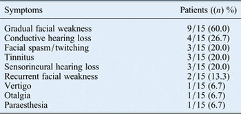
Fifteen patients presented with intrinsic facial nerve tumours over the last 15 years: 8 men and 7 women, aged between 26 and 65 years (mean 38.1 years) at the time of surgery. The most common presenting features were facial symptoms (93.3 per cent of patients), including gradual facial weakness (60.0 per cent), facial spasm and twitching (20.0 per cent) and recurrent weakness (13.3 per cent); and hearing loss (46.7 per cent), including conductive hearing loss (26.7 per cent) and sensorineural hearing loss (20 per cent). Table I summarises the symptomatic findings for all 15 patients.
Symptomatic findings in adult patients with intrinsic facial nerve tumours

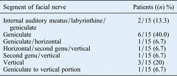
All patients underwent a combination of computed tomography (CT) and post-contrast magnetic resonance imaging (MRI) to delineate the lesion position within the facial nerve course. This revealed that the geniculate ganglion and the vertical portion of the facial nerve were the most common segments involved (in 70.0 per cent and 33.4 per cent of patients, respectively). Multiple segments of the facial nerve were involved in 26.6 per cent of patients. Table II summarises the position of facial nerve lesions from the internal auditory meatus to the stylomastoid foramen.
Position of the lesions on the facial nerve

In all, 12 out of the 15 patients (80.0 per cent) underwent surgery and were followed up for between 17 and 120 months (mean 33.8 months). Of these, 10 underwent tumour removal with interposition gable grafts using the great auricular or surreal nerve and 2 underwent facial nerve decompression. The transmastoid and middle cranial fossa approaches or a combination of both was the most common surgical procedure performed (92.0 per cent of procedures). Hearing was preserved or improved in 66.7 per cent of patients. Histological analysis identified six schwannomas, three haemangiomas and one meningioma.
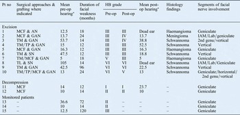
In the excision and grafting group, six patients with a shorter duration of facial symptoms (mean 15.4 months) had House–Brackmann grade III function before surgery. Of these, facial nerve function remained the same for four, and two had House–Brackmann grade IV on the last visit. The remaining four patients, with a longer symptom duration (mean 38 months), had House–Brackmann grade V–VI before surgery. Of these, facial nerve function remained at House–Brackmann grade VI for two and improved to House–Brackmann grade III–V for the other two. The two patients who underwent middle cranial fossa decompression of a lesion involving the geniculate ganglion remained stable at House–Brackmann grade I–II. The remaining three patients with stable facial nerve function (House–Brackmann grade II–III) and no evidence of growth were managed conservatively and followed up for between 14 and 120 months (mean 68.7 months). Table III summarises the conservative and surgical management of all 15 patients.
Patient management, showing hearing and hb facial function grade pre- and post-surgical procedures, tumour type and facial nerve position

*Air conduction at 0.5, 1, 2 and 4 kHz. Pt no = patient number; pre-op = pre-operative; HB = House–Brackmann; post-op = post-operative; MCF = middle cranial fossa; AN = auricular nerve; IAM = internal auditory meatus; Lab = labyrinthine; GAN = great auricular nerve; TM = transmastoid; SN = surreal nerve; TL = translabyrinthine; TP = transparotid.
Discussion
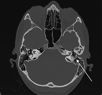
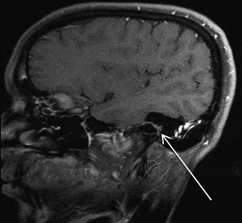
Intrinsic facial nerve tumours are rare; as a result, they can be challenging to diagnose and manage. In any patient presenting with facial spasm or twitching, recurrent facial weakness or weakness that is slow to resolve, a lesion involving the facial nerve should be suspected. Following a thorough assessment, including a pure tone audiogram, patients should undergo high-resolution CT of the temporal bone (Figure 1) and post-contrast MRI of the brain and skull base (Figure 2) to accurately visualise the entire pathway of the facial nerve. If a lesion is discovered within the nerve, these patients should be referred to a dedicated unit with the appropriate expertise in managing these lesions.
Coronal computed tomography scan showing tumour involving the vertical portion of the facial nerve in the left temporal bone (arrow).

Post-contrast magnetic resonance imaging of the patient shown in Figure 1, showing a growing lesion involving the second genu and vertical portion of the facial nerve in the left temporal bone (arrow).

At our institute, all patients with suspected intrinsic lesions of the facial nerve are discussed at a skull base multidisciplinary meeting, followed by review at a dedicated facial palsy clinic by a skull base surgeon, a facial plastic surgeon and a facial physiotherapist. We tend to follow a conservative approach, and only offer definitive treatment to patients with recurrent symptoms of facial weakness or with growing lesions and progressive facial weakness greater than grade III. This approach is well accepted and was described in other large series by McMonagle et al. and Liu and Fagan.Reference McMonagle, Al-Sanosi, Croxson and Fagan5, Reference Liu and Fagan6
Three of our patients (one with neurofibromatosis type 2 (NF2)) with stable facial nerve function (grade II–III) showed no evidence of tumour growth upon yearly imaging over a mean follow up of 68.7 months. These patients continue to be monitored and are provided with additional support by the facial physiotherapists and the NF2 team.
In the surgery group, two patients presented with recurrent facial weakness but excellent residual facial function (grade I–II) and normal hearing. These patients underwent a middle cranial fossa approach and decompression of a lesion involving the geniculate ganglion. A similar method was described by Angeli and Brackmann, as well as others, with good results.Reference Kim, Chang, Oh, Ahn, Hwang and Lee4, Reference Angeli and Brackmann7–Reference Wilkinson, Hoa, Slattery, Fayad, Friedman and Schwartz10 Lesion biopsies were not taken so as to maintain facial nerve integrity and function. Patients were followed up for between 14 and 120 months and have remained asymptomatic with preservation of facial function and hearing.
The remaining 10 patients with progressive symptoms and increasing facial weakness (6 patients with grade III, 1 with grade IV and 3 with grade VI function) had their tumours removed. Decisions on the surgical approach and on lesion removal were made dependent on the tumour size and position and the pre-operative hearing level. The middle cranial fossa approach alone or combined with a transmastoid route was our preferred surgical approach in 92.0 per cent of patients. These approaches allowed excellent exposure of the facial nerve from the internal auditory meatus through to the stylomastoid foramen. (In two patients, the facial nerve was exposed within the parotid gland to facilitate tumour removal and grafting of the proximal end.) The translabyrinthine approach was used in a single patient with poor facial function (grade VI; due to an extensive lesion) and profound hearing loss (mean 105 dB).
The combined middle cranial fossa and transmastoid approach led to hearing preservation or improvement in seven patients. Two patients had ‘dead ears’ as a result of surgery (translabyrinthine approach) and involvement of the otic capsule, and one patient with residual conductive loss went on to receive a bone-anchored hearing aid.
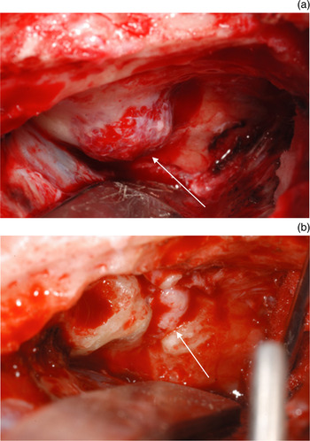
We are aware that small lesions can be removed and cystic components of tumours decompressed to preserve facial nerve continuity, thus preserving facial function.Reference Shirazi, Leonetti, Marzo and Anderson3, Reference Falcioni, Russo, Taibah and Sanna8, Reference Eby, Fisch and Makek11–Reference Sherman, Dagnew, Pensak, van Loveren and Tew20 However, this was not possible in our patient series. To restore continuity, we exposed healthy proximal and distal segments of the facial nerve, excised the tumour and used a great auricular or surreal nerve as a transposition graft, which was secured using Tisseel and 9/0 Prolene (Figure 3). A great auricular graft was used in 80.0 per cent of patients because of ease of access in the neck.
Intra-operative photographs showing (a) a middle cranial fossa approach for a haemangioma involving the geniculate ganglion in the left temporal bone (arrow) and (b) tumour excision, with the great auricular nerve used as a gable graft to restore facial nerve continuity (arrow).

Surgical outcome was dependent not only on facial nerve grading but also on the duration of weakness.Reference Shirazi, Leonetti, Marzo and Anderson3, Reference Kim, Chang, Oh, Ahn, Hwang and Lee4, Reference Falcioni, Russo, Taibah and Sanna8, Reference Dai, Li, Guo and Song21, Reference Saleh, Achilli, Naguib, Taibah, Russo and Sanna22 Six of our patients had grade III function at the time of surgery, with an average symptom duration of between 12 and 24 months (mean 15.4 months). Post-surgery, four patients have unchanged grade III function and two patients have grade IV function after a mean follow up of 33.8 months. Two patients from this group had a gold weight inserted to improve eye closure, along with endoscopic brow lifts. The other patients had botulinum toxin A (Botox®) injections and facial therapy to improve resting symmetry and tone.
The remaining four patients who underwent surgical tumour excision had grade V–VI facial function, with an average duration of facial weakness of 14–96 months (mean 38.8 months). One patient with grade V function secondary to ganglion haemangioma has since recovered nerve function to grade III. The remaining patients stayed at grade VI and have undergone reanimation procedures to improve symmetry and tone.
• Intrinsic facial nerve tumours are rare, with no consensus on their overall management
• Computed tomography and magnetic resonance imaging should be performed for all suspected cases
• Where possible, the native nerve and musculature should be preserved
• Patients with a stable lesion and facial function less than grade III should be managed conservatively
• Symptomatic patients and those with progressive facial weakness greater than grade III should be offered tailored surgery
• Patients with grade VI paralysis and stable lesions should be offered alternative forms of facial reanimation
Stereotactic radiosurgery has recently been described to treat growing tumours in patients with facial nerve function of less than grade III, with favourable results.Reference Wilkinson, Hoa, Slattery, Fayad, Friedman and Schwartz10, Reference Kida, Yoshimoto and Hasegawa23 However, because of limited patient numbers and long-term outcomes we have not used this treatment modality at our institute.
Management protocol
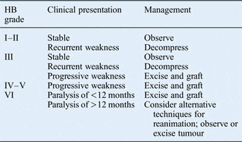
Based on our results and available evidence from the literature, we propose the following protocol. For all clinically stable patients with grade III or better nerve function, we suggest a conservative approach comprising observation, serial imaging and annual electromyography. Patients with recurrent facial nerve weakness and good facial motor recovery should be offered surgical decompression. This patient group may also benefit from stereotactic radiotherapy once further evidence of its long-term benefit has been shown.
For all patients with grade III function and clinical deterioration and for those with grade IV–VI function, we offer surgery individualised according to tumour size and position and the residual hearing level. The surgical aim in these cases is to reanimate the face with minimal morbidity. All patients are informed that they will have residual facial weakness after surgery and can expect grade III function at best. For those with long-standing weakness, emphasis should be placed on facial reanimation; not all will require tumour removal. In all cases, rehabilitation with facial therapy and, in some cases, Botox should also form part of the overall management.
We suggest a transmastoid and/or middle cranial fossa approach for patients with good hearing, and a translabyrinthine approach for those with poor hearing. Table IV summarises our management protocol.
Management guideline for intrinsic facial nerve tumours

HB = House–Brackmann.
Conclusion
Management of facial nerve tumours is complex and influenced by the progression rate and facial weakness grade, weakness duration, the lesion site and residual hearing level. Our approach is to operate when facial motor function is poor or deteriorating, and to use the original nerve and musculature where possible. The treatment decision centres on facial reanimation but our results demonstrate that good outcomes can be achieved, with hearing preservation in most cases.