Introduction
Angiostrongylus vasorum (Baillet, 1866) Kamensky, 1905, is a protostrongylid nematode of cosmopolitan distribution. Adult parasites can be found in the right ventricle and pulmonary arteries and their branches, causing severe consequences to the definitive host. The most common symptoms are cough, breathlessness, exercise intolerance, weight loss, neurological signs, heart failure and death (Ribieri et al., Reference Ribieri, Gottstein, Huber, Welle, Foster and Grosclaude2001; Oliveira et al., Reference Oliveira, Barçante, Barçante, Dias and Lima2005; Saeed et al., Reference Saeed, Maddox-Hyttel, Monrad and Kapel2006). Infection of the definitive host results from the ingestion of infected snails, although paratenic hosts, such as frogs, lizards, mice and rats, may also be ingested by the definitive host (Bolt et al., Reference Bolt, Monrad, Frandsen, Henrikesen and Dietz1993).
Angiostrongylus vasorum has a heteroxenic life cycle and several species of terrestrial and aquatic molluscs serve as intermediate hosts. Egg embryonation occurs in the pulmonary arterioles of the definitive host until the development of the first-stage larvae (L1) that will hatch and penetrate the alveoli and enter the bronchioles and the bronchi. These L1 migrate to the trachea and are either expelled with pulmonary secretions or swallowed and eliminated with faeces (Patteson et al., Reference Patteson, Gibbs, Wotton and Day1993; Cury & Lima, Reference Cury and Lima1995, Reference Cury and Lima2002; Cury et al., Reference Cury, Gumarães, Lima, Caldeira, Couto, Murta, Carvalho and Baptista2002). The L1 remain in the faeces or may reach water collection points. Terrestrial or aquatic molluscs are infected by the ingestion of L1 via the digestive tract or by larval penetration through their soft parts (Thiengo, Reference Thiengo1996). Within mollusc tissues, larvae develop and become infective or third-stage larvae (Guilhon & Cens, Reference Guilhon and Cens1973; Barçante et al., Reference Barçante, Barçante, Dias, Vieira, Lima and Negrão-Corrêa2003a).
Because of the medical importance of angiostrongyliasis in humans and animals, studies have been carried out to determine the efficacy of treatment with albendazole (Eckert & Lämmler, Reference Eckert and Lämmler1972). Most anthelmintic drugs are not efficient against this parasite within the definitive host. For this reason, alternative measures that may be used to combat the environmental spread and the infective forms of these parasites are important; such is the case of natural antagonists. These organisms may include different types of fungi, characterized as predators, endoparasites and opportunists. The advantages of their use include: they are not nutritionally demanding, do not require complex growth media, and concentrate their action on the faecal environment and combat free-living larvae (Araújo et al., Reference Araújo, Mota and Campos2004a).
Until 1964, most fungi were classified as belonging to the genera Arthrobotrys, Dactylis, Dactyella and Trichothecium. Subsequently, several new genera were described, including Duddingtonia, Monacrosporium, Geniculate and Dactylariopsis (Gray, Reference Gray1987). The vast majority of nematophagous fungi are mitosporic and were formerly classified into the division Deuteromycetes, class Hyphomycetes, order Hyphomycetales and family Moliniaceae. Recently, stages of sexual reproduction of these fungi have been observed for some species that are recognized as belonging to the phylum Ascomycota (Griffin, Reference Griffin1994).
Predatory fungi are certainly the most studied groups and show the greatest potential for biological control (Nordbring-Hertz et al., Reference Nordbring-Hertz, Jansson and Tunlid2002; Araújo et al., Reference Araújo, Rodrigues, Silva and Vieira2007). The species Duddingtonia flagrans, Monacrosporium thaumasium, M. sinense and Arthrobotrys robusta are identified as nematophagous fungi and have been studied for their potential as biological control agents for gastrointestinal nematodes of domestic animals (Araújo et al., Reference Araújo, Freitas, Vieira and Campos2006, Reference Araújo, Braga, Araújo, Silva and Tavela2008; Campos et al., Reference Campos, Araújo, Assis, Granda and Guimarães2007).
Duddingtonia flagrans is considered the most promising nematode-trapping species for the control of nematodiosis in domestic animals, due to its plentiful production of chlamydospores. These thick-walled spores caused significant larval reduction in faecal cultures and pastures (Rocha et al., Reference Rocha, Araujo and Amarante2007). It has predatory action by forming traps characterized by a system of simple adhesive hyphae that produce two types of spores – conidia and chlamydospores – interspersed by mature hyphae, which can be used as a biological control agent in the environment (Braga et al., Reference Braga, Araújo, Campos, Carvalho, Silva, Tavela and Maciel2007). Species of the genus Monacrosporium are characterized by producing a single conidium on each conidiophore and production of adhesive networks, forming septate and branched hyphae, while M. sinense produces chlamydospores (Mota et al., Reference Mota, Campos and Araújo2003; Campos et al., Reference Campos, Araújo, Assis, Granda and Guimarães2007). The species A. robusta presents erect conidiophores, sometimes branched, about 300 mm in length, having the extremity increased in size, usually carrying six ovoid-shaped conidia, hyaline, sectioned closely to the intermediate region, 18–27 mm long and 8–12 mm wide, and capable of producing chlamydospores and adhesive networks for preying on nematodes (Araújo et al., Reference Araújo, Gomes and Guimarães1998, Reference Araújo, Stephano and Sampaio2000).
The objective of this work was to compare the in vitro predatory capacity of the fungal species D. flagrans, M. thaumasium, M. sinense and A. robusta on first-stage larvae of A. vasorum.
Material and methods
Fungi
Four nematophagous fungal isolates were used in the experiment: D. flagrans (AC001), M. thaumasium (NF34), M. sinense (SF53) and A. robusta (I31). The isolates were originally obtained from soil located in Viçosa, Minas Gerais, Brazil, 20°45′20″S; 42°52′40″W, 649 m altitude, using the soil sprinkling method of Duddington (Reference Duddington1955), modified by Santos et al. (Reference Santos, Ferraz and Muchove1991). Fungi were kept in test tubes containing 2% cornmeal agar (2% CMA), in the dark, at 4°C for 10 days.
Conidia collection
Culture discs (4 mm in diameter) were removed from the fungal isolates kept in test tubes containing 2% CMA and transferred to 9.0 cm Petri dishes containing 20 ml of 2% potato dextrose agar and kept at 25°C in the dark for 10 days. After growth, new culture discs (4 mm in diameter) were transferred to 9.0 cm diameter Petri dishes containing 20 ml of 2% water-agar (2% WA) and 1 ml of distilled water containing 1000 larvae of Panagrellus sp. was added daily for 21 days to induce conidia formation. When fungal development was complete, 5 ml of distilled water were added to each Petri dish, and the conidial and mycelial fragments were removed as described by Araújo et al. (Reference Araújo, Santos, Ferraz and Maia1993).
L1Angiostrongylus vasorum
The strain used in the assays was originally isolated from faeces of two naturally infected dogs, from the city of Caratinga, Minas Gerais (Lima et al., Reference Lima, Costa, Guimarães and Leite1985). This strain has been maintained by successive passages in dogs. Faeces of infected dogs were collected and placed in a modified Baermann funnel for 12 h, for L1 recovery (Barçante et al., Reference Barçante, Barçante, Dias, Vieira, Lima and Negrão-Corrêa2003a, Reference Barçante, Barçante, Dias and Limab). After this period, the tube was removed and 200 g were centrifuged for 2 min. The supernatant was discarded and the pellet containing A. vasorum L1 was resuspended in 5 ml of 0.85% saline solution. The content was homogenized and three 10 μl aliquots were removed and distributed on 7.5 × 2.5 cm glass slides. Larval counts were carried out under a stereomicroscope (25 × ). The total larval number was estimated by a simple rule of three.
Experimental assay
Treatments consisted of four groups of fungal isolates and a control without fungus plated in 9.0 cm Petri dishes containing 20 ml of 2% WA, with six repetitions each. Petri dishes were previously marked into 4 mm diameter fields. A total of 1000 A. vasorum L1 larvae were plated with 1000 conidia of one of the fungal isolates AC001, NF34, SF53 or I31 in 2% WA. The control group (without fungus) contained 1000 L1 larvae plated with 2% WA only.
Ten random fields (4 mm diameter) were examined per plate of treated and control groups, every 24 h for 7 days under an optical microscope (10 × and 40 × objective lens) for non-predated L1 counts. After 7 days, the non-predated L1 were recovered from the Petri dishes using the Baermann method.
Statistical analysis
Means of recovered A. vasorum L1 were calculated. Data were examined by analysis of variance at significance levels of 1 and 5% probability (Ayres et al., Reference Ayres, Ayres, Ayres and Santos2003). Predation efficiency of L1 relative to the control group was evaluated by the Tukey's test at 1% probability. The reduction percentage of L1 means was calculated according to the following equation: Reduction % = (Average of L1 recovered from control group − Average of L1 recovered from treatment groups) × 100/Average of L1 recovered from the control group.
Results
The tested isolates of predatory fungi D. flagrans (AC001), M. thaumasium (NF34), M. sinense (SF53) and A. robusta (I31) were able to capture A. vasorum L1 in the experimental in vitro test.
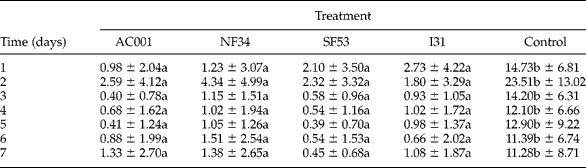
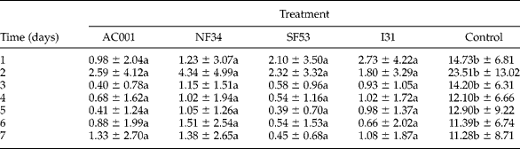
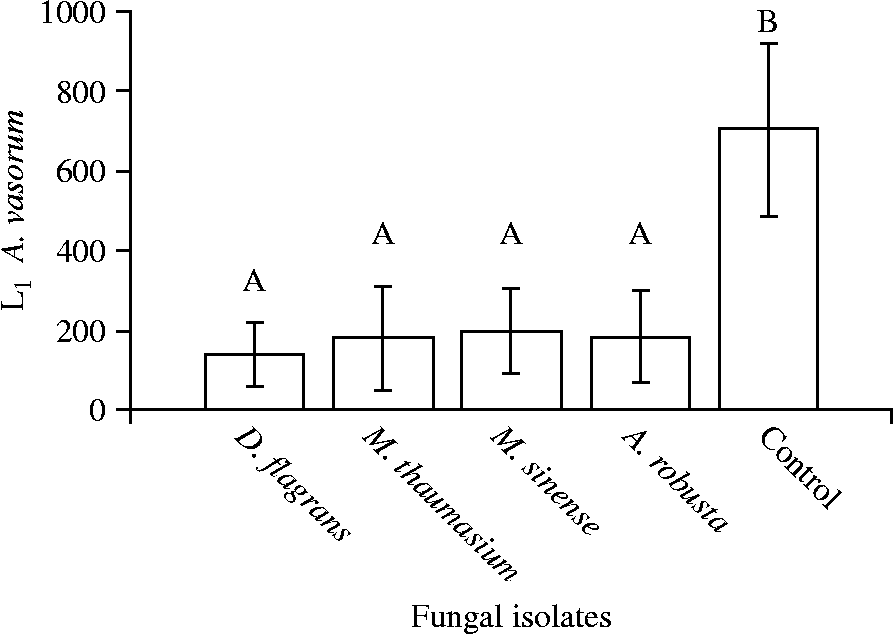
No difference was found (P>0.05) in the comparison between capture and destruction of A. vasorum L1 in Petri dishes of the groups treated with isolates of D. flagrans (AC001), M. thaumasium (NF34), M. sinense (SF53) and A. robusta (I31) during the experimental assay (table 1), but there was difference (P < 0.05) between the means of non-predated A. vasorum L1 per 4 mm diameter field in the Petri dishes of the control group compared to means of L1 recorded in the groups treated with the fungi throughout the experiment. The recorded reduction percentages of A. vasorum L1 were 80.3% (AC001) 74.5% (NF34), 74.2% (SF53) and 71.8% (I31).
Daily means and standard deviations of non-predated first-stage larvae (L1) of Angiostrongylus vasorum per 4 mm diameter field in 2% water-agar during 7 days of treatment with the fungal isolates Duddingtonia flagrans (AC001), Monacrosporium thaumasium (NF34), Monacrosporium sinense (SF53), Arthrobotrys robusta (I31) and control without fungus.
Means followed by the same small letter in the rows were not statistically different (P>0.05).
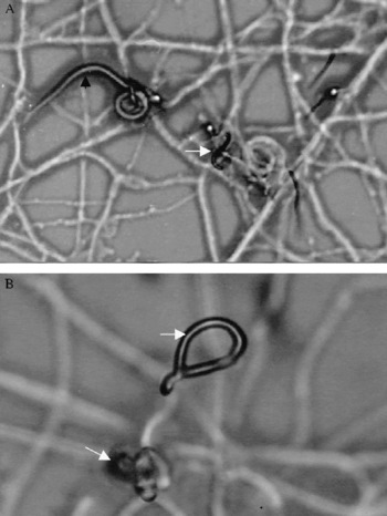
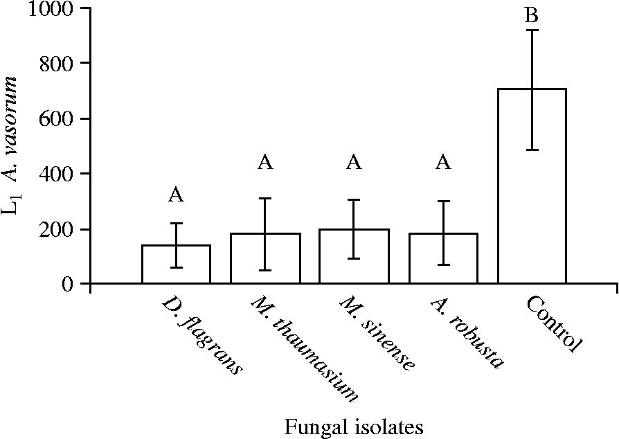
The presence of A. vasorum L1 in Petri dishes containing 2% WA was essential for trap formation by the fungal isolates, since this culture medium is nutritionally poor (fig. 1A and B). The presence of nematophagous fungi was not observed in the control group during the experiment. Evidence of predation was confirmed by the means of recovered A. vasorum L1 using the Baermann method at 7 days post-plating, at the end of the experiment (fig. 2).
Angiostrongylus vasorum first-stage larvae (L1) captured by Duddingtonia flagrans (black arrow) in Petri dishes containing 2% water-agar, and trap formation by the fungal isolate D. flagrans (white arrow). Magnification: (A) 10 × and (B) 40 × objective lens.

Mean number of non-predated Angiostrongylus vasorum first-stage larvae (L1) recovered in 2% water-agar by the Baermann method on the seventh treatment day after interaction with the fungal isolates Duddingtonia flagrans (AC001), Monacrosporium thaumasium (NF34), Monacrosporium sinense (SF53), Arthrobotrys robusta (I31) and control (without fungus). Lines on bars represent standard deviation. Means followed by at least one common capital letter (A) in the row are not significantly different by Tukey's test at a 5% probability level.
The linear regression coefficients calculated by the analysis of A. vasorum L1 means per 4 mm diameter field of the treated and control groups were: − 0.075 (AC001); − 0.190 (NF34); − 0.311 (SF53); − 0.256 (I31) and − 0.282 (control group).
Discussion
The genera Ancylostoma and Toxocara, parasites of dogs and cats, and possibly the species Angiostrongylus vasorum, stand out among helminths with zoonotic potential. Besides showing medical–veterinary importance, A. vasorum as a cardiopulmonary parasite of domestic and wild dogs also requires special attention and investigation because it can also infect humans (Eckert & Lämmler, Reference Eckert and Lämmler1972; Lima et al., Reference Lima, Guimarães and Lemos1994).
Nematophagous fungi, especially the genera Duddingtonia, Monacrosporium and Arthrobotrys, have predatory capacity on infective larvae (L3) of gastrointestinal nematode parasites of domestic animals (Araújo et al., Reference Araújo, Assis, Campos and Mota2004b, Reference Araújo, Freitas, Vieira and Campos2006; Campos et al., Reference Campos, Araújo, Assis, Granda and Guimarães2007). These genera are recognized only as predators, and the predatory capacity of the species D. flagrans and M. thaumasium, M. sinense and A. robusta has been thoroughly discussed and proved in the control of L3 of gastrointestinal nematode parasites of ruminants, horses and small ruminants (Castro et al., Reference Castro, Oliveira, Anjos, Ornellas, Bittencourt, Araújo, Sampaio and Rodrigues2003; Chandrawathani et al., Reference Chandrawathani, Jamnah, Adman, Waller, Larsen and Gillespie2004; Araújo et al., Reference Araújo, Freitas, Vieira and Campos2006, Reference Araújo, Rodrigues, Silva and Vieira2007; Dias et al., Reference Dias, Araújo, Campos, Braga and Fonseca2007). However, the predatory capacity of these species had never been tested on larvae of A. vasorum. This is the first work on the interaction of the fungi D. flagrans, M. thaumasium, M. sinense and A. robusta with A. vasorum L1 larvae. Few studies have mentioned the in vitro predatory activity of different nematophagous fungi on larvae of nematode parasites of dogs (Maciel et al., Reference Maciel, Araújo and Cecon2006a, Reference Maciel, Araújo and Camposb).
In the present work, although the isolate (I31) from A. robusta was effective in reducing A. vasorum L1 with reduction percentage of 71.8%, D. flagrans (AC001) was found to be more efficient compared with the others and at the end of 7 days showed a higher reduction percentage (80.31%). These results showed that D. flagrans (AC001) had higher in vitro predatory activity on A. vasorum L1.
Eren & Pramer (Reference Eren and Pramer1965) reported on the regular supply of nematodes to fungi in nutritionally poor culture media, which reduces fungal saprophytic growth. For this reason, in this study we used only 2% WA, a low nutrient culture medium, to reduce the saprophytic growth of fungal isolates, providing A. vasorum L1 as a single nutritional source. The higher the mobility of nematodes, the greater the stimulus to trap production (Nansen et al., Reference Nansen, Foldager, Hansen, Henriksen and Jorgensen1988). This phenomenon was observed in this study, as trap formation and L1 predation by the fungal isolates were recorded during the first observation, 24 h after interaction (fig. 1). Maciel et al. (Reference Maciel, Araújo and Campos2006b) found that in 2% WA, the isolate A. robusta (I31) showed higher predatory activity on Ancylostoma spp. L3 compared with isolates D. flagrans (CG768) and M. thaumasium (NF34). However, in the present study, the isolate D. flagrans (AC001) in 2% WA showed the highest in vitro predatory activity on A. vasorum L1 at the end of the experiment.
Negative coefficients of correlation indicate a downward behaviour of the regression curves for the treatments with the fungal isolates AC001, NF34, SF53 and I31. This was caused by the reduction in the means of non-predated A. vasorum L1 per 4 mm diameter field during the experimental assay, mainly by the capture of L1 in fungal traps. Reduction in the number of L1 per 4 mm diameter field in the control group, during the study, was caused by larval migration to the periphery of the Petri dishes, where the moisture level was higher, which was also reported by Araújo et al. (Reference Araújo, Freitas, Vieira and Campos2006) in an in vitro assay carried out in Petri dishes.
The results of this study confirm previous work on the efficiency of nematophagous fungi in the control of larvae of nematode parasites of dogs. This allows us to conclude that the fungi D. flagrans, M. thaumasium, M. sinense and A. robusta have predatory activity on first-stage larvae of A. vasorum and that this is a likely alternative method for biological control of A. vasorum larvae.