Aortopulmonary window is a rare congenital defect caused by the incorrect formation of the wall dividing the arterial trunk into the aorta and the pulmonary artery and accounts for less than 1% of all CHDs. Percutaneous closure of the Aortopulmonary window remains a rare therapeutic option, usually giving ground to surgical approach. Several successful attempts have been reported since 1995 using a variety of devices, including specifically designed “umbrellas”,Reference Stamato, Benson, Smallhorn and Freedom 1 Amplatzer Duct Occluders,Reference Atiq, Rashid, Kazmi and Qureshi 2 the Cardio-O-Fix Occluder,Reference Bialkowski, Akdeniz and Celebi 3 the Amplatzer Muscular VSD Occluder,Reference Kosmač, Eicken, Kühn, Heinrich and Ewert 4 , Reference Trehan, Nigam and Tyagi 5 and Amplatzer Duct Occluders II.Reference Noonan, Desai and Degiovanni 6 To date, however, there are no reported cases of Amplatzer Duct Occluder II: Additional Sizes use. We report a case of percutaneous closure of an aortopulmonary window using Amplatzer Duct Occluder II: Additional Sizes in a 4.5-month-old patient.
Case report
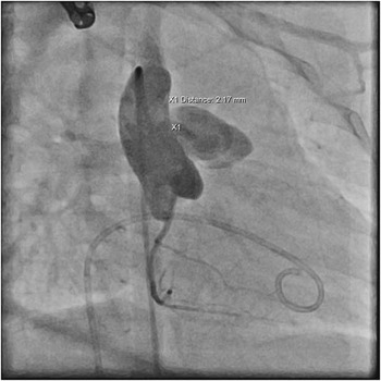
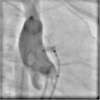
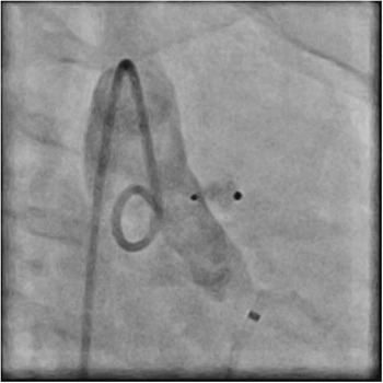
A 4.5-month-old child weighing 6 kg, with a continuous heart murmur detected at day 3 after birth, was admitted to the paediatric cardiology department. Initially, at 1 month of age, the suspected diagnosis was right coronary artery-to-main pulmonary artery fistula and the patient was administered spironolactone and captopril. At 3 months of age, a tachypnoea and signs of heart failure appeared accompanied by viral upper respiratory tract infection complicated by pneumonia. At the current admission, the patient presented with mild dyspnoea but proper weight gain. The family history was positive for CHD – the patient’s 15-year-old brother had undergone surgical closure of an aortopulmonary window and atrial septal defect type II. On physical examination, there was a loud continuous murmur and a 1.5-cm liver enlargement. An echocardiogram revealed an aortopulmonary window with a point of exit in the main pulmonary artery slightly above the pulmonary valve (peak gradient 80 mmHg), as well as borderline enlargement of the left atrium and left ventricle (left ventricle end-diastolic diameter [LVEDD]: 2.5 cm, z-score 1.12). Informed consent was obtained for diagnostic catheterisation and potential window closure. The procedure was performed under general anaesthesia. After establishing venous and arterial access, haemodynamics were obtained (aorta: 103/54/69 mmHg, pulmonary artery: 35/20/27 mmHg, Qp:Qs 1.6), and an aortogram was performed in a right anterior oblique 40º projection (Fig 1) as well as in posterior–anterior projections. The aortogram revealed a left-to-right shunt between the ascending aorta and the main pulmonary artery (window diameter: 2.17 mm, distance between aorta and main pulmonary artery: 2.3 mm, measurements in right anterior oblique 40º projection). The distance between the defect and coronary ostia was 7.5 mm (Fig 2). Subsequently, the aortopulmonary window was crossed from the main pulmonary artery using a 4-F internal mammary artery catheter and a hydrophilic guidewire. Thereupon, the internal mammary artery catheter was exchanged for a 4-F delivery catheter, through which an Amplatzer Duct Occluder II: Additional Sizes was introduced (waist diameter: 4 mm, implant length: 2 mm). The device was deployed under fluoroscopic guidance, and the implant position was confirmed by aortic angiography before and after release (Figs 2 and 3). An echocardiogram performed 12 hours after the procedure confirmed that the implant remained in good position with no residual shunting and no obstruction to aortic or pulmonary artery flow. The distance between the implant and the pulmonary valve measured 2.5 mm leaving the valve fully functional. Owing to a decreased pulse and diminished flow in the cannulated femoral artery in the Doppler examination, the patient was temporarily treated with heparin infusion. On a follow-up visit 4 months later, the patient presented in good general health with satisfactory weight gain (3.8 kg). On echocardiographic examination, the LVEDD was 2.7 cm (z-score: 0.26), and neither aortic nor pulmonary insufficiency were observed.
Aortogram (right anterior oblique 40º projection) showing a left-to-right shunt between the ascending aorta and the main pulmonary artery (MPA) – aortopulmonary window (APW). APW diameter: 2.17 mm, distance between aorta and MPA: 2.3 mm.

Aortogram demonstrating distance from the left coronary artery to the device (7.5 mm).

Aortogram (right anterior oblique 40º projection) performed after the device’s release, confirming the correct implant position.

Discussion
Percutaneous closure of the aortopulmonary window remains a rare therapeutic option that is almost exclusively reserved for patients with restrictive, small, and type 4 (intermediate; classification by The Society of Thoracic SurgeonsReference Jacobs, Quintessenza, Gaynor, Burke and Mavroudis 7 ) defects. There are, however, exceptions to the above. Trehan et alReference Trehan, Nigam and Tyagi 5 described transcatheter closure of non-restrictive aortopulmonary windows in three infants. Similarly, the size of the defect should not constitute an absolute limitation, given the variety of devices and sizes available in the market. The scope of reported sizes of aortopulmonary windows closed percutaneously varies from 3Reference Stamato, Benson, Smallhorn and Freedom 1 to 10 mm.Reference Trehan, Nigam and Tyagi 5 The only feature of an aortopulmonary window that seems to be of vital importance in terms of percutaneous closure is the presence of adequate superior and inferior rims – a quality that is found only in type 4 (intermediate) defects.
Various techniques for crossing the defect have been described. Trehan et alReference Trehan, Nigam and Tyagi 5 reported an approach with arteriovenous loop formation, with the window crossed from the aortic side using either a Swan–Ganz catheter or a cobra catheter. Other researchers have also used arteriovenous loop, crossing the defect with help of a Judkins right catheter.Reference Noonan, Desai and Degiovanni 6 , Reference Marini, Calcagni, Ou, Bonnet and Agnoletti 8 Only a handful of articles have described crossing the defect from the pulmonary artery.Reference Kosmač, Eicken, Kühn, Heinrich and Ewert 4 , Reference Trehan, Nigam and Tyagi 5 , Reference Viswanathan, Vaidyanathan and Kumar 9 This is most likely due to difficulties in introducing the guidewire into the aortopulmonary window, which is often located in proximity to the pulmonary valve. In our case, the use of an internal mammary artery catheter and hydrophilic wire allowed us to easily cross the defect and deploy the implant from the venous side using a 4-F vascular sheath.
The potential risks of percutaneous closure of aortopulmonary windows include damage to the pulmonary valve caused by the implant’s discs and the possibility of aortic or pulmonary artery narrowing due to the implant’s protrusion into the arterial lumen. Therefore, the choice of the most suitable device is crucial. In most cases after the year 2000, Amplatzer Duct Occluders were utilised; however, in a few cases Amplatzer Muscular VSD Occluder and Amplatzer Septal Occluder were used. These implants could not be used in our case because of the patient’s low weight (6 kg) and their relative stiffness generating a high risk of protrusion to the aortic or the pulmonary lumen, which, consequently, could lead to a significant aortic or pulmonary artery obstruction. The Amplatzer Duct Occluder II, a device made from thin nitinol mesh wires with two discs connected with the waist in a flexible manner, appeared to be a more suitable and gentler solution. There is, however, a serious disadvantage with this implant. Both retention discs are proportionally bigger than the device’s waist, which in the case of small children could lead to suboptimal disc configuration in the aorta or the pulmonary artery and cause significant pressure gradient. Similar problems were found in patients with patent ductus arteriosus closed with Amplatzer Duct Occluder II, where inadequate aortic disc positions produced serious aortic obstructions.Reference Thanopoulos, Eleftherakis, Tzannos, Stefanadis and Giannopoulos 10 These considerations led us to the idea of using the Amplatzer Duct Occluder II: Additional Sizes. These devices, which were originally designed for the closure of patent ductus arteriosus of <4 mm accompanied by small or shallow ductal ampulla, were certified and released onto the Polish market in 2011. They are characterised by low-profile retention discs, minimising protrusion into the vessel lumen, thus reducing the risk of aortic or pulmonary artery obstruction. The implant fulfilled our expectations – the soft waist filled the aortopulmonary connection and both discs, pulmonary and aortic, were properly shaped and pressed against the vessel walls. In addition, the small retention discs did not influence pulmonary valve function, despite the window’s proximity to the valve. In our opinion, this outcome may suggest that Amplatzer Duct Occluder II: Additional Sizes implants could prove to be particularly suitable for the closure of small aortopulmonary window, especially in young children.
Conclusions
The Amplatzer Duct Occluder II: Additional Size implant is a safe and useful device for percutaneous closure of an aortopulmonary window in a carefully selected group of patients.
Acknowledgements
None.
Financial Support
This research received no specific grant from any funding agency or from commercial or not-for-profit sectors.
Conflicts of Interest
None.
Ethical Standards
The authors assert that all procedures contributing to this work comply with the ethical standards of the relevant national guidelines on human experimentation in Poland and with the Helsinki Declaration of 1975, as revised in 2008, and has been approved by the institutional committee of Silesian Center for Heart Diseases.