




No CrossRef data available.
Tick–host interactions: saliva-activated transmission
Published online by Cambridge University Press: 19 April 2005
Abstract
The skin site at which ticks attach to their hosts to feed is the critical interface between the tick and its host, and tick-borne pathogens. This site is highly modified by the pharmacologically active molecules secreted in tick saliva. For pathogens, it is an ecologically privileged niche that many exploit. Such exploitation is referred to as saliva-activated transmission (SAT) – the indirect promotion of tick-borne pathogen transmission via the actions of bioactive tick saliva molecules on the vertebrate host. Here we review evidence for SAT and consider what are the most likely candidates for SAT factors among the tick pharmacopoeia of anti-haemostatic, anti-inflammatory and immunomodulatory molecules identified to date. SAT factors appear to differ for different pathogens and tick vector species, and possibly even depend on the vertebrate host species. Most likely we are searching for a suite of molecules that act together to overcome the redundancy in host response mechanisms. Whatever they turn out to be, the quest to identify the tick molecules that mediate SAT is an exciting one, and offers new insights to controlling ticks and tick-borne diseases.
Information
- Type
- Research Article
- Information
- Copyright
- © 2004 Cambridge University Press
TICK–HOST–PATHOGEN INTERACTIONS
The relationships between tick-borne pathogens, their tick vectors and diverse vertebrate hosts, can be represented by a triangle of parasitic interactions (Fig. 1). The interactions are between (1) pathogen–tick, (2) pathogen–host, and (3) tick–host. In (1) the pathogen interacts with its vector, infecting and replicating within tick cells or extracellular spaces (including those of the gut, haemocoel and salivary glands). The ability of a particular tick species to act as a vector depends on whether the pathogen can survive and overcome several ‘barriers’ within the tick, e.g. the environment within the midgut where the pathogen is initially taken up in the blood meal, the gut infection barrier, and the salivary gland infection barrier. The role of the tick immune system in controlling infections is largely unknown, as is the nature of any conspecific or heterospecific interactions between strains or genotypes of these pathogens during multiple infections of an individual tick.
Interaction (2) also involves infection: the pathogen interacts with its vertebrate host, infecting and replicating extracellularly or within cells. The outcome of infection depends on the tropism of the pathogen for specific host cell or tissue types, or locations (e.g. nervous tissue, joints), and its pathogenic phenotype, and on the age and immune status of the host, and genetic background.

Fig. 1. The triangle of interactions between tick-borne pathogens, their tick vectors and vertebrate hosts. (1) pathogen–tick interactions; (2) pathogen–host interactions; and (3) tick–host interactions. The vertical arrow represents interactions of a pathogen with tick–host interactions at the skin site of tick attachment and blood-feeding (the tick–host–pathogen interface) and the double-headed arrow represents the cross-roads for pathogens at which they pass from infected tick vector to vertebrate host, or from infected host to tick vector.
The third component of the triangle (Fig. 1, interaction 3) is the interaction between the tick and its host. Although this is the non-infective face of the triangle, the skin site at which ticks attach to feed is the cross-roads for pathogens. It is the initial site at which pathogens gain access to either their host or vector: the tick–host–pathogen interface. This cross-roads is particularly busy for tick-borne pathogens because blood feeding of ticks is such a complex and comparatively long and highly ordered process, especially in ixodid species. Attachment and feeding takes several days to complete (minutes to hours for argasids), and involves sawing through the epidermis by means of toothed chelicerae, inserting the mouthparts (barbed hypostome and chelicerae) into the resulting wound site which are then (for ixodids) cemented in place, followed by the formation of a feeding pool resulting from tick and host activities. For ixodid species, the majority of the blood meal is not taken up until the last day of attachment (Kemp, Stone & Binnington, 1982). Such a profound physical and chemical assault on the host should provoke strong haemostatic, inflammatory and immune responses. However, despite the host's armoury of rejection mechanisms, the tick manages to remain attached and achieve engorgement. Successful feeding of ticks relies on a pharmacy of chemicals located in their complex salivary glands and secreted, in tick saliva, into the feeding pool (see chapters by Brossard & Wikel and Valenzuela in this Supplement).
Increasing evidence indicates that the survival of tick-borne pathogens depends on their ability to exploit the pharmacological activities of tick saliva molecules. This is depicted in Fig. 1 by the broad vertical arrow representing the interaction of the pathogen with the modified tick–host interface. Why do we use such a forceful representation? Because it demonstrates our view of pathogen adaptation to the very specific environment created by the intimate and dramatic interplay between host and vector, and concentrated in the tick feeding site. By exploiting this unique ecological niche – the feeding site – tick-borne pathogens are making an easier living for themselves, increasing their chances of survival in nature.
SALIVA-ACTIVATED TRANSMISSION (SAT)
Promotion of tick-borne pathogen transmission, via the actions of tick saliva components on the host, has been termed saliva-activated transmission (SAT). It represents the pathogen's quest for saliva-mediated host modulation, represented by the large vertical arrow in Fig. 1. The term SAT was first coined to describe the promotion of Thogoto virus transmission by SGE of Rhipicephalus appendiculatus (Nuttall & Jones, 1991). Since then, the phenomenon has been demonstrated for a number of tick-borne pathogens (Tables 1 and 3). Direct and indirect evidence of SAT has also been reported for several insect-borne pathogens including Leishmania spp. and their sandfly (Lutzomyia, Phlebotomus) vectors (Titus & Ribeiro, 1988; Belkaid et al. 1998), La Crosse virus and Aedes triseriatus mosquitoes (Osorio et al. 1996), Cache Valley virus and Aedes aegypti, Aedes triseriatus and Culex pipiens mosquitoes (Edwards, Higgs & Beaty, 1998), vesicular stomatitis virus and Simulium vittatum blackflies (Mead et al. 2000), and Orientia tsutsugamushi (the aetiological agent of scrub typhus) and Leptotrombidium deliense and Blankaartia acuscutellaris mite larvae (Frances et al. 2000).

Experimentally, SAT is demonstrated by enhanced transmission of infectivity when the pathogen plus salivary gland extract (SGE) is syringe-inoculated into a host, compared with the level of infectivity when the pathogen alone is injected. For example, comparison of pooled experimental data for Thogoto virus infection reveals the attenuating effect of SGE (Table 2). Experimental inoculation of guinea pigs with a mixture of the virus plus SGE of partially fed Rhipicephalus appendiculatus or Amblyomma variegatum female ticks increased the number of nymphs that became infected approximately 10-fold compared with inoculation with virus alone. Similar direct evidence of SAT has been reported for tick-borne encephalitis virus (TBEV), the Lyme disease spirochaetes Borrelia afzelii, B. burgdorferi sensu stricto, and B. lusitaniae, and Franciscella tularensis (Table 1).

Indirect evidence of SAT is provided by observations of efficient non-systemic transmission between infected and uninfected ticks co-feeding on the same host (Table 3). The first unequivocal example was demonstrated by the non-viraemic transmission of Thogoto virus (Jones et al. 1987). Remarkably, virus transmission from infected to uninfected ticks co-feeding on non-viraemic guinea pigs was more efficient than transmission on hamsters that exhibited high levels of viraemia. Non-viraemic transmission has been shown for several other tick-borne viruses, including TBEV, Crimean-Congo haemorrhagic fever virus, Louping ill virus, Bhanja virus, Palma virus and West Nile virus (Table 3). A possible example exists for Kyansanur forest disease virus transmitted by Argas persicus feeding on a domestic chick (Singh, Goverdhan & Bhat, 1971). Comparable non-systemic transmission has been reported for B. burgdorferi ss, B. garinii and B. afzelii (Table 3). Transmission of Ehrlichia ruminantium, the causative agent of heartwater in cattle, between infected and uninfected ticks co-feeding on tortoises, is another possible example of non-systemic transmission (Bezuidenhout, 1987). By contrast, Anaplasma marginale, the tick-borne rickettsial pathogen of cattle, and the agent of human granulocytic ehrlichiosis (Anaplasma phagocytophilum), do not appear to utilize co-feeding non-systemic transmission (Hodzic et al. 2001; Kocan & de la Fuente, 2003).

Results of co-feeding experiments using pathogen-immune natural rodent hosts also support the concept of SAT (Labuda et al. 1997b). These animals supported TBEV transmission in the presence of virus-specific neutralizing antibodies. In sharp contrast, immunization with specific tick-derived antigens significantly diminished TBEV transmission and surprisingly, also increased the survival of mice following an otherwise lethal infective I. ricinus tick bite (Labuda, unpublished observation). The protective effect of anti-tick immunity against TBEV infection (which has been observed with other pathogen-vector systems) underpins the concept of transmission-blocking vaccines (see chapter by Willadsen in this Supplement).
Further indirect evidence of SAT is based on in vitro studies in which SGE is shown to promote infections by tick-borne pathogens. For the Lyme disease spirochaete, SGE of unfed female I. ricinus stimulated growth of B. garinii cultures in vitro while SGE of unfed female D. reticulatus, a non-competent vector of the Lyme disease spirochaete, did not (Rudolf & Hubalek, 2003). In vitro studies with murine macrophages showed that killing of B. afzelii spirochaetes was inhibited by SGE of I. ricinus females fed for 5 days (Kuthejlova et al. 2001). As macrophages represent one of the the first lines of host defence against this spirochaete, suppression of their antimicrobial activity may well contribute to SAT. A stimulatory chemotactic effect of SGE from I. scapularis on B. burgdorferi ss was observed using a modified U-tube chemotaxis assay (Shih, Chao & Yu, 2002). This apparent chemotactic potential of SGE may contribute to the acquisition of Lyme disease spirochaetes by feeding ticks, which also can be considered a form of SAT. Thus the effect of saliva at the feeding site of uninfected ticks may act as a magnet, drawing spirochaetes to the site. The mechanism for this, if it occurs, is unknown.
In vitro treatment of bovine lymphocytes with SGE of R. appendiculatus enhanced their susceptibility to infection by Theileria parva sporozoites (Shaw, Tilney & McKeever, 1993). Curiously, the effect was observed with SGE from unfed ticks as well as ticks that had fed for 4 days, and it occurred within only 90 min of treatment. Studies are needed to determine whether the apparent stimulating effect on T. parva invasion of host cells reflects an ability of the tick vector to promote transmission of this important pathogen. Tick SGE has even been shown to accelerate replication of vesicular stomatitis virus, an arthropod-borne virus (arbovirus) not transmitted by ticks (see Cytokine inhibitors, below).
An interesting approach to investigating whether tick-induced immunomodulation affects tick-borne pathogen infection was reported for Anaplasma phagocytophilum, the aetiological agent of human granulocytic ehrlichiosis (Borjesson et al. 2002). Mice were infected via syringe inoculation and then infested with uninfected Ixodes scapularis nymphs. At the same time as the infestation, a suture was placed through the skin, distant from the ticks. The suture served as a non-specific inflammatory source and acted as a positive control for the specific inflammation induced by the feeding ticks. A marked increase in bacteraemia and rate of A. phagocytophilum infection of neutrophils were observed following tick feeding. The increased bacteraemia was not explained in terms of a specific tick-induced modulatory mechanism, nevertheless, this approach warrants further investigation.
All the direct and indirect evidence for SAT involves ixodid tick species, with one clear exception (Table 3). Whether this indicates a greater capacity of ixodid species for SAT remains to be determined, as comparatively few studies have been reported with argasid species. The one exception is the reported non-viraemic transmission of West Nile virus (WNV) between co-feeding infected and uninfected Ornithodoros moubata (Lawrie et al. 2004). This report is unusual because WNV is regarded as a mosquito-borne virus although epidemiological evidence indicates it can be maintained in nature by argasid species (see chapter by Labuda & Nuttall in this Supplement).
DETERMINANTS OF SAT
For tick-borne viruses, the SAT factor promotes transmission through its activity on the host rather than by a direct effect on the virus. The most compelling evidence for this is from studies with Thogoto virus. When Thogoto virus was mixed with SAT-active SGE and then assayed in cell culture and mice, viral infectivity was unchanged (Jones, Hodgson & Nuttall, 1989, 1990a). This suggests that the enhancing factor is not a proteolytic enzyme that cleaves a viral surface protein to expose a more infectious virus particle, as occurs with some insect-borne viruses (Borucki et al. 2002; Takamatsu et al. 2003). However, more compelling is the observation that SAT was observed when SGE and Thogoto virus were inoculated at different times into guinea pigs (Jones, Kaufman & Nuttall, 1992b). When SGE was inoculated into the skin of tick-infested guinea pigs, and then Thogoto virus was inoculated in the same site at the same time or 1, 2, 3 or 4 days later, the number of nymphs infected was increased compared with the controls inoculated with virus alone. In fact, yields of infected ticks were highest when the virus was inoculated 2 or 3 days after inoculation of SGE. Similar results were obtained in the converse experiment, in which SGE was inoculated after the virus, although the window for enhancement appeared to be shorter. The key to successful enhancement in these experiments was the inoculation of virus and SGE into the same skin site; when inoculated at different sites, the numbers of infected nymphs fell to levels observed when guinea pigs were inoculated with virus alone (Jones et al. 1989). Immunomodulatory effects of tick saliva that may mediate SAT and explain these observations are considered in the next section (Candidate SAT Factors).
Tick-borne transmission studies undertaken with laboratory animals suggest a correlation between pathogen, vector competence and SAT. Thus, SAT was demonstrated with TBEV and SGE from Ixodes ricinus and Rhipicephalus appendiculatus; both tick species can transmit the virus (although R. appendiculatus is not a natural vector). However, Thogoto virus showed SAT with SGE or saliva of R. appendiculatus (its natural vector species) but not with SGE from I. ricinus, a non-competent vector species that is unable to transmit Thogoto virus following per os infection (Jones et al. 1989, 1992a; Labuda et al. 1993b). A similar observation has been made for the Lyme disease spirochaete. SGE of I. ricinus enhanced the spirochaete load of B. lusitaniae but not B. burgdorferi ss, whereas I. scapularis SGE specifically increased levels of B. burgdorferi ss, but did not affect the dissemination of the related B. lusitaniae (Zeidner et al. 2002).
The apparent correlation between SAT and vector competence is somewhat contradicted by the fact that tick SGE accelerates the replication of vesicular stomatitis virus, in vitro (Hajnicka et al. 1998). This insect-borne virus has no known tick vector. By contrast, attempts to show SAT with Dugbe virus and Kadam virus, using SGE from their natural tick vector species and a variety of mammalian species as hosts, have been unsuccessful (Steele & Nuttall, 1989; L. D. Jones personal communication).
Together, these observations on the determinants of SAT demonstrate that the interface between vector and host, with which the pathogen interacts (indicated by the vertical arrow, Fig. 1), is highly complex, remarkably specific and considerably variable. Both the vector and the vertebrate host species of the pathogen, and possibly even the strain of the pathogen, have roles to play in determining whether SAT occurs.
The implications of SAT and non-systemic transmission for the survival of tick-borne pathogens in nature are not considered in this review. However, this important aspect of tick–host–pathogen interactions has been discussed in other publications (see Randolph et al. 1996, 1999; Nuttall & Labuda, 2003).
CANDIDATE SAT FACTORS
To date, the SAT factor has not been identified for any of the pathogens shown in Tables 1 and 3. Indeed, it is not known whether the SAT factor of a particular tick-borne pathogen comprises one or more than one saliva molecule. Studies with Thogoto virus demonstrated that the SAT factor is not present in the salivary glands of unfed ticks but that it accumulates in the salivary glands and is secreted in saliva as feeding progresses. Maximal SAT activity was shown by SGE and saliva of uninfected female A. variegatum that had been feeding for 5 to 8 days, after which SAT activity declined (Jones et al. 1992b). For adult female R. appendiculatus, maximum SAT activity was shown by SGE of ticks that had been feeding for 6 days, whereas SGE from adult female Boophilus microplus, collected at a feeding weight range of 3 to 250 mg, all showed SAT and with no obvious peak (Jones et al. 1989; Jones, Matthewson & Nuttall, 1992c). Whatever the one-host tick species, B. microplus, produces that promotes Thogoto virus transmission appears to be present for most, if not all, of the adult female feeding period. The dynamics of SAT activity for Thogoto virus and its tick vector, R. appendiculatus, suggest that the active saliva ingredient is probably not an anti-haemostatic, anti-inflammatory or anti-complement factor, as these activities are expressed early during the feeding period, in parallel with activation of the matching host responses. Physicochemical analysis indicates that the SAT factor for Thogoto virus is one or more proteins or peptides (Jones et al. 1990a).
As mentioned in the preceding section, the limited evidence to date indicates that the SAT factor differs for different pathogens and vector species. The SAT factor may even differ with different vertebrate host species, a possibility that has not been explored. Nevertheless, all the evidence for SAT factors points to the bioactive molecules present in the salivary glands and secreted in saliva. These saliva molecules are responsible for countering host haemostatic, inflammatory and immune responses. Although their precise role and mode of action in tick-borne pathogen transmission is unknown, a number of these bioactive molecules are likely to affect the host in a way that is beneficial to pathogen transmission (Table 4). Their isolation and activities are described in this Supplement (see chapters by Brossard & Wikel and Valenzuela). Here we consider certain tick saliva molecules and immunomodulatory activities as SAT factor candidates for promoting tick-borne pathogen transmission.

Anti-haemostatic molecules
Tick anticoagulant and antiplatelet proteins and peptides seem unlikely candidates for SAT factors. However, a study of the acquisition of Anaplasma phagocytophilum by Ixodes scapularis nymphs feeding on infected mice identified a role for the formation of small haemorrhages within the feeding pool (Borjesson et al. 2002). Furthermore, the interface between coagulation and inflammation (involving serine protease receptors) suggests opportunities for a potential role (Cirino et al. 2000). Vasodilators are more obvious candidates but most vasodilatory activity of ticks has been attributed to prostaglandins, for which a role in pathogen transmission has not been identified. The nature of vasoactive compounds in the salivary glands of Rhipicephalus appendiculatus and Dermacentor reticulatus has not been identified (Rajská and Labuda, unpublished observations).
Anti-inflammatory molecules
Histamine-binding proteins
Histamine is a key mediator of inflammation and also affects immune functions. It is secreted by mast cells, basophils and (in some species) blood platelets. In mast cells, histamine is stored within granules and released following activation of the cells by crosslinking of IgE receptors. Apart from its role in the brain, this biogenic amine is believed to function as a primitive means of controlling parasites, hence its abundance in the gastrointestinal tract and in skin (Stebbings, 1974). Following extracellular release, histamine binds to its receptors, triggering a cascade of inflammatory and immune responses that result in swelling (oedema), redness (erythema) and irritation. The importance of histamine as a mediator in successful anti-tick responses, particularly in previously exposed hosts that have developed an immune (anamnestic) response, has been demonstrated in several host species, including cattle, rabbits and guinea pigs (Willadsen, Wood & Riding, 1979; Brossard, 1982; Wikel, 1982).
Histamine plays a role in allergies and other inflammatory diseases. Most drugs that control the action of histamine are receptor antagonists, acting on one of the histamine receptors. Ticks have adopted a different strategy to control the effects of histamine: they produce saliva molecules that bind histamine directly (Paesen et al. 1999, 2000). Tick histamine-binding proteins are structurally related to a large family of barrel-shaped ligand-binding proteins known as lipocalins. They have been named histacalins. Their high affinity for histamine enables them to out-compete histamine receptors and thereby prevent a histamine-mediated inflammatory response. Histacalins have been isolated from several ixodid tick species including Rhipicephalus appendiculatus and Ixodes hexagonus. However, they have not been detected in the important vector species, I. ricinus (although anti-histamine activity has been detected) and O. moubata (G. C. Paesen, personal communication). Homologues of the R. appendiculatus histacalins have been identified in the salivary glands of I. scapularis (see the chapters by Brossard & Wikel and Valenzuela in this Supplement) but it remains to be determined whether they bind histamine. In addition, a histamine release factor homologue is secreted by at least one ixodid tick species, suggesting ticks have a multifaceted control mechanism for histamine (Mulenga et al. 2003).
The apparent absence of histacalins from I. ricinus and O. moubata, if confirmed, suggests that they are not SAT factor candidates for tick-borne pathogens transmitted by these two important vector species. However, this does not preclude a role for histacalins in SAT or a role for other anti-histamine molecules. A site of inflammation is a hostile environment for invading pathogens. Anti-histamines are likely to benefit tick-borne pathogens even if they are not the key factor in facilitating their transmission. Histamine upregulates certain cytokines such as tumour necrosis factor alpha (TNFα), a suppressor of B. burgdorferi infections, and activates natural killer (NK) cells, which have antiviral activity. Through the H4 receptor, histamine is involved in leukocyte trafficking, including chemotaxis of eosinophils and mast cells, and recruitment of neutrophils (Takeshita et al. 2003). In a mouse model of allergic asthma, a histacalin of R. appendiculatus largely abrogated this inflammatory disease (Couillin et al. 2004). If ticks can effectively manipulate the many functions of histamine, all tick-borne pathogens would probably benefit to some degree.
Complement inhibitors
The complement system is the principal effector arm of the humoral immune system in vertebrates, involved in inflammation and innate immunity. The two activation pathways (classical and alternative) comprise parallel cascades that converge on the C3 protein resulting in complement activation via the formation of similar catalytic C3 and C5 convertases (Law & Reid, 1995). Complement activation results in the release of the anaphylatoxins, C3a and C5a, by the C3 and C5 convertases, respectively. These acute inflammatory peptides induce damage by recruiting and stimulating granulocytes to release proinflammatory mediators, tissue degradative enzymes, and oxygen free radicals (Ember, Jagels & Hugli, 1998). The anaphylatoxins also increase adhesion molecule and inflammatory cytokine expression (Kohl, 2001). The terminal membrane attack complex of the complement system can disrupt pathogens or infected host cells and has proinflammatory activity. Additional effects of complement activation include opsonisation and phagocytosis of pathogens, clearance of immune cell complexes by recruitment of macrophages, and increased efficiency of antigen presentation to B cell receptors. Clearly, complement is as much a threat to tick-borne pathogens as it is to ticks.
Activation of host complement occurs when ticks feed, contributing to resistance to tick infestation (Wikel & Allen, 1977; Allen, Khalil & Graham, 1979). Not surprisingly, ticks have evolved complement inhibitors. Saliva or SGE of several Ixodes spp. (I. scapularis, I. ricinus, I. hexagonus and I. uriae) inhibit the alternative complement pathway (Ribeiro & Spielman, 1986; Ribeiro, 1987; Astigarraga et al. 1997; Lawrie, Randolph & Nuttall, 1999) while Ornithodoros spp. target both the classical and alternative pathways (Astigarraga et al. 1997; Nunn et al. 2004). The complement inhibitor of I. scapularis is a 18·5 kD protein that appears to destabilise the active convertase, C3bBb (Valenzuela et al. 2000). Attempts to isolate an homologous protein from I. ricinus have been unsuccessful but activity was characterized as strongly inhibiting generation of the C3a anaphylatoxin (Lawrie, Sim & Nuttall, 2004). The complement inhibitor of O. moubata is a 17 kD lipocalin-like protein, which prevents production of the C5a but not the C3a anaphylatoxin (Nunn et al. 2004). Characterization of complement inhibitors from other tick species is needed to determine whether the different strategies (targeting the alternative pathway alone or inhibiting both alternative and classical pathways) is a feature of ixodid compared with argasid species, and its impact on pathogen transmission.
For I. ricinus, anti-complement activity is demonstrated by SGE from unfed as well as feeding ticks (Lawrie et al. 1999). This reflects the fact that activation of the alternative complement cascade is one of the first events following tick attachment, and a constant threat throughout tick feeding. However, the existence of significant anti-complement activity in unfed I. ricinus SGE indicates the complement inhibitor is not the SAT factor of TBEV, as SGE from unfed adult female I. ricinus does not demonstrate SAT activity (Labuda et al. 1993b). Nevertheless, complement inhibitors should be explored in relation to transmission of Lyme disease spirochaetes.
Complement sensitivity is a major influence in Lyme disease ecology and pathogenicity (Kurtenbach et al. 1998; van Dam, 2002; see also chapter by Piesman & Gern in this Supplement). When different members of the Borrelia burgdorferi sensu lato complex were incubated in vitro in serum from different vertebrate species, a striking correlation was observed with host susceptibility. Thus B. garinii strain ZQ 1 survives in avian serum but is rapidly lysed by rodent serum. By contrast, B. afzelii survives rodent serum but is killed by avian serum (Kurtenbach et al. 1998). Similar observations have been made for B. bissettii (Ullmann et al. 2003). The borreliacidal effect is due to the activity of the alternative complement pathway, and specific mechanisms appear to mediate resistance (Kurtenbach et al. 1998; Hellwage et al. 2001; Pausa et al. 2003). Several B. garinii strains are sensitive to human serum (van Dam et al. 1997). Despite this sensitivity, B. garinii is able to infect humans and cause neuroborreliosis. This may well be a property of particular B. garinii strains. But could it be that anti-complement factors in tick saliva provide sufficient protection of B. garinii from the borreliacidal effects of human complement, enabling the tick-transmitted borreliae to seek out an immunoprotected site within the tick-bitten human host?
If tick complement inhibitors promote transmission and aid survival of Lyme disease spirochaetes (and other tick-borne pathogens), another influential factor could be the host specificity of anti-complement activity (Lawrie et al. 1999). For example, the cosmopolitan I. ricinus demonstrates anti-complement activity against a range of mammalian (including human) and avian hosts. By contrast, the hedgehog tick, I. hexagonus, has high levels of activity against hedgehog serum, but no anti-complement activity against serum from avian species. However, complement inhibitors cannot be the only SAT factor for B. burgdorferi sl as I. ricinus (adult females) SGE can inhibit red deer (Cervus scoticus) serum complement and yet deer (including red deer) are not hosts for the Lyme disease spirochaete although they are important hosts for the principal Ixodes vector species (reviewed by Piesman & Gern in this Supplement).
Immunomodulators
Immunoglobulin-binding proteins
When ticks feed, a small proportion of host plasma proteins escape digestion and pass through the gut wall, into the haemocoel. These host proteins include immunoglobulin G (IgG) molecules, some of which may be pathogen-specific antibodies (Fujisaki, Kamio & Kitaoka, 1984; Jasinskas, Jaworski & Barbour, 2000). The fate of host immunoglobulins that enter the tick haemocoel was unknown until the discovery that adult female R. appendiculatus excrete intact IgG in their saliva (Wang & Nuttall, 1994). Further investigations revealed a family of immunoglobulin G-binding proteins (IGBPs) in the haemolymph and salivary glands of adult R. appendiculatus, and proteins with similar activity in other ixodid species, including I. ricinus and Amblyomma variegatum, and a sequence homologue in I. scapularis (Wang & Nuttall, 1999; Packila & Guilfoile, 2002). Although IGBPs have not been reported for argasid tick species, circumstantial evidence suggests they may occur (Minoura, Chinzei & Kitamura, 1985).
The prevalence and abundance of IGBPs indicate that they play an important role in blood feeding. Possibly they provide a Tick Immunoglobulin Excretion System (TIES) that enables ticks to ferry potentially damaging antibodies safely through the haemocoel to their salivary glands from where they are excreted (Wang & Nuttall, 1999). If such a TIES exists, it may benefit tick-borne pathogens by protecting them (and possibly the infected tick) from pathogen-specific antibodies taken up in the bloodmeal. An indication of such protection was obtained in studies of Thogoto virus infection of R. appendiculatus ticks in the presence of antibody to Thogoto virus (Jones & Nuttall, 1989).
The difficulty with the TIES hypothesis is that it does not explain why ticks have such an elaborate system for transporting biologically active immunoglobulins through their body; a simpler solution would be to ensure that all immunoglobulins in the blood-meal are degraded. The answer to this apparent conundrum is that the tick appears to use the excreted immunoglobulins against its host. Thus when guinea pigs were immunised with IGBPMC, a secreted IGBP from adult male R. appendiculatus, the feeding performance of its female mate was impaired. In this particular case, the male tick seems to use the host's immunoglobulins to induce local immunosuppression for the benefit of the female, a novel form of ‘mate guarding’ (Wang et al. 1998). Whether such an effect aids in the transmission or acquisition of tick-borne pathogens by feeding ticks remains to be explored.
Cytokine inhibitors
Cytokines are the chemical mediators of inflammation and immunity. The type 1 interferons are a cytokine superfamily comprising four subfamilies of which IFNα and IFNβ are induced by viral infections. Most cell types produce them. A common inducer is dsRNA, a product of viral replication. Infected cells synthesise and release IFN, which then binds to IFN receptors of neighbouring cells, triggering more that 50 genes via the JAK/STAT signalling pathway (Stark et al. 1998). Several factors are activated (including dsRNA-dependent protein kinase R, the 2′–5′ oligoadenylate synthestase, and the Mx proteins) that create an antiviral state in which inhibition occurs at different stages of viral replication and assembly (Goodbourn, Didcock & Randall, 2000).
Arboviruses are not generally recognised as strong inducers of IFNα/β and there have been few (if any) studies of IFN induction following vector-borne virus transmission. A notable exception as a strong IFN inducer is vesicular stomatitis virus, an insect-borne rhabdovirus. This virus was used to study the effect of tick SGE on viral replication in vitro, although it is not tick-borne. Cultures of mouse L cells were treated with SGE from partly fed adult R. appendiculatus or D. reticulatus and then infected with low doses of vesicular stomatitis virus (Hajnicka et al. 1998). Viral yield was increased by 100- to 1000-fold 16–23 h post-infection compared with untreated cultures, and corresponded with the earlier appearance of vesicular stomatitis virus nucleocapsid protein. This was the first published evidence that tick SGE can promote virus replication in vitro. A follow-up study showed that the effect was likely to result from inhibition of the antiviral effect of IFN by salivary gland products (Hajnicka et al. 2000). The suppressive effect was most apparent when cells were treated with SGE prior to virus infection, suggesting that the effect may be directed against the IFNα/β receptor rather than a direct interaction with IFN.
IFNα/β-induced viral resistance is mediated by antiviral factors such as Mx gene products that are active against viruses of several different families including the Orthomyxoviridae, Bunyaviridae and Togaviridae. The interferon-induced mouse Mx1 protein has intrinsic antiviral activity against influenza A and B viruses, and the tick-borne orthomyxovirus, Thogoto virus (Haller et al. 1995). Mice carrying the Mx1 gene are resistant to Thogoto virus infection by needle injection. However, they are susceptible to tick-borne virus challenge (non-viraemic transmission from infected to uninfected co-feeding ticks) and, to a lesser degree, injection of virus mixed with tick SGE (Dessens & Nuttall, 1998). These data are consistent with the ability of tick SGE to interfere with the antiviral action of IFNα/β. Cells exposed to tick saliva in the skin of Mx1+ mice may not respond to released interferon (through the putative effect of the saliva on the IFNα/β receptor) thus preventing induction of the Mx1 protein (a large nuclear GTPase) and the resulting antiviral state. The biological significance of these observations is unknown, particularly as Thogoto virus produces a potent IFNα/β antagonist of its own (Hagmaier et al. 2003). However, it is curious that Mx1-based resistance is found in approximately 75% of wild mice and yet no murine influenza viruses have been recorded, begging the question why mice should have a potent defence mechanism against influenza A and B viruses (Haller, Acklin & Staehli, 1987; Haller et al. 1995). Inhibition of IFNα/β appears to be a good candidate for the SAT factor of Thogoto virus. However, studies to date have been confined to D. reticulatus and need to be extended to the vector of Thogoto virus, R. appendiculatus.
In addition to the action on IFN (described above), several studies have reported effects of tick feeding or tick salivary gland products on cytokine expression or activities (see chapter by Brossard & Wikel in this Supplement). Many of the effects on cytokine activities appear to be due to a wealth of cytokine binding molecules produced by ixodid tick species and secreted in their saliva (Gillespie et al. 2001; Hajnická et al. 2001 and unpublished data). Cytokine binders react with IL-2, IL-4, IL-8, MCP-1, MIP-1α, RANTES and eotaxin, and probably more. The IL-2 binding protein of I. scapularis provides a mechanism for suppressing T cell proliferation and other IL-2-stimulated immune responses (Gillespie et al. 2001). The IL-8 binder of D. reticulatus out-competes IL-8 receptors on neutrophils, inhibiting IL-8-induced chemotaxis of neutrophils (Hajnická et al. 2001). Biological activities of other putative cytokine binders have not yet been investigated. Nevertheless, the apparent strategy of manipulating the cytokine network most likely overcomes redundancy in this innate immune system, and should greatly facilitate blood feeding.
By binding different cytokines, ixodid ticks provide a potential gateway for tick-borne pathogens. To what extent this gateway is exploited remains to be determined. The only indication to date is that inoculation of C3H/HeJ mice with a mixture of TNF-α, IFN-γ and IL-2 at the time of tick feeding suppressed B. burgdorferi transmission by I. scapularis, suggesting that cytokine manipulation by ticks might aid borrelia transmission (Zeidner et al. 1996). Clearly, the extent to which ticks manipulate the cytokine network, and the consequences for tick-borne pathogen transmission, need to be explored.
Leukocyte modulators
Numerous effects of tick saliva or SGE on T cell and macrophage function have been described in vitro (see chapter by Wikel & Brossard in this Supplement). Many of them are mediated through the effects on cytokines. The potential benefits to tick-borne pathogens are obvious but the real benefits of leukocyte modulators have not been elucidated. For example, neutrophils phagocytize B. burgdorferi hence the ability of their tick vector's saliva to inhibit neutrophil function could be highly significant (Ribeiro, Weis & Telford, 1990; Suhonen, Hartiala & Viljanen, 1998). Langerhans cells are thought to play a key role in non-viraemic transmission of TBEV (see ‘The “red herring” hypothesis’ in Nuttall & Labuda, 2003). Obviously the effect of SGE on Langerhans cells needs to be examined in the search for the SAT factor of TBEV. The function of the homologue of Macrophage Migration Inhibitory Factor (MIF, a mammalian pro-inflammatory cytokine) is unknown. It has been detected in both salivary glands and midgut tissues of unfed and feeding A. americanum adult females and, in vitro, inhibited macrophage migration (Jaworski et al. 2001). Potentially it may reduce macrophage microbicidal activity.
Tick infestation is characterized by polarization towards a Th2 response. This appears to benefit tick-borne pathogens such as B. burgdorferi and Babesia bovis (Zeidner et al. 1996, 1997; Christe, Rutti & Brossard, 2000; Goff et al. 2003). Indirect evidence of the potential benefit for TBEV was demonstrated by the reduction of co-feeding transmission in laboratory mice in which a Th1-like response was induced by immunization with a recombinant tick salivary gland protein (Labuda and Lickova, unpublished data). Mice repeatedly infested with I. scapularis nymphs, and showing a Th2-polarized response, became resistant to B. burgdorferi transmission indicating that the benefits of such polarization may be overcome by the host (Wikel et al. 1997).
Direct immunosuppression of B cells by I. ricinus SGE (Hannier et al. 2003) may explain the reduced ability of tick-infested hosts to produce antibodies (Wikel, 1985; Christe et al. 2000). Such immunosuppression is also likely to affect B cell production of immune regulatory cytokines such as IFNγ. B cells play a crucial role in antimicrobial immunity (Ochsenbein et al. 1999; Baumgarth, 2000). Tick-borne pathogens such as B. burgdorferi express surface B cell mitogens (Ma & Weis, 1993). Obviously, secretion of a B cell inhibitor in tick saliva, through inhibiting T cell-dependent and T cell-independent B cell activation, could provide a safe haven in the skin feeding site for tick-borne pathogens. As yet, the B cell inhibitor has not been characterized.
The role of natural killer (NK) cells includes control of viral infections through the killing of cells that express viral antigens on their surface. They may even kill extracellular bacteria. NK cells secrete cytokines, providing an important means by which innate immunity communicates with the acquired immune system (Lanier, 2000). Direct evidence of a role for NK cells during tick feeding is lacking, nevertheless tick salivary glands contain a protein that suppresses NK cell activity (Kubeš et al. 1994). Such activity was demonstrated with SGE from partially fed Dermacentor reticulatus, Amblyomma variegatum and Haemaphysalis inermis, but not from Ixodes ricinus or Rhipicephalus appendiculatus (Kubeš et al. 2002). The apparent absence of activity for the latter two important vectors suggests that control of NK cells does not play an important role in promoting tick-borne pathogen transmission, at least for these two tick species. However, a suppressive effect on NK cell activity was observed in a mouse model with SGE from partially fed female I. ricinus (Kopecky & Kuthejlova, 1998). Interactions between dendritic cells and NK cells, during the early stages of B. burgdorferi infection, influence development of a protective humoral response in mice (Mbow et al. 2001). Suppression of NK cell activity might thus affect B. burgdorferi, although possibly at a later stage than SAT.
CONCLUSIONS
The hunt for tick saliva molecules that promote pathogen transmission (the SAT factors) goes on. Most probably we are searching for a suite of molecules that act cooperatively. For ticks, cooperative salivary activity is the only way they can overcome redundancy in host protective systems to a degree that allows them to complete their luxurious meal. Cooperation balances the need, for example, to control blood flow and cell recruitment so that ticks have plenty to feed on but they do not have excessive host factors to fight against. Tick-borne pathogens apparently have evolved to exploit a combination of immunomodulatory activities in order to establish a crucial toehold in the site of tick feeding. Anti-tick vaccines may have to neutralize the suite of activities to be effective in blocking tick-borne pathogen transmission.
One specific difficulty in the quest for SAT factors is that, in most cases, SGE or saliva is taken from adult females. However, for many tick-borne pathogens, epidemiological data implicate nymphs as the key transmitters of these pathogens, and larvae as the acquirers of infections. The epidemiological role of adult females is to lay eggs and produce more acquirers (larvae) and transmitters (nymphs) of tick-borne pathogens. Thus more studies are needed on immature stages, to determine how their pharmacological prowess compares with that of their mothers. Isolation of candidate SAT factors, and testing their effects (as natural or synthetic chemicals, or recombinant proteins) on pathogen transmission (singly or as cocktails of different molecules), should help identify the key ingredients in the tick pharmacopoeia that promote pathogen transmission. The challenge then will be to determine how this information can be used to control ticks and tick-borne diseases.
References
REFERENCES
ALEKSEEV, A. N. &
CHUNIKHIN, S. P. (
1990).
Exchange of tick-borne encephalitis virus between Ixodidae simultaneously feeding on animals with subthreshold levels of viraemia.
Meditsinskaya Parazitologiya i Parazitarnye Bolezni
2, 48–50.Google Scholar
ALEKSEEV, A. N., CHUNIKHIN, S. P., RUKHKYAN, M. Y. &
STEFUTKINA, L. F. (
1991).
Possible role of Ixodidae salivary gland substrate as an adjuvant enhancing arbovirus transmission.
Meditsinskaya Parazitologiya i Parazitarnye Bolezni
1, 28–31.Google Scholar
ALLEN, J. R., KHALIL, H. A. &
GRAHAM, J. E. (
1979).
The location of tick salivary antigens, complement and immunoglobulin in the skin of guinea-pigs infested with Dermacentor andersoni larvae.
Immunology
38, 467–472.Google Scholar
ASTIGARRAGA, A., OLEAGA-PEREZ, A., PEREZ-SANCHEZ, R., BARANDA, J. A. &
ENCINAS-GRANDES, A. (
1997).
Host immune response evasion strategies in Ornithodoros erraticus and O. moubata and their relationship to the development of an antiargasid vaccine.
Parasite Immunology
19, 401–410.Google Scholar
BAUMGARTH, N. (
2000).
A two-phase model of B-cell activation.
Immunology Reviews
176, 171–180.Google Scholar
BELKAID, Y., KAMHAWI, S., MODI, G., VALENZUELA, J., NOBEN-TRAUTH, N., ROWTON, E., RIBEIRO, J. &
SACKS, D. (
1998).
Development of a natural model of cutaneous leishmaniasis: powerful effects of vector saliva and saliva pre-exposure on the long-term outcome of Leishmania major infection in the mouse ear dermis.
Journal of Experimental Medicine
188, 1941–1953.CrossRefGoogle Scholar
BEZUIDENHOUT, J. D. (
1987).
Natural transmission of heartwater.
Onderstepoort Journal of Veterinary Medicine
54, 349–351.Google Scholar
BORJESSON, D. L., SIMON, S. I., HODZIC, E., DECOCK, H. E. V., BALLANTYNE, C. M. &
BARTHOLD, S. W. (
2002).
Roles of neutrophil β2 integrins in kinetics of bacteremia, extravasation, and tick acquisition of Anaplasma phagocytophila in mice.
Blood
101, 3257–3264.Google Scholar
BORUCKI, M. K., KEMPF, B. J., BLITVICH, B. J., BLAIR, C. D. &
BEATY, B. J. (
2002).
La Crosse virus: replication in vertebrate and invertebrate hosts.
Microbes and Infection
4, 341–350.CrossRefGoogle Scholar
BROSSARD, M. (
1982).
Rabbits infested with adult Ixodes ricinus L.: effects of mepyramine on acquired resistance.
Experientia
38, 702–704.Google Scholar
CHRISTE, M., RUTTI, B. &
BROSSARD, M. (
2000).
Cytokines (IL-4 and IFN-gamma) and antibodies (IgE and IgG2a) produced in mice infected with Borrelia burgdorferi sensu stricto via nymphs of Ixodes ricinus ticks or syringe inoculations.
Parasitology Research
86, 491–496.CrossRefGoogle Scholar
CIRINO, G., NAPOLI, C., BUCCI, M. &
CICALA, C. (
2000).
Inflammation-coagulation network: are serine protease receptors the knot?
Trends in Pharmacological Science
21, 170–172.Google Scholar
COUILLIN, I., VARGAFTIG, B. B., JACOBS, M., PAESEN, G. C., NUTTALL, P., MAILLET, I., LEFORT, J., MOSER, R., WESTON-DAVIES, W. &
RYFFEL, B. (
2004).
Arthropod-derived histamine binding protein prevents murine allergic asthma.
Journal of Immunology (in press).CrossRefGoogle Scholar
DESSENS, J. T. &
NUTTALL, P. A. (
1998).
Mx1-Based resistance to Thogoto virus in A2G mice is bypassed in tick-mediated virus delivery.
Journal of Virology
72, 8362–8364.Google Scholar
EDWARDS, J. F., HIGGS, S. &
BEATY, B. J. (
1998).
Mosquito feeding-induced enhancement of Cache Valley virus (Bunyaviridae) infection in mice.
Journal of Medical Entomology
35, 261–265.CrossRefGoogle Scholar
EMBER, J. A., JAGELS, M. A. &
HUGLI, T. E. (
1998).
Characterisation of complement anaphylatoxins and their biological responses. In
The Human Complement System in Health and Disease
(ed. Volanakis, J. E. & Frank, M. M.), pp.
241–284.
New York, Marcel Dekker.CrossRef
FRANCES, S. P., WATCHARAPICHAT, P., PHULSUKSOMBATI, D. &
TANSKUL, P. (
2000).
Transmission of Orientia tsutsugamushi, the aetiological agent of scrub typhus, to co-feeding mites.
Parasitology
120, 601–607.CrossRefGoogle Scholar
FUJISAKI, K., KAMIO, T. &
KITAOKA, S. (
1984).
Passage of host serum components, including antibodies specific for Theileria sergenti, across the digestive tract of argasid and ixodid ticks.
Annals of Tropical Medicine and Parasitology
78, 449–450.CrossRefGoogle Scholar
GERN, L. &
RAIS, O. (
1996).
Efficient transmission of Borrelia burgdorferi between cofeeding Ixodes ricinus ticks (Acari: Ixodidae).
Journal of Medical Entomology
33, 189–192.CrossRefGoogle Scholar
GILLESPIE, R. D., DOLAN, M. C., PIESMAN, J. &
TITUS, R. G. (
2001).
Identification of an IL-2 binding protein in the saliva of the Lyme disease vector tick, Ixodes scapularis.
Journal of Immunology
166, 4319–4327.CrossRefGoogle Scholar
GOFF, W., JOHNSON, W., HORN, R., BARRINGTON, G. &
KNOWLES, D. (
2003).
The innate response in calves to Boophilus microplus tick transmitted Babesia bovis involves type-1 cytokine induction and NK-like cells in the spleen.
Parasite Immunology
25, 185–188.CrossRefGoogle Scholar
GOODBOURN, S., DIDCOCK, L. &
RANDALL, R. E. (
2000).
Interferons: cell signalling, immune modulation, antiviral responses and virus countermeasures.
Journal of General Virology
81, 2341–2364.CrossRefGoogle Scholar
GORDON, S. W., LINTHICUM, K. J. &
MOULTON, J. R. (
1993).
Transmission of Crimean-Congo hemorrhagic fever virus in two species of Hyalomma ticks from infected adults to cofeeding immature forms.
American Journal of Tropical Medicine and Hygiene
48, 576–580.CrossRefGoogle Scholar
HAGMAIER, K., JENNINGS, S., BUSE, J., WEBER, F. &
KOCHS, G. (
2003).
Novel gene product of Thogoto virus segment 6 codes for an interferon antagonist.
Journal of Virology
77, 2747–2752.CrossRefGoogle Scholar
HAJNICKÁ, V., FUCHSBERGER, N., SLOVAK, M., KOCAKOVA, P., LABUDA, M. &
NUTTALL, P. A. (
1998).
Tick salivary glands extracts promote virus growth in vitro.
Parasitology
116, 533–538.CrossRefGoogle Scholar
HAJNICKÁ, V., KOCÁKOVÁ, P., SLÁVIKOVÁ, M., SLOVÁK, M., GAšPERÍK, J., FUCHSBERGER, N. &
NUTTALL, P. A. (
2001).
Anti-interleukin-8 activity of tick salivary gland extracts.
Parasite Immunology
23, 483–489.CrossRefGoogle Scholar
HAJNICKÁ, V., KOCÁKOVÁ, P., SLOVÁK, M., LABUDA, M., FUCHSBERGER, N. &
NUTTALL, P. A. (
2000).
Inhibition of the antiviral action of interferon by tick salivary gland extract.
Parasite Immunology
22, 201–206.CrossRefGoogle Scholar
HALLER, O., ACKLIN, M. &
STAEHLI, P. (
1987).
Influenza virus resistance in wild mice: wild-type and mutant Mx alleles occur at comparable frequencies.
Journal of Interferon Research
7, 647–656.CrossRefGoogle Scholar
HALLER, O., FRESE, M., ROST, D., NUTTALL, P. A. &
KOCHS, G. (
1995).
Tick-borne Thogoto virus infection in mice is inhibited by the orthomyxovirus resistance gene product Mx 1.
Journal of Virology
69, 2596–2601.Google Scholar
HANNIER, S., LIVERSIDGE, J., STERNBERG, J. M. &
BOWMAN, A. S. (
2003).
Ixodes ricinus tick salivary gland extract inhibits IL-10 secretion and CD69 expression by mitogen-stimulated murine splenocytes and induces hyporesponsiveness in B lymphocytes.
Parasite Immunology
25, 27–37.CrossRefGoogle Scholar
HELLWAGE, J., MERI, T., HEIKKILA, T., ALITALO, A., PANELIUS, J., LAHDENNE, P., SEPPALA, I. J. T. &
MERI, S. (
2001).
The complement regulator factor H binds to the surface protein OspE of Borrelia burgdorferi.
Journal of Biological Chemistry
276, 8427–8435.CrossRefGoogle Scholar
HODZIC, E., BORJESSON, D. L., FENG, S. &
BARTHOLD, S. W. (
2001).
Acquisition dynamics of Borrelia burgdorferi and the agent of human granulocytic ehrlichiosis at the host-vector interface. Vector Borne Zoonotic Disease
1, 149–158.CrossRefGoogle Scholar
JASINSKAS, A., JAWORSKI, D. C. &
BARBOUR, A. G. (
2000).
Amblyomma americanum: specific uptake of immunoglobulins into tick hemolymph during feeding.
Experimental Parasitology
96, 213–221.CrossRefGoogle Scholar
JAWORSKI, D. C., JASINSKAS, A., METZ, C. N., BUCALA, R. &
BARBOUR, A. G. (
2001).
Identification and characterization of a homologue of the pro-inflammatory cytokine Macrophage Migration Inhibitory Factor in the tick, Amblyomma americanum.
Insect Molecular Biology
10, 323–331.CrossRefGoogle Scholar
JONES, L. D., DAVIES, C. R., STEELE, G. M. &
NUTTALL, P. A. (
1987).
A novel mode of arbovirus transmission involving a nonviraemic host.
Science
237, 775–777.CrossRefGoogle Scholar
JONES, L. D., DAVIES, C. R., WILLIAMS, T., CORY, J. &
NUTTALL, P. A. (
1990 b).
Non-viraemic transmission of Thogoto virus: vector efficiency of Rhipicephalus appendiculatus and Amblyomma variegatum.
Transactions of the Royal Society of Tropical Medicine and Hygiene
84, 846–848.Google Scholar
JONES, L. D., GAUNT, M., HAILS, R. S., LAURENSON, K., HUDSON, P. J., REID, H., HENBEST, P. &
GOULD, E. A. (
1997).
Transmission of louping-ill virus between infected and uninfected ticks co-feeding on mountain hares.
Medical and Veterinary Entomology
11, 172–176.CrossRefGoogle Scholar
JONES, L. D., HODGSON, E. &
NUTTALL, P. A. (
1989).
Enhancement of virus transmission by tick salivary glands.
Journal of General Virology
70, 1895–1898.CrossRefGoogle Scholar
JONES, L. D., HODGSON, E. &
NUTTALL, P. A. (
1990 a).
Characterization of tick salivary gland factor(s) that enhance Thogoto virus transmission.
Archives of Virology (Suppl.)
1, 227–234.Google Scholar
JONES, L. D., HODGSON, E., WILLIAMS, T., HIGGS, S. &
NUTTALL, P. A. (
1992 a).
Saliva activated transmission (SAT) of Thogoto virus: relationship with vector potential of different haematophagous arthropods.
Medical and Veterinary Entomology
6, 261–265.Google Scholar
JONES, L. D., KAUFMAN, W. R. &
NUTTALL, P. A. (
1992 b).
Modification of the skin feeding site by tick saliva mediates virus transmission.
Experientia
48, 779–782.Google Scholar
JONES, L. D., MATTHEWSON, M. &
NUTTALL, P. A. (
1992 c).
Saliva-activated transmission (SAT) of Thogoto virus: dynamics of SAT activity in the salivary glands of Rhipicephalus appendiculatus, Amblyomma variegatum, and Boophilus microplus.
Experimental and Applied Acarology
13, 241–248.Google Scholar
JONES, L. D. &
NUTTALL, P. A. (
1989).
The effect of virus-immune hosts on Thogoto virus infection of the tick, Rhipicephalus appendiculatus.
Virus Research
14, 129–140.CrossRefGoogle Scholar
KEMP, D. H., STONE, B. F. &
BINNINGTON, K. C. (
1982).
Tick attachment and feeding: Role of the mouthparts, feeding apparatus, salivary gland secretions and host response. In
Physiology of Ticks
(ed. Obenchain, F. D. & Galun, R.), pp.
119–168.
Oxford, Pergamon Press.CrossRef
KOCAN, K. M. &
DE LA FUENTE, J. (
2003).
Co-feeding studies of ticks infected with Anaplasma marginale.
Veterinary Parasitology
112, 295–305.CrossRefGoogle Scholar
KOHL, J. (
2001).
Anaphylatoxins and infectious and non-infectious inflammatory diseases.
Molecular Immunology
38, 175–187.CrossRefGoogle Scholar
KOPECKY, J. &
KUTHEJLOVA, M. (
1998).
Suppressive effect of Ixodes ricinus salivary gland extract on mechanisms of natural immunity in vitro.
Parasite Immunology
20, 169–174.Google Scholar
KROCOKA, Z., MACELA, A., HERNYCHOVA, L., KROCA, M., PECHOVA, J. &
KOPECKY, J. (
2003).
Tick salivary gland extract accelerates proliferation of Franciscella tularensis in the host.
Journal of Parasitology
89, 14–20.CrossRefGoogle Scholar
KUBEš, M., FUCHSBERGER, N., LABUDA, M., ZUFFOVA, E. &
NUTTALL, P. A. (
1994).
Salivary gland extracts of partially fed Dermacentor reticulatus ticks decrease natural killer cell activity in vitro.
Immunology
82, 113–116.Google Scholar
KUBEš, M., KOCÁKOVÁ, P., SLOVÁK, M., SLÁVIKOVÁ, M., FUCHSBERGER, N. &
NUTTALL, P. A. (
2002).
Heterogenity in the effect of different ixodid tick species on human natural killer cell activity.
Parasite Immunology
24, 23–28.CrossRefGoogle Scholar
KURTENBACH, K., SEWELL, H., OGDEN, N., RANDOLPH, S. E. &
NUTTALL, P. A. (
1998).
Serum complement sensitivity as a key factor in Lyme disease ecology.
Infection and Immunity
66, 1248–1251.Google Scholar
KUTHEJLOVA, M., KOPECKY, J., STEPANOVA, G. &
MACELA, A. (
2001).
Tick salivary gland extract inhibits killing of Borrelia afzelii spirochaetes by mouse macrophages.
Infection and Immunity
69, 575–578.CrossRefGoogle Scholar
LABUDA, M., ALVES, M. J., ELECKOVA, E., KOZUCH, O. &
FILIPE, A. R. (
1997 a).
Transmission of tick-borne bunyaviruses by cofeeding ixodid ticks.
Acta Virologica
41, 325–328.Google Scholar
LABUDA, M., JONES, L. D., WILLIAMS, T., DANIELOVA, D. &
NUTTALL, P. A. (
1993 a).
Efficient transmission of tick-borne encephalitis virus between cofeeding ticks.
Journal of Medical Entomology
30, 295–299.Google Scholar
LABUDA, M., JONES, L. D., WILLIAMS, T. &
NUTTALL, P. A. (
1993 b).
Enhancement of tick-borne encephalitis virus transmission by tick salivary gland extracts.
Medical and Veterinary Entomology
7, 193–196.Google Scholar
LABUDA, M., KOZUCH, O., ZUFFOVA, E., ELECKOVA, E., HAILS, R. S. &
NUTTALL, P. A. (
1997 b).
Tick-borne encephalitis virus transmission between ticks co-feeding on specific immune natural rodent hosts.
Virology
235, 138–143.Google Scholar
LABUDA, M., NUTTALL, P. A., KOZUCH, O., ELECKOVA, E., WILLIAMS, T., ZUFFOVA, E. &
SABO, A. (
1993 c).
Non-viraemic transmission of tick-borne encephalitis virus: a mechanism for arbovirus survival in nature.
Experientia
49, 802–805.Google Scholar
LANIER, L. (
2000).
The origin and functions of natural killer cells.
Clinical Immunology
95, S14–S18.CrossRefGoogle Scholar
LAW, S. K. &
REID, K. B. M. (
1995).
Complement.
New York, Oxford University Press.
LAWRIE, C. H., RANDOLPH, S. E. &
NUTTALL, P. A. (
1999).
Ixodes ticks: serum species sensitivity of anti-complement activity.
Experimental Parasitology
93, 207–214.CrossRefGoogle Scholar
LAWRIE, C. H., SIM, R. B. &
NUTTALL, P. A. (
2004).
Investigation of the mechanisms of anti-complement activity in Ixodes ticks.
Molecular Immunology (in press).Google Scholar
LAWRIE, C. H., UZCATEGUI, N. Y., GOULD, E. A. &
NUTTALL, P. A. (
2004).
Ixodid and argasid ticks and West Nile virus.
Emerging Infectious Diseases
10, 653–657.CrossRefGoogle Scholar
MA, Y. &
WEIS, J. (
1993).
Borrelia burgdorferi outer surface lipoproteins OspA and OspB possess B-cell mitogenic and cytokine-stimulatory properties.
Infection and Immunity
61, 3843–3853.Google Scholar
MBOW, M. L., ZEIDNER, N. S., GILMORE, R. D. J., DOLAN, M., PIESMAN, J. &
TITUS, R. G. (
2001).
Major histocompatibility complex class II-independent generation of neutralizing antibodies against T-cell-dependent Borrrelia burgdorferi antigens presented by dendritic cells: regulation by NK and γδ T cells.
Infection and Immunity
69, 2407–2425.CrossRefGoogle Scholar
MEAD, D. G., RAMBERG, F. B., BESSELSEN, D. G. &
MARE, C. J. (
2000).
Transmission of vesicular stomatitis virus from infected to uninfected black flies co-feeding on nonviremic deer mice.
Science
287, 485–487.CrossRefGoogle Scholar
MINOURA, H., CHINZEI, Y. &
KITAMURA, S. (
1985).
Ornithodoros moubata: host immunoglobulin G in tick haemolymph.
Experimental Parasitology
60, 355–363.CrossRefGoogle Scholar
MULENGA, A., MACALUSO, K. R., SIMSER, J. A. &
AZAD, A. F. (
2003).
The Amercian dog tick, Dermacentor variabilis, encodes a functional histamine release factor homolog.
Insect Biochemistry and Molecular Biology
33, 911–919.CrossRefGoogle Scholar
NUNN, M. A., SHARMA, A., PAESEN, G. C., ADAMSON, S., WILLIS, A. C. &
NUTTALL, P. A. (
2004).
Complement inhibitor of C5 activation from the soft tick Ornithodoros moubata.
Journal of Immunology (in press).Google Scholar
NUTTALL, P. A. (
1998).
Displaced tick-parasite interactions at the host interface.
Parasitology
116 (Suppl.), S65–S72.CrossRefGoogle Scholar
NUTTALL, P. A. &
JONES, L. D. (
1991).
Non-viraemic tick-borne virus transmission: mechanism and significance. In
Modern Acarology
(ed. Dusbabek, F. & Bukva, V.), pp.
3–6.
Prague and The Hague, Academia and SPB Academic Publishing bv.
NUTTALL, P. A. &
LABUDA, M. (
2003).
Dynamics of infection in tick vectors and at the tick-host interface.
Advances in Virus Research
60, 233–272.CrossRefGoogle Scholar
OCHSENBEIN, A. F., FEHR, T., LUTZ, C., SUTER, M., BROMBACHER, F., HENGARTNER, H. &
ZINKERNAGEL, R. (
1999).
Control of early viral and bacterial distribution and disease by natural antibodies.
Science
286, 2156–2159.CrossRefGoogle Scholar
OSORIO, J. E., GODSEY, M. S., DEFOLIART, G. R. &
YUILL, T. M. (
1996).
La Crosse viremias in white-tailed deer and chipmunks exposed by injection or mosquito bite.
American Journal of Tropical Medicine and Hygiene
54, 338–342.CrossRefGoogle Scholar
PACKILA, M. &
GUILFOILE, P. G. (
2002).
Mating, male Ixodes scapularis express several genes including those with sequence similarity to immunoglobulin-binding proteins and metalloproteases.
Experimental and Applied Acarology
27, 151–160.CrossRefGoogle Scholar
PAESEN, G. C., ADAMS, P. L., HARLOS, K., NUTTALL, P. A. &
STUART, D. I. (
1999).
Tick histamine-binding proteins: isolation, cloning, and three-dimensional structure.
Molecular Cell
3, 661–671.CrossRefGoogle Scholar
PAESEN, G. C., ADAMS, P. L., NUTTALL, P. A. &
STUART, D. L. (
2000).
Tick histamine-binding proteins: lipocalins with a second binding cavity.
Biochimica et Biophysica Acta
1482, 92–101.CrossRefGoogle Scholar
PATRICAN, L. (
1997).
Acquisition of Lyme disease spirochetes by cofeeding Ixodes scapularis ticks.
American Journal of Tropical Medicine and Hygiene
57, 589–593.CrossRefGoogle Scholar
PAUSA, M. P. V., CINCO, M., GIULIANINI, P. G., PRESANI, G., PERTICARARI, S., MURGIA, R. &
TEDESCO, F. (
2003).
Serum-resistant strains of Borrelia burgdorferi evade complement-mediated killing by expressing a CD59-like complement inhibitory molecule.
Journal of Immunology
170, 3214–3222.CrossRefGoogle Scholar
PECHOVA, J., STEPANOVA, G., KOVAR, L. &
KOPECKY, J. (
2002).
Tick salivary gland extract-activated transmission of Borrelia afzelii spirochaetes.
Folia Parasitologica
49, 153–159.CrossRefGoogle Scholar
RANDOLPH, S. E., GERN, L. &
NUTTALL, P. A. (
1996).
Co-feeding ticks: epidemiological significance for tick-borne pathogen transmission.
Parasitology Today
12, 472–479.CrossRefGoogle Scholar
RANDOLPH, S. E., MIKLISOVA, D., LYSY, J., ROGERS, D. J. &
LABUDA, M. (
1999).
Incidence from coincidence: patterns of tick infestations on rodents facilitate transmission of tick-borne encephalitis virus.
Parasitology
118, 177–186.CrossRefGoogle Scholar
RIBEIRO, J. M. C. (
1987).
Ixodes dammini: salivary anti-complement activity.
Experimental Parasitology
64, 347–353.CrossRefGoogle Scholar
RIBEIRO, J. &
SPIELMAN, A. (
1986).
Ixodes dammini: salivary anaphylatoxin inactivating activity.
Experimental Parasitology
62, 292–297.CrossRefGoogle Scholar
RIBEIRO, J. M. C., WEISS, J. J. &
TELFORD, S. R. III (
1990).
Saliva of the tick Ixodes dammini inhibits neutrophil function.
Experimental Parasitology
70, 382–388.CrossRefGoogle Scholar
RICHTER, D., ALLGOWER, R. &
MATUSCHKA, F.-R. (
2002).
Co-feeding transmission and its contribution to the perpetuation of the Lyme disease spirochaete Borrelia afzelii.
Emerging Infectious Diseases
8, 1421–1425.CrossRefGoogle Scholar
RUDOLF, I. &
HUBALEK, Z. (
2003).
Effect of the salivary gland and midgut extracts from Ixodes ricinus and Dermacentor reticulatus (Acari: Ixodidae) on the growth of Borrelia garinii in vitro.
Folia Parasitologica
50, 159–160.CrossRefGoogle Scholar
SATO, Y. &
NAKAO, M. (
1997).
Transmission of the Lyme disease spirochaete, Borrelia garinii, between infected and uninfected Ixodes persulcatus during cofeeding on mice.
Journal of Parasitology
83, 547–550.CrossRefGoogle Scholar
SHAW, M. K., TILNEY, L. G. &
McKEEVER, D. J. (
1993).
Tick salivary gland extract and interleukin-2 stimulation enhance susceptibility of lymphocytes to infection by Theileria parva sporozoites.
Infection and Immunity
61, 1486–1495.Google Scholar
SHIH, C.-M., CHAO, L. L. &
YU, C. P. (
2002).
Chemotactic migration of the Lyme disease spirochete (Borrelia burgdorferi) to salivary gland extracts of vector ticks.
American Journal of Tropical Medicine and Hygiene
66, 616–621.CrossRefGoogle Scholar
SINGH, K. R. P., GOVERDHAN, M. K. &
BHAT, U. K. M. (
1971).
Transmission of Kyasanur forest disease virus by soft tick, Argas persicus (Ixodoidea: Argasidae).
Indian Journal of Medical Research
59, 213–218.Google Scholar
STARK, G. R., KERR, I. M., WILLIAMS, B. R., SILVERMAN, R. H. &
SCHREIBER, R. D. (
1998).
How cells respond to interferons.
Annual Review of Biochemistry
67, 227–254.CrossRefGoogle Scholar
STEBBINGS, J. H. J. (
1974).
Immediate hypersensitivity: a defense against arthropods?
Perspectives in Biology and Medicine
17, 233–239.Google Scholar
STEELE, G. M. &
NUTTALL, P. A. (
1989).
Difference in vector competence of two species of sympatric ticks, Amblyomma variegatum and Rhipicephalus appendiculatus, for Dugbe virus (Nairovirus, Bunyaviridae).
Virus Research
14, 73–84.CrossRefGoogle Scholar
SUHONEN, J., HARTIALA, K. &
VILJANEN, M. K. (
1998).
Tube phagocytosis, a novel way for neutrophils to phagocytize Borrelia burgdorferi.
Infection and Immunity
66, 3433–3435.Google Scholar
TAKAMATSU, H., MELLOR, P. S., MERTENS, P. P., KIRKHAM, P. A., BURROUGHS, J. N. &
PARKHOUSE, R. M. (
2003).
A possible overwintering mechanism for bluetongue virus in the absence of the insect vector.
Journal of General Virology
84, 227–235.CrossRefGoogle Scholar
TAKESHITA, K., SAKAI, K., BACON, K. B. &
GANTNER, F. (
2003).
Critical role of histamine H4 receptor in leukotriene B4 production and mast cell-dependent neutrophil recruitment inuced by zymosan in vivo.
Journal of Pharmacology and Experimental Therapeutics
307, 1–7.Google Scholar
TITUS, R. G. &
RIBEIRO, J. C. (
1988).
Salivary gland lysates from the sandfly Lutzomyia longipalpis enhance Leishmania infectivity.
Science
239, 1306–1308.CrossRefGoogle Scholar
ULLMANN, A. J., LANE, R. S., KURTENBACH, K., MILLER, M., SCHRIEFER, M. E., ZELDNER, N. &
PIESMAN, J. (
2003).
Bacteriolytic activity of selected vertebrate sera for Borrelia burgdorferi sensu stricto and Borrelia bissettii.
Journal of Parasitology
89, 1256–1257.CrossRefGoogle Scholar
VALENZUELA, J. G., CHARLAB, R., MATHER, T. N. &
RIBEIRO, J. M. C. (
2000).
Purification, cloning, and expression of a novel salivary anticomplement protein from the tick, Ixodes scapularis.
Journal of Biological Chemistry
275, 18717–18723.CrossRefGoogle Scholar
VAN DAM, A. P. (
2002).
Diversity of Ixodes-borne Borrelia species: clinical, pathogenetic, and diagnostic implications and impact on vaccine development. Vector Borne Zoonotic Disease
2, 249–254.CrossRefGoogle Scholar
VAN DAM, A. P., OEI, A., JASPARS, R., FIJEN, C., WILSKE, B., SPANJAARD, L. &
DANKERT, J. (
1997).
Complement-mediated serum sensitivity among spirochetes that cause Lyme disease.
Infection and Immunity
65, 1228–1236.Google Scholar
WANG, H. &
NUTTALL, P. A. (
1994).
Excretion of host immunoglobulin in tick saliva and detection of IgG-binding proteins in tick haemolymph and salivary glands.
Parasitology
109, 525–530.CrossRefGoogle Scholar
WANG, H. &
NUTTALL, P. A. (
1999).
Immunoglobulin binding proteins in ticks: new target for vaccine development against a blood-feeding parasite.
Cellular and Molecular Life Sciences
56, 286–295.CrossRefGoogle Scholar
WANG, H., PAESEN, G. C., NUTTALL, P. A. &
BARBOUR, A. G. (
1998).
Male ticks help their mates to feed.
Nature
391, 753–754.CrossRefGoogle Scholar
WIKEL, S. K. (
1982).
Histamine content of tick attachment sites and the effects of H1 and H2 histamine antagonists on the expression of resistance.
Annals of Tropical Medicine and Parasitology
76, 179–185.CrossRefGoogle Scholar
WIKEL, S. K. (
1985).
Effect of tick infestation on the plaque-forming cell response to a thymic dependent antigen.
Annals of Tropical Medicine and Parasitology
79, 195–198.CrossRefGoogle Scholar
WIKEL, S. K. &
ALLEN, J. R. (
1977).
Acquired resistance to ticks. III. Cobra venom factor and the resistance response.
Immunology
32, 457–465.Google Scholar
WIKEL, S. K., RAMACHANDRA, R. N., BERGMAN, D. K., BURKOT, T. R. &
PIESMAN, J. (
1997).
Infestation with pathogen-free nymphs of the tick Ixodes scapularis induces host resistance to transmission of Borrelia burgdorferi by ticks.
Infection and Immunity
65, 335–338.Google Scholar
WILLADSEN, P., WOOD, G. M. &
RIDING, G. A. (
1979).
The relation between skin histamine concentration, histamine sensitivity and the resistance of cattle to the tick Boophilus microplus.
Zeitschrift für Parasitenkunde
59, 87–93.CrossRefGoogle Scholar
ZEIDNER, N., DREITZ, M., BELASCO, W. &
FISH, D. (
1996).
Suppression of acute Ixodes scapularis-induced Borrelia burgdorferi infection using tumour necrosis factor-alpha, interleukin-2 and interferon-gamma.
Journal of Infectious Diseases
173, 187–195.CrossRefGoogle Scholar
ZEIDNER, N., MBOW, M., DOLAN, M., BACCA, E., MASSLING, R. &
PIESMAN, J. (
1997).
Effects of Ixodes scapularis and Borrelia burgdorferi on modulation of the host immune response: Induction of the Th2 cytokine response in Lyme disease-susceptible (C3H/HeJ) mice but not in disease-resistant (BALB/c) mice.
Infection and Immunity
65, 3100–3106.Google Scholar
ZEIDNER, N. S., SCHNEIDER, B. S., NUNCIO, M. S., GERN, L. &
PIESMAN, J. (
2002).
Coinoculation of Borrelia spp. with tick salivary gland lysate enhances spirochaete load in mice and is tick species-specific.
Journal of Parasitology
88, 1276–1278.Google Scholar
ZELLER, H. G., CORNET, J.-P. &
CAMICAS, J.-L. (
1994).
Experimental transmission of Crimean-Congo hemorrhagic fever virus by West African wild ground-feeding birds to Hyalomma marginatum rufipes ticks.
American Journal of Tropical Medicine and Hygiene
50, 676–681.CrossRefGoogle Scholar
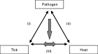
Fig. 1. The triangle of interactions between tick-borne pathogens, their tick vectors and vertebrate hosts. (1) pathogen–tick interactions; (2) pathogen–host interactions; and (3) tick–host interactions. The vertical arrow represents interactions of a pathogen with tick–host interactions at the skin site of tick attachment and blood-feeding (the tick–host–pathogen interface) and the double-headed arrow represents the cross-roads for pathogens at which they pass from infected tick vector to vertebrate host, or from infected host to tick vector.
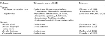
Table 1. Tick-borne pathogens showing direct evidence of saliva-activated transmission
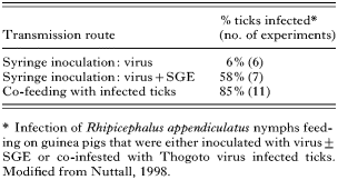
Table 2. Thogoto virus infection of ticks via different transmission routes
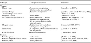
Table 3. Tick-borne pathogens showing indirect evidence of saliva-activated transmission (non-systemic transmission)
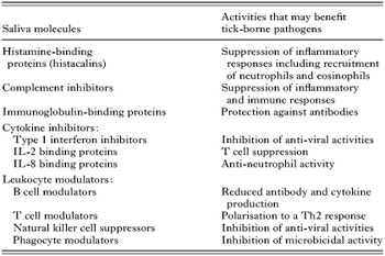
Table 4. Candidate SAT factors
 You have
Access
- 164
- Cited by