Introduction
The onset of schizophrenia occurs before the age of 25 years in 62% of affected males and 47% of affected females (Häfner et al. Reference Häfner, Riecher-Rössler, Anderheiden, Maurer, Fätkenheuer and Löffler1993). The peak in age of onset during puberty and early adulthood suggests a relationship between the expression of psychopathology and the changes in the brain and body that take place during this dynamic maturational period (Keshavan, Reference Keshavan1999). One of the major changes includes a dramatic increase in circulating oestrogens and androgens from the gonads (Veldhuis, Reference Veldhuis1996; Spear, Reference Spear2000). There is ample evidence pointing to adolescence as a sensitive period for hormone-dependent brain organization. Converging lines of evidence suggest that hormones play a key role in neuromaturational processes. Gonadal hormones, as well as adrenal hormones, have both an organizational and activational effect on the brain. In other words, they influence anatomical maturation of the brain over the course of development, as well as have more immediate effects on functional neural circuits to direct information processing and behaviour (Sisk & Zehr, Reference Sisk and Zehr2005). Animal research has shown that hormones can also trigger the expression of genes directing post-pubertal neuromaturational processes (McEwen, Reference McEwen1994; Ojeda & Ma, Reference Ojeda and Ma1999; Watson & Gametchu, Reference Watson and Gametchu1999). Hence, the behavioural changes elicited by hormones may be partially mediated by their effects on gene expression in addition to hormones directly affecting neurodevelopment.
Variation in maturational trajectories of brain development and variation in the timing and amplitude of a pubertal wave of hormones might help explain individual differences in behavioural adaptation and risk of sex-biased psychopathologies (Sisk & Zehr, Reference Sisk and Zehr2005). Indeed, it has been argued that some of the neural changes associated with schizophrenia, for which the onset in adolescence appears to be sex-specific (Häfner et al. Reference Häfner, an der Heiden, Behrens, Gattaz, Hambrecht, Löffler, Maurer, Munk Jørgensen, Nowotny, Riecher-Rössler and Stein1998), may result from the effects of gonadal steroids on the expression of genes that direct such maturational processes (Keshavan & Hogarty, Reference Keshavan and Hogarty1999; Walker & Bollini, Reference Walker and Bollini2002). There is also evidence to suggest that dysfunction of the hypothalamic–pituitary–gonadotropin (HPG) axis might contribute to the pathophysiology of schizophrenia, with several studies pointing to lower testosterone associated with more negative symptoms (Shirayama et al. Reference Shirayama, Hashimoto, Suzuki and Higuchi2002; Goyal et al. Reference Goyal, Sagar, Ammini, Khurana and Alias2004, Akhondzadeh et al. Reference Akhondzadeh, Rezaei, Larijani, Nejatisafa, Kashani and Abbasi2006; Ko et al. Reference Ko, Jung, Joe, Lee, Jung, Jung, Kim and Lee2007). Studies on putative abnormalities in gonadal hormones in schizophrenia have, to our best knowledge, been confined to adulthood rather than adolescence. This is surprising, considering that adolescence represents a stage of development at which individual differences in gonadal hormone levels are most prominent. Also, the study of dysfunctions in the HPG axis in adolescents at imminent clinical risk for psychotic disorders may point to the role of gonadal hormone dysfunction in the aetiology of psychotic symptoms, rather than being the result of chronic illness factors or medication effects which play a role in studies with adult, chronic patients. However, the study of hormonal profiles in adolescence has been hampered by the fact that it is a difficult challenge to identify those that are at imminent risk for developing a psychotic disorder such as schizophrenia, but not yet met criteria for a full-threshold psychotic disorder.
A solution for this may lie in the use of clinical high-risk strategies, based on the identification of adolescents with prepsychotic ‘prodromal’ symptoms of the disorder (Olsen & Rosenbaum, Reference Olsen and Rosenbaum2006;McGorry et al. Reference McGorry, Killackey and Yung2008; Correll et al. Reference Correll, Hauser, Auther and Cornblatt2010) using specific clinical screening protocols and ‘ultra-high-risk’ (UHR) criteria. Follow-up studies have reported high conversion rates to psychosis in adolescent samples defined by such UHR criteria (Olsen & Rosenbaum, Reference Olsen and Rosenbaum2006). In the present study, we assessed levels of testosterone and oestradiol in adolescents with prodromal symptoms aged 12–18 years. Studying this clinical population in this particular age range may help in formulating hypotheses with regard to mechanisms underlying increased levels of prodromal symptoms during puberty.
Method
Subjects
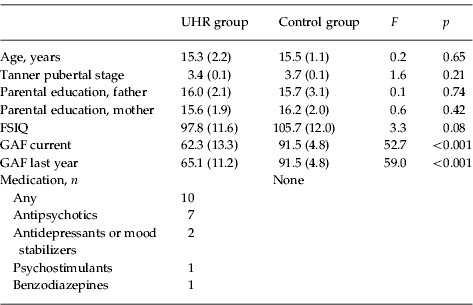
We included 21 male adolescents with prodromal symptoms and 21 male non-clinical controls in the age range of 12–18 years. The groups were matched on age, Tanner (pubertal) stage, IQ and socio-economic status indicated by average parental education (see Table 1). For both groups, mental retardation and neurological disorders were exclusion criteria, and age between 12 and 18 years was an inclusion criterion.
Group characteristics with regard to age, pubertal stage, socio-economic status, intelligence level, global functioning and medication
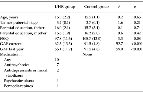
Data are given as mean (standard deviation) or number.
UHR, Ultra-high-risk; FSIQ, full-scale intelligence quotient; GAF, global assessment of functioning.
The UHR group was derived from a referred sample at the Department of Child Psychiatry at the University Medical Center Utrecht. In order to identify UHR adolescents, inclusion criteria and clinical instruments were used that in other studies have been proved to be successful in identifying those at UHR for psychosis (Olsen & Rosenbaum, Reference Olsen and Rosenbaum2006). The Structured Interview for Prodromal Syndromes (SIPS) (McGlashan et al. Reference McGlashan, Miller, Woods, Rosen, Hoffman and Davidson2001; Miller et al. Reference Miller, McGlashan, Rosen, Cadenhead, Ventura, McFarlane, Perkins, Pearlson and Woods2003) is a structured diagnostic interview and includes the Scale of Prodromal Symptoms, the Schizotypal Personality Disorder Checklist, a family history questionnaire and a Global Assessment of Functioning (GAF) scale. The semi-structured Bonn Scale for the Assessment of Basic Symptoms–Prediction List (BSABS-P) interview was used to rate subjective disturbances that have been found to be predictive for psychosis (Schultze-Lutter & Klosterkotter, Reference Schultze-Lutter and Klosterkotter2002). More specifically, adolescents were included because they met at least one of the four European Prediction of Psychosis Study (EPOS) inclusion criteria (see Klosterkotter et al. Reference Klosterkotter, Ruhrmann and Schultze-Lutter2005; Sprong et al. Reference Sprong, Becker, Schothorst, Swaab, Ziermans, Dingemans, Linszen and van Engeland2008):
(1) Brief, limited or intermittent psychotic symptoms (‘BLIPS’ assessed with the SIPS): a history of psychotic symptoms with duration of less than 1 week and spontaneous remission.
(2) Presence of at least one of: ideas of reference, odd beliefs or magical thinking, perceptual disturbance, odd thinking and speech, paranoid ideation, odd behaviour or appearance, as defined in Diagnostic and Statistical Manual of Mental Disorders, fourth edition (DSM-IV) and assessed with the SIPS. These symptoms should occur at least several times per week and should have been present for at least 1 week.
(3) Individuals with a first- or second-degree relative with a history of any DSM-IV psychotic disorder or a DSM-IV schizotypal personality disorder and a change in mental state or functioning leading to a reduction of 30 points or more on the GAF scale (assessed with the SIPS).
(4) At least three self-perceived deficiencies of cognition or perception from the list of basic symptoms as assessed with the BSABS-P.
In this study, none of the adolescents met criterion 1 (BLIPS), 85.7% met criterion 2 (attenuated positive symptoms), none met criterion 3 (family history and reduced functioning) and 56.7% met criterion 4 (basic symptoms). Overall, 47.6% of the UHR adolescents met one criterion and 52.4% met two criteria. Information with regard to functioning (GAF) and medication can be found in Table 1.
The control group was recruited from local secondary schools. Using identical screening instruments as for the UHR group, none of the controls belonged to one of the four above-mentioned groups and hence did not meet UHR criteria. They were also excluded if they had a history of any psychiatric illness themselves, or in a first-degree relative, or a second-degree relative with a psychotic disorder. The study was approved by the local Ethics Committee at the University Medical Center Utrecht. Informed consent was obtained from all adolescents and parents, according to the declaration of Helsinki.
Prodromal symptoms
The semi-structured SIPS interview has been designed to assess a broad spectrum of prodromal signs and symptoms. It has four subscales: Positive (five items), Negative (six items), Disorganization (four items), and General symptoms (four items), and also includes a GAF scale with well-defined anchor points (McGlashan et al. Reference McGlashan, Miller, Woods, Rosen, Hoffman and Davidson2001). Symptoms are scored on a seven-point scale from 0 (absent) through to 6 (extreme/psychotic intensity). The distinction between a score of 5 (severe) and a score of 6 (psychotic) for the positive symptoms is mainly determined by ‘the lack of conviction regarding the externally generated, ‘real’ nature of the symptom as well as the maintenance of insight regarding the sense that the experience is, in fact, a symptom' (Miller et al. Reference Miller, McGlashan, Rosen, Cadenhead, Ventura, McFarlane, Perkins, Pearlson and Woods2003). The semi-structured BSABS-P interview was used to rate subjective disturbances that have been found to be predictive for psychosis (Schultze-Lutter & Klosterkotter, Reference Schultze-Lutter and Klosterkotter2002). The 33 items are summarized in three subscales, i.e. Cognitive (11 items), Perceptual (19 items) and Motor disturbances (three items). In line with the SIPS, a seven-point scoring scale has been developed to rate the presence and severity of the symptoms (0=absent, 6=frequent/extreme).
Salivary testosterone and oestradiol
All samples were collected between 12.00 and 13.00 hours in order to minimize diurnal variation as much as possible. On the saliva-collecting day, participants were asked not to eat, brush their teeth or drink for 1 h prior to collection in order to prevent sample dilution or contamination. Then saliva was collected by unstimulated passive drool. Samples were stored at −20°C within 2 h. Free testosterone in saliva was measured in duplicate by the EiAsy™ ELISA kit (CAN-TE-300; Diagnostic Biochemicals Canada, Canada) using 2×100 μl saliva; the sensitivity of the testosterone saliva kit was 1.0 pg/ml. Free 17β-oestradiol in saliva was measured in duplicate by the 17β-Estradiol Saliva ELISA kit (RE52601; IBL International GmbH, Germany) using 2×100 μl saliva; the sensitivity of the 17β-Estradiol Saliva kit was 0.4 pg/ml.
Tanner pubertal stage
Stage of pubertal development was assessed by self-ratings of schematic drawings of secondary sex characteristics associated with the five standard Tanner stages of pubertal development, with stage 1 corresponding to infantile and stage 5 to complete puberty (Tanner, Reference Tanner1962). Self-reports of schematic drawings are generally considered an acceptable way to assess physical development, and have demonstrated adequate validity (Coleman & Coleman, Reference Coleman and Coleman2002).
Statistical analyses
Data were analysed using SPSS version 17.0 (SPSS, Inc., USA). Analysis of variance (ANOVA) was used to test for group differences in age, pubertal stage, IQ, parental education and GAF scores. Between-group differences in levels of prodromal symptoms were tested using multivariate ANOVA (MANOVA) with ‘group’ (control, clinical) as a fixed factor and the psychopathology subscales as independent factors. Only if the MANOVA resulted in a significant multivariate effect, did we proceed to the univariate group effects on specific measures. ANOVA was also used to test for group differences in levels of testosterone and oestradiol, with the fixed factor ‘group’ (control, UHR) and the dependent variables ‘testosterone’ (pg/ml) and ‘oestradiol’ (pg/ml). Spearman's r was used for correlational analysis. Effect sizes were calculated using Cohen's d, representing the difference between two means expressed in standard deviations (mean of the pooled standard deviations in the two groups). Level of significance was set at p=0.05 (two-tailed).
Results
Prodromal symptoms
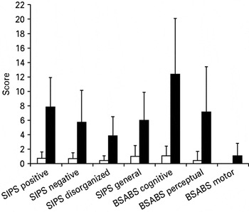
As expected, based on the inclusion procedure, the UHR group showed substantially higher levels of prodromal symptoms as compared with the control group. The multivariate effect of ‘group’ was significant [F(7, 32)=4.9, p=0.001]. Univariate group effects on specific subscales showed that on all subscales of the SIPS and BSABS, scores were significantly higher in the clinical group (see Fig. 1).
Levels of prodromal symptoms in the ultra-high-risk (▪) and control (□) groups. Values are means, with standard deviations represented by vertical bars. SIPS, Structured Interview for Prodromal Syndromes; BSABS, Bonn Scale for the Assessment of Basic Symptoms.

Salivary testosterone and oestradiol
Within-subject duplicate analysis of the saliva samples (across all subjects) revealed a variation of 8.0 (s.d.=7.4) % for the testosterone analysis and 5.1 (s.d.=35.6) % for the oestradiol analysis. As a validation of the testosterone and oestradiol measurements, we tested the hypotheses that levels of testosterone and oestradiol correlate with age and/or measures of pubertal development. Indeed, testosterone levels (across all subjects) were significantly correlated with Tanner pubertal stage (r=0.58, p<0.001) as well as age (r=0.50, p=0.001). No significant correlations were founds with oestradiol. Although there was a correlation between testosterone levels and age, this could not affect any group effects on testosterone levels as the clinical group and control group were carefully matched on age.
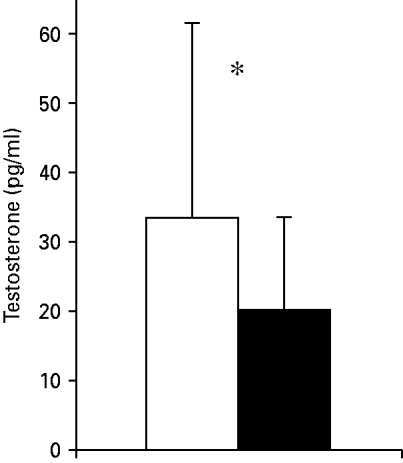
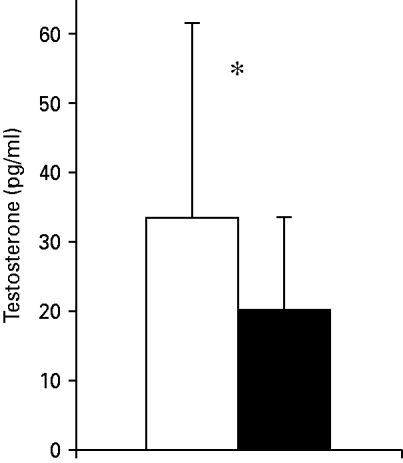
ANOVA was used to test for group differences, with the fixed factor ‘group’ (control, UHR) and the dependent variables ‘testosterone’ (pg/ml) and ‘oestradiol’ (pg/ml). For testosterone, a significant main effect of group was found [F(1, 40)=3.9, p=0.05], with levels of testosterone lower in UHR subjects (mean=20.0, s.d.=13.6) as compared with controls (mean=33.6, s.d.=28.2) (see Fig. 2). The effect size was 0.7. For oestradiol, we did not observe a significant main effect of group. All analyses were re-run with the subjects taking medication (n=10) left out of the analysis, but this did not change the pattern and significance of the results [F(1, 32)=4.3, p=0.45 for group effect on testosterone levels]. No significant correlations were found between levels of testosterone and degree of prodromal symptoms.
Levels of testosterone in control (□) and ultra-high-risk (▪) adolescents. Values are means, with standard deviations represented by vertical bars. * Mean value was significantly different from that of the control group (p⩽0.05).
Discussion
This study examined levels of testosterone and oestradiol in male adolescents aged 12–18 years with prodromal symptoms and meeting criteria for UHR for psychosis. Levels of testosterone were significantly lower in UHR adolescents as compared with non-clinical controls, with an effect size of 0.7. No group differences in oestradiol were found. In the total sample, level of testosterone was significantly correlated with age and Tanner pubertal stage, indicating that the levels of testosterone in this study have biological and maturational relevance and are closely related to pubertal developmental mechanisms.
Our findings of lower testosterone point to a dysfunction in the HPG axis in male adolescents with prepsychotic, prodromal symptoms. As these dysfunctions are observed in adolescents in a prodromal state, not meeting criteria for a psychotic disorder, we propose that HPG axis dysfunctions may be related to the aetiology of psychotic symptoms, rather than arising as a consequence of chronic illness factors or medication. In line with this, excluding UHR adolescent using any medication (n=10) did not have any effect on the pattern and significance of our findings.
Our findings of low testosterone levels in adolescents with prodromal symptoms complements other studies that have shown androgen dysfunctions in patients with psychotic disorders such as schizophrenia. Huber et al. (Reference Huber, Tettenborn, Leifke and Emrich2005) found lower levels of testosterone in 34 acutely admitted men with schizophrenia. In a group of 56 men with schizophrenia, only those with lower dosages of medication had lower levels of testosterone in a study by Kaneda & Fujii (Reference Kaneda and Fujii2000) . In a study of 10 patients, Goyal et al. (Reference Goyal, Sagar, Ammini, Khurana and Alias2004) found lower testosterone levels in schizophrenia patients with predominantly negative symptoms. Similarly, in a study of 28 schizophrenia patients, those with negative symptoms showed lower levels of testosterone as compared with controls (Shirayama et al. Reference Shirayama, Hashimoto, Suzuki and Higuchi2002). Levels of testosterone were also correlated with negative symptoms. In a larger study of 54 schizophrenia patients, lower levels of testosterone were found in the patient group as a whole, and lower levels of testosterone were associated with more negative symptoms (Akhondzadeh et al. Reference Akhondzadeh, Rezaei, Larijani, Nejatisafa, Kashani and Abbasi2006). Ko et al. (Reference Ko, Jung, Joe, Lee, Jung, Jung, Kim and Lee2007) also found a correlation between lower testosterone and negative symptoms in 35 schizophrenia patients, even after covarying for depressive symptoms. However, other studies have found no androgen dysfunctions (Oades, Reference Oades1994; Ceskova et al. Reference Ceskova, Prikryl and Kasparek2007), or found effects of medication on the HPG axis (Rinieris et al. Reference Rinieris, Hatzimanolis, Markianos and Stefanis1989; Kaneda & Fujii, Reference Kaneda and Fujii2000; Konarzewska et al. Reference Konarzewska, Wolczynski, Szulc, Galinska, Poplawska and Waszkiewicz2009). The effect of medication has generally limited the interpretation of testosterone dysfunctions in studies with adult, chronic patients. Interestingly, data pointing to early effects of testosterone on neurodevelopment in relation to risk for schizophrenia come from studies on the ratio between finger length of the second digit as compared with the fourth digit (2D:4D ratio). The 2D:4D ratio is a well-known marker of the amount of prenatal circulating testosterone, as empirically shown by significant correlations between high 2D:4D ratio and low levels of fetal testosterone (and vice versa) (Lutchmaya et al. Reference Lutchmaya, Baron-Cohen, Raggatt, Knickmeyer and Manning2004). Abnormal 2D:4D ratios have been identified in individuals with schizophrenia (Arató et al. Reference Arató, Frecska, Beck, An and Kiss2004; Collinson et al. Reference Collinson, Lim, Chaw, Verma, Sim, Rapisarda and Chong2010) and adolescents with schizotypal personality disorder (Walder et al. Reference Walder, Andersson, McMillan, Breedlove and Walker2006), pointing to possible early organizational effects of testosterone on brain development.
Hypotheses with regard to the mechanisms by which androgen dysfunctions might contribute to psychotic symptoms have been put forward. These hypotheses are based on the influence of testosterone on neurodevelopment. Concerning the putative mechanism of action of testosterone, only 1–2% of testosterone is free and bioavailable, with the rest largely bound to plasma proteins (Dunn et al. Reference Dunn, Nisula and Rodbard1981). In its free form testosterone can cross the blood–brain barrier and bind to androgen receptors within target cells distributed in the brain (Bialek et al. Reference Bialek, Zaremba, Borowicz and Czuczwar2004). Salivary levels of testosterone are shown to be strongly correlated with free, bioavailable testosterone in serum (Wang et al. Reference Wang, Plymate, Nieschlag and Paulsen1981), and hence may reveal androgen dysfunctions that have an impact on brain development and brain functioning. Androgen receptors are concentrated in the hypothalamus and limbic system, influencing perception, emotion, cognition and behaviour (Stevens, Reference Stevens2002). Androgen receptors are situated in the nucleus as well as on the surface of the neuron (McEwen, Reference McEwen1994; Watson & Gametchu, Reference Watson and Gametchu1999). Those on the surface of the neuron are important for (non-genomic) short-term modulation of perception, emotion, cognition and behaviour. Receptors in the nucleus are involved in gene expression and allow androgens to have genomic effects, influencing neuronal structure and function, including regulation of neurotransmitter systems such as dopamine (Häfner et al. Reference Häfner, Behrens, De Vry and Gattaz1991). Hence, androgens may also contribute to the pathophysiology of schizophrenia by modulating the action of various neurotransmitters and neuropeptides through non-genomic or genomic mechanisms. As testosterone has been assigned neuroprotective functions (Bialek et al. Reference Bialek, Zaremba, Borowicz and Czuczwar2004), Ko et al. (Reference Ko, Jung, Joe, Lee, Jung, Jung, Kim and Lee2007) have posited that lower testosterone in schizophrenia may negatively affect brain development. Shirayama et al. (Reference Shirayama, Hashimoto, Suzuki and Higuchi2002) have related their findings of lower testosterone in patients with more negative symptoms to stress-vulnerability models of schizophrenia, arguing that the typical increase in testosterone in response to stress, where testosterone has inhibitory effects on cortisol responses to stress (Viau, Reference Viau2002), is blunted in schizophrenia patients with negative symptoms.
Our findings need replication in larger studies, preferably including the assessment of other neuroendocrines as well. Besides the limited range of neuroendocrine measures in this study, other limitations are the relatively small sample size and the lack of longitudinal data (such as transition to a psychotic disorder). It will be interesting to assess in future follow-up studies whether levels of androgens have predictive value for transition to psychosis, although transition to a full-fledged disorder is probably best predicted by a combination of several neurodevelopmental processes, in interaction with environmental factors, that have an additive effect.
In summary, our finding of lower levels of testosterone in male adolescents with prepsychotic, prodromal symptoms is in line with current hypotheses stressing the role of neuroendocrine factors during adolescence in brain developmental processes involved in the expression of psychotic symptoms. From a developmental perspective, susceptibility to psychotic disorders increases during adolescence. Our data suggest that testosterone might, in part, mediate this increased vulnerability, although the mediating neural mechanisms by which testosterone has an impact on psychotic symptoms should be further investigated. To illustrate, Peper et al. (Reference Peper, Brouwer, Schnack, van Baal, van Leeuwen, van den Berg, Delemarre-Van de Waal, Boomsma, Kahn and Hulshoff Pol2009) have recently shown that lower serum levels of testosterone are significantly correlated with smaller grey matter volume in pubertal boys. The effects of testosterone on brain development are not limited to activational effects of pubertal gonadal steroids, but may also involve organizational effects during puberty, further having an impact on the development of cognition, behaviour and psychopathology into adulthood (Romeo, Reference Romeo2003). In the search for early risk markers for psychosis, studying neuroendocrine factors in relation to neurobehavioural trajectories might increase our understanding of ‘at-risk’ developmental pathways.
Acknowledgements
This work was supported by a VernieuwingsImpuls grant (no. 016.026.027 to A.A.) and a Veni grant (no. 016.095.060 to S.v.R.) from the Netherlands Organization for Scientific Research (NWO). We thank Dr J. van Pelt (Eperitos, Leiden) for analysis of the saliva samples.
Declaration of Interest
None.