Introduction
Pseudorabies, also known as Aujeszky’s disease, is a viral disease caused by the pseudorabies virus (PrV). The disease causes serious reproductive failure followed by neonatal disease. In newborn pigs, it progresses rapidly, and usually results in the death of some or the entire litters. Many laboratory tests have already been developed including virus isolation, polymerase chain reaction (PCR), Enzyme-Linked Immunosorbent Assay (ELISA) (Corn et al., Reference Corn, Stallknecht, Mechlin, Luttrell and Fischer2004). For general diagnostic purposes it has been widely replaced by ELISA because of its suitability for large-scale testing. The tests can be performed on meat juice as well as serum.
Pseudorabies is a viral disease characterized by inflammation. It has been reported that some candidate genes have relationship with immune and inflammatory responses, and developing central nervous system (CNS) of early embryogenesis. For instance, interleukin 10 (IL-10) regulates many aspects of the immune and inflammatory responses, and its critical role in protection against autoimmunity has been highlighted by in vivo studies on animal models (Kamali et al., Reference Kamali, Kiany, Gharesi and Robati2006); The chemokine stromal cell-derived factor 1 (SDF-1, CXCL12) plays an important role in both the developing brain and adult CNS (Lu et al., Reference Lu, Grove and Miller2002). The euchromatic histone-lysine N-methyltransferase 2 (EHMT2) acts as a co-regulator for specific transcription factors essential in early embryogenesis (Fang et al., Reference Fang, Miao, Xiang, Ponugoti, Treuter and Kemper2007); The protein HLA-B-associated transcript 2 (BAT2) gene has been showed specific expression, especially in central nervous tissue, and suggests BAT2 as a novel differentiation marker (Schneidersa et al., Reference Schneidersa, Thiela, Winklera, Mollerb and Koch2005).
The mutations of some functional genes, e.g. the mutation in the exonic loci that change amino acids or splicing of candidate genes, can lead to diseases. For example, TNF (tumor necrosis factor) genes polymorphisms are associated with autoimmune thyroid diseases including Graves disease and autoimmune hypothyroidism (Maha et al., Reference Maha, Noura, Kaouthar, Mouna, Joumaa, Mohamed, Ahmed and Hammadi2008). These mutations might provide a direct association of genotype with phenotype (Stenson et al., Reference Stenson, Ball, Mort, Phillips, Shiel, Thomas, Abeysinghe, Krawczak and Cooper2003). Therefore, the mutations (single-nucleotide polymorphisms (SNPs)) from the coding region of the mediators of pro-inflammatory response or other candidate genes, e.g. IL10, CXCL12, EHMT2 and BAT2 (Klipper et al., Reference Klipper, Wasserman, Braunspiegel, Borstein, Peleg, Assa, Karp, Benjamini, Hochberg and Laron1995; Jiwakanon et al., Reference Jiwakanon, Persson and Dalin2006; Kamali et al., Reference Kamali, Kiany, Gharesi and Robati2006), in pigs could indicate their potential involvement in susceptibility or resistance to PrV infection.
So far there is no previous association study performed on the effect of pig-host gene mutations with PrV phenotype data. Thus our study is the first to address this issue.
To find out whether there is genetic variation that correlates with susceptibility or resistance to PrV infection, the genotypes of SNPs from the genes that mediate pro-inflammatory response, IL10, CXCL12, EHMT2 and BAT2, were determined in pigs using high throughput microarray method, and then association analysis with genotypes and antibody were performed.
Material and methods
Samples and antibody test
A total of 178 sows were selected randomly from a pool of commercial healthy L × LW (Landrace × Large White) sows. These sows were vaccinated with gene deletion TK−/gE− PrV vaccine 6 weeks after the start of pregnancy. Two blood samples were collected from each sow 2 months after vaccination, one taken into ethylenediaminetetraacetic acid for DNA extraction using Qiagen reagents according to the manufacturer’s instructions (Qiagen, Hilden, Germany) and the other untreated blood for serology tests for antibodies against PrV by pseudorabies virus (PrV)-ELISA and glycoprotein E (gE)-ELISA. The antibody elicited during vaccination is some evidence that is protective against PrV, as antibody response is typically ‘delayed’ under PrV infection to target serum neutralization (SN) titers. As we know that PrV gE is an envelope glycoprotein consisting of 588 amino acids. It is one of PrV virulence determinants and a non-essential protein for in vitro replication as well (Van et al., Reference Van, Gielkens, Moormann and Berns1990). When the gE gene is deleted, the propagation and antigenicity of PrV are not affected, but its virulence is reduced greatly (Jacobs et al., Reference Jacobs, Mulder, Van Oirschot, Gielkens and Kimman1993; Nauwynck, Reference Nauwynck1997). On the other hand, gE is present in almost all examined PrV field isolates, and the natural infection always gives rise to detectable antibodies against gE. The PrV gE-ELISA procedure followed as Ao et al. (Reference Ao, Wang, Chen, Wang and Long2003).
Microarray-based genotyping of SNP
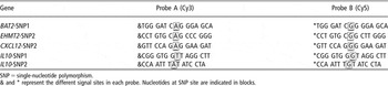
Genotyping of 178 samples were performed using microarray technology. The alleles defining the SNP were detected using allele-specific oligonucleotides labelled with either Cy3 or Cy5 dyes. The ratio of Cy3 and Cy5 then indicated the genotype. The homozygous genotype yielded strong fluorescence of Cy3 (green fluorescence) or Cy5 (red fluorescence), whereas the heterozygous genotype yielded fluorescence of both dyes Cy3 and Cy5 that give a yellow spot. Primers and Cy3 or Cy5 5′ prime labelled oligonucleotide probes used in this study to detect SNPs were synthesized and purified by Sigma (Sigma, St. Louis, MO, USA). And each SNP was genotyped on a single slide. Each sample was printed on slides in triplicates. The sequence of the labelled probes and primers were shown in Tables 1–3.
The primers sequence of cSNP identified from the candidate genes by sequencing

SNP = single-nucleotide polymorphism; cSNP = SNP in cDNA.
The primers sequence of cSNP for microarray

SNP = single-nucleotide polymorphism; cSNP = SNP in cDNA.
The sequences of the probes used for microarray genotype

SNP = single-nucleotide polymorphism.
& and * represent the different signal sites in each probe. Nucleotides at SNP site are indicated in blocks.
Microarray-based genotyping of the samples was performed according to the protocol (Ji et al., Reference Ji, Hou, Li, He and Lu2004) with some modification. Briefly, samples were amplified in 96-well plate using primers that cover the SNP. The average length of PCR product was 100 bp. PCR was carried out in 10-μl reaction volumes containing 10 to 30 ng of porcine genomic DNA, 1 μl dNTPs (250 μmol/l each), 10 pmol of each primer, 1 μl of 10× buffer and 0.1 μl of dynazyme DNA polymerase (Dynazyme, Finnzymes, Woburn, MA, USA). Amplification conditions were 5 min at 94°C, followed by first to 22nd cycles of 30 s at 94°C, 30 s at 65°C, 30 s at 72°C and touchdown to 52°C by decreasing the annealing temperature by 1°C every cycle, and final extension of 10 min at 72°C. In order to identify each PCR product, we tested by electrophoresis (data were not shown).
The PCR products were purified using QIAquick columns (Qiagen), then the product was resuspended in 3× SSC (standard saline citrate) to a final concentration of approximately 300 ng/μl and transferred to 384-well plates (4309849; Applied Biosystems, Foster City, CA, USA). After that, the purified products were printed on GAP slides (40005; Corning, Coming, NY, USA) and baked for 2 h at 80°C. After baking the slides, the bound DNA was denatured by 2-min incubation in distilled water at 95°C. The slides were then transferred into a bath of 95% ethanol at room temperature, rinsed and then spun dry in a clinical centrifuge. Slides were stored in a closed box at room temperature until used.
Hybridization
Hybridization mixture consisted of Cy3 and Cy5 labelled probes capable of detecting the two different alleles of SNP mixed in equivalent amounts, 2 μl of salmon testis DNA, 10 μl of 20× SSC, 6 μl each of labelled probes (1 μM each), 2 μl of 10% SDS and 14 μl distilled water in 40 μl volume. Hybridization was conducted in a moist-sealed hybridization chamber under a cover slip at 37°C for 30 min. After hybridization, the slides were, respectively, rinsed and washed at room temperature with 5× SSC and 0.1% SDS together, 5× SSC and distilled water, 5 min for each wash and dried by centrifugation at 600 r.p.m. for 5 min.
Image scanning and data analysis
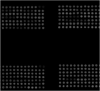
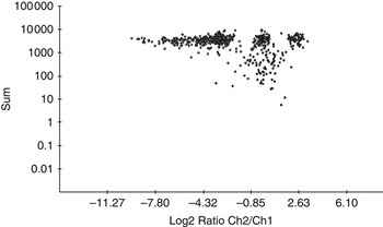
The hybridized slides were scanned using Axon 4100A scanner (Molecular Devices, Sunnyvale, CA, USA). Analysis and genotype calling of the images acquired by the scanner were done using Blue fuse software (Bluegnome, Cambridge, UK) (see Figures 1 and 2). In addition, the signal was normalized at the beginning.
The images of microarray genotyping.
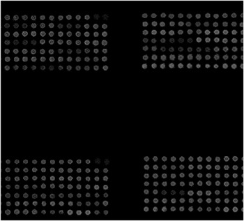
The image of microarray genotyping and the three genotype groups of single-nucleotide polymorphisms from EHMT2 gene.

Allelic frequency determination
Allelic frequencies of the SNPs were calculated using the following formula. Allelic frequency (A) = [(2 × AA) + AB]/[(2 × (AA + AB + BB)], allelic frequency (B) = [(2 × BB) + AB]/[(2 × (AA + AB + BB)].
The χ 2 analysis and Fisher’s exact test were carried out to identify statistical significance. All properties correlation analysis adopted the model showed below:

where Y is the observed antibody, X the effects of genotype, W the effects of farms, e the effects of random; the data analysis was performed according to the protocol (Yuan et al., Reference Yuan, Moaeen-ud-Din, Gong, Peng, Yang, Feng, Liu, Hu, Affara, Jafer and Zhang2007a and Reference Yuan, Jafer, Affara, Gong, Yang, Liu, Moaeen-ud-Din, Li and Zhang2007b).
Results
Genotyping and allele frequency determination
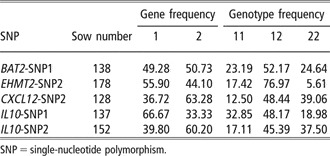
Genotyping of 178 samples were performed using microarray technology. Figure 1 showed the three different colors for the different homozygous and heterozygous genotypes. The allele and genotype frequency of the SNP were summarized in Table 4. There were three genotypes in the pig population in CXCL12-SNP2, IL10-SNP1, EHMT2-SNP2, IL10-SNP2 and BAT2-SNP1.
The genotypic and allelic frequencies of five SNPs

SNP = single-nucleotide polymorphism.
The association of SNP genotypes and antibody
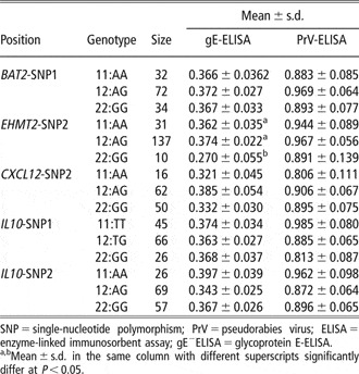
There was a significant difference of gE-ELISA antibody in 178 LLW between genotypes 11 (AA), 12 (AG) and 22 (GG) in EHMT2-SNP2. Table 5 showed that the gE-ELISA antibody of pigs with genotypes 11 (AA) or 12 (AG) was significantly higher than genotype 22 (GG) (P < 0.05).
Association with different SNP and PrV antibody in LLW sows

SNP = single-nucleotide polymorphism; PrV = pseudorabies virus; ELISA = enzyme-linked immunosorbent assay; gE−ELISA = glycoprotein E-ELISA.
a,bMean ± s.d. in the same column with different superscripts significantly differ at P < 0.05.
Discussion
Pseudorabies disease is caused by PrV, an alphaherpes, DNA virus. The economic losses are estimated at 60 to 120 Yuan (RMB of Chinese Yuan) infected pig. Therefore the impact of the disease in infected sows and commercial pig herds has become serious and very significant and it has become an important and urgent task to prevent, control and eradicate this disease.
For eradication of pseudorabies, vaccines marked by deletion of non-necessary genes encoding immunogenic viral proteins have been constructed and used widely. Attenuated vaccines, which generally induce long-lasting immunity, present the risk of insufficient attenuation and/or genetic instability. Inactivated vaccines are less efficient and required several injections. Immunization with inactivated vaccines may induce post-vaccinal allergic reaction such as lack of appetite, hyperthermia or abortion in pregnant sows. Moreover, a serious consequence would be made if the virus is not inactivated completely. In my opinion the best way is to breed good breeds, which have the resistance to pseudorabies, with the help of good molecular markers.
Previous studies have shown that there is a distinct difference in susceptibility to PrV between the Large White and Meishan breeds. At least two of the quantitative trait loci are linked to the interferon-gamma locus and at the interferon receptor locus (Reiner et al., Reference Reiner, Melchinger and Kramarova2002). Genetic differences in the immune response of pigs to antigens or vaccines have been reported for vaccination with pseudorabies vaccine (Rothschild et al., Reference Rothschild, Hill, Christian and Warner1984). So we want to do some work to find some useful and effective genetic markers to select well performance sows which have lower susceptibility to PrV.
We chose four candidate genes IL10, CXCL12, EHMT2 and BAT2 to test. Interleukin 10 (IL-10), secreted by macrophages and lymphocytes, regulates many aspects of the immune and inflammatory responses, and its critical role in protection against autoimmunity has been highlighted by in vivo studies on animal models. For instance, IL-10-deficient mice spontaneously develop chronic inflammatory bowel disease (Kamali et al., Reference Kamali, Kiany, Gharesi and Robati2006). The chemokine stromal cell-derived factor 1 (SDF-1, CXCL12), a member of the CXC chemokine family, plays an important role in both the developing brain and adult CNS. Both in vivo and in vitro studies suggest that CXCL12 and its ligand CXCR4 are involved in neurogenesis processes such as neural progenitor/stem cell migration, proliferation and differentiation (Lu et al., Reference Lu, Grove and Miller2002; Peng et al., Reference Peng, Kolb, Kennedy and Zheng2007). In addition, it is a key regulator of B-cell lymphopoiesis, hematopoietic stem cell mobilization and leukocyte migration. The euchromatic histone-lysine N-methyltransferase 2 (EHMT2) is responsible for mono- and di-methylation of H3 Lys-9 (H3-K9) in euchromatin. It acts as a co-regulator for specific transcription factors. Studies in EHMT2-null mice showed that euchromatic H3K9 methylation by EHMT2 is essential in early embryogenesis and is critically involved in transcriptional silencing of developmentally regulated genes (Fang et al., Reference Fang, Miao, Xiang, Ponugoti, Treuter and Kemper2007). The protein HLA-B-associated transcript 2 (BAT2), which contains four Arg-Gly-Asp motifs typical of the integrin receptor family, is very rich in praline and includes short tracts of polyproline, polyglycine and charged amino acids. It does not appear to be a member of any known family of proteins (Baner et al., Reference Baner, Sands, Storominger and Spiest1990). The BAT2 gene lies within the class III region of the major histocompatibility complex. Immunostaining of embryonic tissue sections showed specific expression, especially in central nervous tissue, and suggests BAT2 as a novel differentiation marker (Schneidersa et al., Reference Schneidersa, Thiela, Winklera, Mollerb and Koch2005).
In our paper, we have genotyped 178 samples for five SNPs from four candidate genes IL10, CXCL12, EHMT2 and BAT2 determined the allelic and genotype frequencies for each SNP (see Table 4). We have also determined the level of antibodies of gE by gE-ELISA. It was interesting to note that the association of EHMT2-SNP2 genotype, localized within the major histocompatibility complex class III region, with PrV antibody was significant (P < 0.05). The antibody elicited during vaccination was protective against PrV, as antibody response is typically ‘delayed’ under PrV infection to target SN titers.
The animal’s immune response system helps to provide a defence against disease. The important candidate genes are assumed to play crucial roles in host defence against PrV. Therefore it is possible that the EHMT2 mutation could have an effect in pig susceptibility or resistance to PrV infection. Further study of the causes and mechanism of EHMT2 on antibody response is needed.
Acknowledgements
Financial assistance from 863 High Technology and Development Project of China (2006AA10Z195, 2007AA10Z152), Chinese projects (2006BAD14B08-02, 2006BAD04A02-11), Hubei project (2003AA211041, 2006CA023), Wuhan project (20067003111-06) and National Project of China (04EFN214200206) are greatly appreciated. We like to thank the research group of Molecular Genetics and Mammalian Genome at University of Cambridge for providing the microarray facility.