Introduction
Airway obstruction secondary to haemorrhage following neck surgery is a rare but life-threatening condition that all ENT surgical trainees are taught to recognise and manage. At the level of the vocal folds, the airway is well protected from external compression by the cricoid and thyroid cartilages, leaving two possible causes for such airway obstruction: direct compression by a haematoma onto the trachea below the cricoid level; or supraglottic oedema secondary to venous compression in the neck, due to the haematoma.
The maximum possible pressure achievable within the neck due to a post-operative haemorrhage is systolic blood pressure. Blood flows across a pressure gradient, and thus when the pressure in the neck reaches systolic blood pressure the blood will stop flowing from the vascular system into the neck space.
In this study, we created an in vitro model enabling the application of varying pressures to whole tracheas. This enabled investigation of whether the maximum possible pressure produced by a haematoma in the neck would be sufficient to directly compress the trachea to such an extent that it caused airway obstruction.
Materials and methods
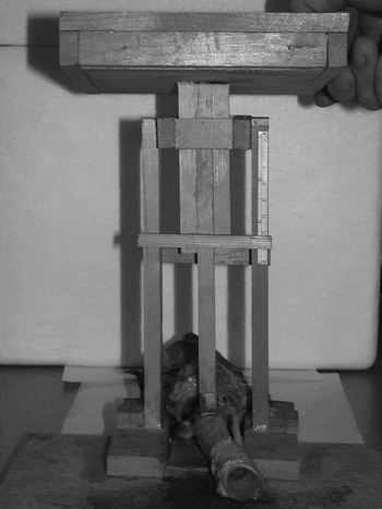
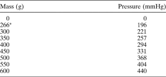
A platform device was constructed (Figure 1) with which varying degrees of force could be applied over a 1 cm2 area to pig tracheas, by placing different masses on top of the platform, thus reproducing the possible pressures present in haematomas developing after neck surgery (Table I). Using this platform, increasing pressures were applied to 10 pig tracheas in vitro over the second and third cartilaginous rings, in an anterior–posterior direction. The pig tracheas were from approximately one-year-old pigs obtained from an organic butcher, and were used in the study within 24 hours of slaughter. The tracheas used were attached to the larynx superiorly and were cut at the level of the tracheal bifurcation inferiorly. Anterior–posterior and lateral diameters were measured before compression; anterior–posterior diameters were then measured under compression.
Platform device used to apply varying pressures to the region of the second and third cartilaginous rings of the pig trachea.

Pressures exerted by different mass applied over a 1 cm2 area of trachea

* Weight of platform.
Results
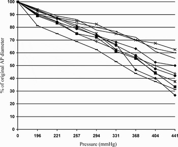
The average pre-compression anterior–posterior and lateral diameters in the region of the second and third tracheal rings were 17.2 and 17.0 mm, respectively. The compression results are shown in Figure 2. The average tracheal compression observed with 257 mmHg of pressure was 20.8 per cent of the original anterior–posterior diameter.
Results for anterior–posterior (AP) compression of 10 pig tracheas with increasing pressures.

Discussion
Traditionally, it was believed that the airway obstruction encountered in the presence of haematoma developing after neck surgery was caused by direct compression of the trachea by the haematoma.Reference Lahey and Hoover1 In 1979, Wade stated in the Cecil Joll Lecture that he believed that the pressures present in neck haematomas were not enough to compress what is often a hard, calcified trachea, and that the obstruction was probably due to supraglottic oedema caused by laryngeal venous obstruction.Reference Wade2 However, to date, no objective evidence has been supplied to support this assertion.
In this study, we demonstrated that the maximum pressure achievable due to a haematoma developing after neck surgery, i.e. systolic blood pressure, was not enough to cause direct obstruction of the airway. In our study, the average tracheal compression achieved at 257 mmHg (i.e. 350 g over a 1 cm2 area), which approximates to maximal systolic blood pressure, was only 20.8 per cent of the original anterior–posterior diameter.
The pressure required to cause venous compression in the neck is far less than systolic blood pressure. The normal pressure in the internal jugular vein is less than 10 cm of water, which equates to 7.35 mmHg. We believe that it is compression of the venous system which leads to supraglottic oedema, which in turn causes airway obstruction.
This study suffers from the limitation of any in vitro study, in that the in vivo situation cannot be exactly replicated. However, we believe that, in humans with a haematoma developing after neck surgery, the trachea would be even more resistant to tracheal compression, compared with our in vitro model, because: (1) in vivo, the trachea receives extra support from surrounding soft tissues; and (2) the tracheas we used were from pigs of around one year of age, approximating to a human young adult; haematoma developing after neck surgery normally occurs in an older population with more tracheal calcification and therefore a more rigid, stronger trachea.
It is possible that the trachea would undergo changes post mortem that would affect the strength of the structure. Such changes would be difficult to assess, as testing the strength of the trachea may alter the integrity of the structure and thus bias future testing of the same specimen. Drobnic et al. assessed post mortem changes in cartilage chondrocytes and found viable chondrocytes in cartilage samples stored in vitro at day nine post mortem.Reference Drobnic, Mars, Alibegovic, Bole, Balazic and Grubic3 The chondrocyte count reduced with time and was significantly greater when the cartilage was stored at 4°C compared with 35°C. We believe we minimised any post mortem changes in our specimen tracheas by using them within 24 hours of slaughter and storing them at 4°C.
None of the pigs involved in the study had significant enlargement of the thyroid. In humans, it is possible that an enlarged thyroid may have an effect on the strength of the trachea. However, as airway obstruction due to neck surgery induced haematoma is encountered in other types of head and neck surgery apart from thyroid surgery, this clearly is not the most important factor in the development of airway obstruction.
We believe the pig to be a good animal model for the human trachea, due to the gross anatomical, cellular and sub-cellular similarities seen between the human and pig larynx and trachea.Reference Jiang, Raviv and Hanson4–Reference Gorti, Birchall, Haverson, Macchiarini and Bailey7 In addition, previous anatomical studies have shown that human tracheas are of a similar size to the pig tracheas used in this study.Reference Randestad, Lindholm and Fabian8
In this study, we applied force in an anterior to posterior direction. This was because the trachea's resistance to a force applied from a lateral direction is more reliant on its posterior soft tissue attachments, compared with an anterior force. This is due to the ‘c’ shape of the cartilaginous rings. Force applied from a lateral direction will be directly transferred along the line of the open part of the cartilaginous ring, and the trachea's ability to resist this force will be dependent on its soft tissue attachments posteriorly. Such a situation is more difficult to replicate in an in vitro model. In vivo, it is possible that the trachea would be more sensitive to pressure applied laterally. Although anecdotally during surgery there does not appear to be a significant difference to the strength of the trachea when pressure is applied anteriorly or laterally.
In this study, we applied pressure over the second and third cartilaginous rings. The cricoid cartilage is a solid ring and will therefore afford some protection to the upper trachea. We applied pressure over a 1 cm2 area, and it is therefore possible that the cricoid could have a disproportionate effect on the compression resulting from the force applied. We minimised this possible effect by not applying pressure to the first cartilaginous ring.
There is no published evidence of neck surgery induced haematomas causing direct compression of the trachea; however, there are some reported cases in which supraglottic oedema has been seen in conjunction with airway compromise caused by such haematomas. In Shaha and Jaffe's series of 600 thyroidectomy cases, in which seven patients developed airway problems due to post-operative haematoma, they described supraglottic and pharyngeal oedema at reintubation.Reference Shaha and Jaffe9 Savargaonkar also described a case of post-thyroidectomy airway compromise due to a haematoma, in which gross oedema of the larynx and pharynx was observed.Reference Savargaonkar10
Another situation in which venous drainage of the head and neck is severely compromised is seen in patients undergoing bilateral neck dissections in which both internal jugular veins are sacrificed. When this is necessary, the neck dissection is often staged in the hope that collateral venous drainage will develop in the interim period, and a tracheostomy is often used to prevent supraglottic obstruction.Reference McGuirt and McCabe11 In Frazell and Moore's series of 467 staged bilateral neck dissections, six of 467 patients developed airway obstruction which was attributed to obstruction of venous return.Reference Frazell and Moore12
Other situations in which pressures are applied to the neck include hanging and strangulation. In short drop hanging, where the force is not enough to fracture the cervical spine and transect the spinal cord (as opposed to long drop hanging), and the pressures involved are greater than those seen in neck haematomas. The force produced from the weight of the human body is enough to compress both the arterial and venous systems as well as sometimes causing fractures of the laryngeal cartilages.Reference Maier, Malatskey, Fradis and Krebs13–Reference Feigin15 In strangulation, the pressures applied to the neck vary considerably depending on the intent, from the higher pressures seen where there is homicidal intent, to lower pressures seen where no permanent injury is intended (such as in auto-erotic asphyxiation). Where there is homicidal intent, the pressure applied will also vary depending on the strength of the assailant. Depending on the force applied, again, venous and arterial obstruction are possible as well as fracture of the laryngeal cartilages. Kuriloff and Pincus described a case of failed strangulation in which oropharyngeal and supraglottic oedema was seen.Reference Kuriloff and Pincus16 We hypothesise that if the carotid arteries were completely obstructed as well as the venous system, loss of consciousness from brain ischaemia would occur and the back pressure in the venous system would be insufficient to cause laryngeal oedema.
• Airway obstruction due to haematoma developing after neck surgery is a rare but serious complication of neck surgery
• No published research is available on the causes of such airway obstruction
• Direct compression of the airway by such a haematoma is unlikely to cause airway obstruction
• Such airway obstruction is more likely to be caused by supraglottic oedema secondary to venous obstruction due to the pressure of the haematoma
It is also possible that supraglottic lymphoedema is responsible for some of the supraglottic oedema observed, as the lymphatic system is also a low pressure system which will be prone to obstruction with any increase in pressure within the neck. However we do not feel that this is the main cause of the supraglottic oedema because in bilateral neck dissections where the internal jugular is sacrificed on both sides there is an increase in venous pressure and airway obstruction is more likely compared with a bilateral neck dissection where the internal jugular is not sacrificed on both sides and the venous pressure is lower.Reference McGuirt and McCabe11, Reference Frazell and Moore12
Our literature search revealed no evidence of direct tracheal compression in either strangulation or hanging where higher pressures were involved.
We believe that we have produced enough evidence to show that the airway obstruction associated with neck surgery induced haematoma cannot be caused by direct compression of the trachea and therefore must be caused by supraglottic oedema from venous compression within the neck. This may be a similar mechanism to the airway obstruction seen in bilateral neck dissections and in some cases of strangulation. We believe that this knowledge will help surgeons pre-operatively to assess patient risk more accurately, and will enable increased post-operative monitoring of those patients at greater risk, as well as encouraging surgeons to preserve venous drainage where possible and appropriate in high risk patients.