INTRODUCTION
Advances in anatomical and functional imaging have provided increasingly valuable tools in radiotherapy (RT) treatment planning and deliveryReference Schmidt and Payne 1 for the reduction of dose to Organs at Risk (OAR) and improvements to dose conformity. Computed tomography (CT) remains the most widely used imaging modality in RT. It is widely available, provides electronic densities of tissues for RT dose calculation and does not suffer from geometric distortion.Reference Dirix, Haustermans and Vandecaveye 2 In recent decades,Reference Liney and Moerland 3 the increased soft tissue differentiation of magnetic resonance imaging (MRI) has brought additional benefits to RT planning. MRI provides superior contrast resolution to CT and reduces a patient’s exposure to radiation. It is unsurprising, therefore, that an increasing number of studies report attempts to integrate both imaging modalities and, in many cases, substitute CT with MRI.Reference Liney and Moerland 3 , Reference Djan, Petrović, Erak, Nikolić and Lučić 4
Emerging research in RT suggests a valuable role of MRI in image-guided radiotherapy (IGRT). IGRT optimises the precision and accuracy of dose delivery by decreasing positioning uncertainty, shrinking margins and thus reducing radiation dose to normal tissue. A range of IGRT technology is being tested clinically, using both CT and MRI principles. The increased soft tissue contrast arising from MRI, coupled with the reduced ionising radiation dose, makes MRI an attractive option for image guidance.Reference Kupelian and Sonke 5 , Reference Lagendijk, Raaymakers and Raaijmakers 6 In combination with ultra-fast planning technology, MRI-guided IGRT (MRIGRT) is ideal for the initiation of daily plan adaptation based upon anatomical changes.Reference Vestergaard, Hafeez and Muren 7 In addition, new research into functional MRI, response assessment and motion tracking could see adaptive RT become more focussed upon exploiting individualised tumour characteristics. Despite the potential advantages of MRI over CT, there are still challenges to its exclusive use in RT planning. The lack of electron density information and presence of geometric distortions are challenges to both calculation of dose distribution and accurate localisation.Reference Dirix, Haustermans and Vandecaveye 2 , Reference Djan, Petrović, Erak, Nikolić and Lučić 4 In addition, addressing the deficiency of detailed MRI knowledge among therapeutic radiographers may require substantial training, particularly in those sites not traditionally associated with MRI use.Reference Dirix, Haustermans and Vandecaveye 2 This review aims to evaluate the current skill base in MRI among therapeutic radiographers by investigating the body of relevant research. This, in turn, may help to identify the requirements for additional MRI interpretation skills and thus establish the future training needs of the RT profession.
METHODS
A review of the literature was performed using the University of Liverpool ‘DISCOVER’ database search engine. This covered the most relevant databases for RT including MEDLINE, Scopus and ScienceDirect. Relevant evidence was identified using the terms ‘radiotherapy’ and ‘magnetic resonance imaging’ combined with ‘outlining’, ‘target’, ‘planning’ and ‘image-guided radiotherapy’. Sources were also limited by the subject ‘radiotherapy’ and ‘magnetic resonance imaging’. Articles were excluded if they solely concerned diagnosis, follow-up or staging. Non-English or Portuguese articles and duplicate articles were also excluded.
The evidence was subjected to abstract analysis before being grouped into three categories: outlining, planning and IGRT. ‘Outlining’ articles comprised those which concerned delineation of tumour volumes and localisation. ‘Planning’ articles addressed registration, MRI dose calculation, synthetic CT (sCT) and MRI-based planning. IGRT articles focussed on the use of MRI in IGRT including recent uses, trends, feasibility and safety.
Once articles were grouped, full text analysis affirmed group allocation (outlining, planning or IGRT) and further grouped evidence by sub-themes for analysis. Sub-themes concerned individual tumour sites or ‘technical’ aspects; the latter subgroup comprised articles that addressed the principles of MRI use for outlining, planning or IGRT. These included: ‘dose calculation using MRI’, ‘synthetic CT (sCT)’, ‘issues and challenges for using MRI in RT planning’ and ‘registration’. Any articles failing to fit in any of these categories were grouped as ‘others’.
RESULTS
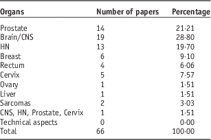
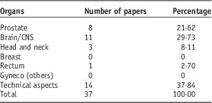
The literature search and filtering resulted in 66 articles related to ‘Outlining’, 37 to ‘Planning’ and 20 to ‘IGRT’. As expected, most papers were related to ‘Outlining’ (Table 1) where most papers focussed on the brain and central nervous system (CNS) (28·8%), prostate (21·21%) or head and neck (HN) tumours (19·7%). A similar picture was seen in the ‘Planning’ branch (Table 2) with brain/CNS tumours comprising 29·7% of the papers, followed by prostate (21·6%) and HN tumours (8·1%). The ‘Planning’ and ‘IGRT’ groups included ‘Technical aspects’ articles, whereas in ‘Outlining’ there were none. All the ‘IGRT’ papers were related to technical aspects and not specific tumour sites.
Papers related to magnetic resonance imaging and ‘Outlining’

Abbreviations: HN, head and neck; CNS, central nervous system.
Papers related to magnetic resonance imaging and ‘Planning’

Abbreviations: CNS, central nervous system.
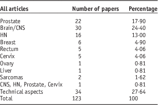
It is clear from the results that some disease sites remain poorly studied in relation to MRI and RT. Gynaecological tumours were the focus for ~18% of the ‘Outlining’ papers (Table 1), comprising 9·1% breast, 7·5% cervix and 1·5% ovary tumours. Otherwise in the ‘Planning’ branch there was just one paper outside the three main MRI regions of brain, prostate and HN. An overview of all articles obtained in this search can be seen in Table 3.
All papers including magnetic resonance imaging and image-guided radiotherapy

Abbreviations: HN, head and neck; CNS, central nervous system.
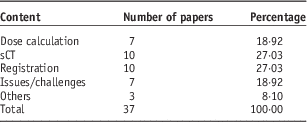
More than half of the ‘Planning’ papers concerned sCT (techniques and its uses) and registration (54%), whereas dose calculation comprised 38% (as seen in Table 4).
Technical aspects of ‘Magnetic Resonance Imaging Planning’ papers

Abbreviations: sCT, synthetic computerised tomography
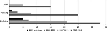
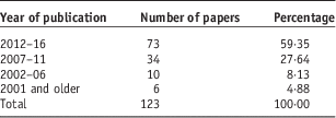
Table 5 shows the publication chronology of the retrieved papers; it can be seen that the majority of publications occurred within the last decade. Approximately 90% of papers were published after 2007, and 60·1% of papers from 2012 onwards (Table 5). It is also interesting to note the growth in research across the different themes (Figure 1). MRIGRT is clearly the most rapidly growing development, along with ‘Planning’, which has accounted for 70% of the publications since 2012. The latter explosion in publication rate is associated with ongoing efforts to overcome the lack of electron density information in MRI, including use of pseudo-CT scans.
Year of publication by theme. IGRT, image-guided radiotherapy.

All articles year of publication percentage

There were three articles that did not fit in any category of ‘MRI Planning’ in technical aspects (Table 4). One concerned the use of MRI in RT simulation, another addressed the use of functional MRI in intensity modulated RT treatment planning, and the last one concerned the use of fluid-attenuated inversion recovery (FLAIR) imaging in planning.
DISCUSSION
Limitations
This paper aimed to evaluate the current MRI skill base in RT as evidenced from a review of the literature. The greatest limitation of the study was that the findings are solely derived from published research, and thus may not correlate directly to the every-day clinical uses of MRI in RT. Topics that have been published are related to innovative practice or identification of issues, with an emphasis on reporting findings that could be implemented into clinical practice. This does not necessarily demonstrate that the findings have been implemented. A more valuable method to address the aim would be an international audit of MRI practice in RT, but the large scope of this severely limits the feasibility.
Site-specific MRI use
RT is used in many tumour sites as adjuvant therapy, and sometimes as primary therapy. A single-day census of RT treatment in Australia showed that the most common sites are breast, prostate, HN, skin, lung, brain and rectum.Reference Bridge, Dempsey and Giles 8 In addition, the same study identified that the most common indications and usage of MRI fusion were prostate, brain, HN and rectum.Reference Bridge, Dempsey and Giles 8 This supports the evidence within this paper with research papers related to prostate and intracranial tumours comprising over half of the sources. These sites clearly dominated the research papers with 24·4% of papers related to brain, 17·9% to prostate and 13% to HN tumours. The prevalence of these sites is explained by the superior soft-tissue visualisation offered by CT–MR fusion. This significantly enhances localisation of intracranial,Reference Djan, Petrović, Erak, Nikolić and Lučić 4 , Reference Tsien, Cao and Chenevert 9 HNReference Dirix, Haustermans and Vandecaveye 2 and prostate tumours.Reference Villeirs and De Meerleer 10
As with many other aspects of RT practice, research into prostate RT dominates this topic. MRI offers excellent soft tissue contrast on T2-weighted images and allows direct multi-planar image acquisition of prostate tumours without loss of spatial resolution.Reference Khoo, Padhani, Tanner, Finnigan, Leach and Dearnaley 11 Consequently, MRI outperforms CT in demonstration of internal prostatic anatomy, margins and the extent of tumours.Reference Villeirs and De Meerleer 10 Use of diffusion-weighted imaging and dynamic contrast-enhanced images in RT delineation,Reference Bridge, Dempsey and Giles 8 and multi-parametric MRIReference Steenbergen, Haustermans and Lerut 12 are key areas of ongoing research aiming to maximise this potential. Ongoing work into alternative MRI sequencesReference Lips, Heide and Haustermans 13 also suggests the need for a rapidly evolving and adaptable MRI-based skill mix within the workforce.
The need for precise determination of target volumes in RT planning arises from the consequences of a geographical miss of a tumour. Intracranial lesions are particularly well delineated on MRIReference Djan, Petrović, Erak, Nikolić and Lučić 4 due to the increased contrast between white and grey matter.Reference Dirix, Haustermans and Vandecaveye 2 However, data obtained by CT is still currently required for treatment planning as it provides the electron density for dose calculation.Reference Nyholm and Jonsson 14 Another exciting research theme seen in many of the review papers related to overcoming this problem through use of pseudo-CTReference Dowling, Lambert and Parker 15 or synthetic CT. Efforts to move towards an MRI-based planning process will vastly increase the range of tumour sites that could benefit from MRI. MRI is already considered the gold standard for rectal imagingReference Group 16 , Reference Gwynne, Mukherjee and Webster 17 as it addresses many of the key CT limitations, such as definition of depth of invasion through the rectal wall into local structures.Reference Gwynne, Mukherjee and Webster 17 , Reference Khoo and Joon 18 The superior soft-tissue contrast provided by MRI also plays a vital role in the RT planning of HN tumours.Reference Chung, Ting, Lui, Hsu and Wang 19 The research evidence certainly demonstrates the value of MRI for these specific sites in relation to outlining and planning. Some of the new developments, however, point to future use of MRI across a wider range of tumour sites. What is not known yet is which tumour sites will gain the most value from these developments and the implementation of MRIGRT.
Training implications: MRI image interpretation
This study has provided an overview of current MRI expertise in RT as evidenced by research studies. The areas of research evidenced by the study suggest that skills in MRI interpretation among the RT professional community primarily relate to brain, HN and prostate anatomy. As discussed previously, the imminent adoption of MRIGRT represents a significant departure from traditional MRI practice in RT. Current use is mainly restricted to sites where MRI offers a considerable imaging advantage over CT, where technical adoption is straightforward. With MRIGRT, usage will not be dictated solely by image quality, but more by the gains arising from real-time image guidance. Introduction of new disease sites is likely to be linked to increased technical proficiency in organ motion tracking and advanced functional MRI sequences; particularly in support of adaptive planning techniques. It is likely, therefore, that the indications for MRIGRT will be radically different from current practice, which will clearly require an extension of the current MRI interpretation skill base. The emerging MRI-cogent workforce will not only require training in use of MRI equipment and techniques, but also a sound understanding of MRI image interpretation across a wider range of anatomical regions than is currently required.
Training implications: relational anatomy
There is another potential aspect of practice that more widespread adoption of MRIGRT may influence. Historically since the adoption of CT-based RT planning, there has been a reliance on axial, or transverse, imaging. The advent of cone-beam CT, which also required a large-scale upskilling of the workforce, still relied predominantly on interpretation of transverse images with limited use of orthogonal planes. Although volumetric acquisition and image reconstruction technology is capable of generating alternative imaging planes, this has largely been restricted to orthogonal images. Reliance on transverse images has, of course, arisen from the physical configuration of the CT scanner and linear accelerator. With MRI, there is no such restriction with an unlimited range of imaging planes available for use. In diagnostic imaging, it is common practice within some regions to select a non-axial, or even non-orthogonal, plane to best highlight the required anatomy or pathology. Findings from a recent 3D outlining evaluationReference Bridge, Fielding, Pullar and Rowntree 20 suggested that RT clinicians were less comfortable with sagittal and coronal images than with transverse. The extent to which new imaging planes will be adopted in MRIGRT is still uncertain, but it seems likely that the RT workforce will require training in non-transverse image interpretation. This is a significant paradigm shift that will require a more rigorous understanding of relational anatomy than a purely axial approach. Pre-registration courses, as well as continuing professional development (CPD) provision, will need to address this in the future if the workforce is to be adequately prepared for the MRI revolution.
CONCLUSION
It is clear that recent research related to MRI in RT is focussed on disease sites in which MRI offers an advantage over CT. Brain, prostate and HN tumours show important gains from the superior soft-tissue contrast of MRI. RT professional MRI expertise in other tumour sites is not evidenced strongly by recent research publications. The advent of MRIGRT and MR-based planning may extend the indications for MRI use to a wide range of tumour sites, and it seems likely that the therapy radiography profession is currently under-prepared for this skill requirement. Future training in MRI image interpretation should not only build on the existing expertise, but also cover a wider range of tumour sites and focus on non-axial planar interpretation. A national audit of current MRI expertise among clinical therapeutic radiographers would build on this review and further help to identify potential training needs.
Acknowledgements
The authors wish to acknowledge the financial and logistical support of Conselho Nacional de Desenvolvimento Científico e Tecnológico (the Brazilian National Counsel of Technological and Scientific Development) for facilitating this collaborative project.
Financial support
A. M. M. studied at the University of Liverpool under the Ciencias sem Fronteiras programme, a Brazilian Government-sponsored student mobility scheme.