Introduction
Matrix metalloproteinases (MMPs) have been implicated in the pathogenesis of the blood–brain barrier breakdown (Rosenberg et al., Reference Rosenberg, Kornfeld, Estrada, Kelley, Liotta and Stetler-Stevenson1992; Leppert et al., Reference Leppert, Leib, Grygar, Miller, Schaad and Holländer2000). Increased levels of MMP-9 are detectable in cerebrospinal fluid samples from mice with eosinophilic meningitis caused by Angiostrongylus cantonensis in BALB/c (Lai et al., Reference Lai, Twu, Jiang, Hsu, Chen, Chiaing, Wang, Tseng, Shyu and Lee2004) and ICR (Lee et al., Reference Lee, Chou, Chen and Lai2004) mice. Janus kinase (JAK)-signal transducer and activator of transcription (STAT) signalling, originally identified as the signalling pathway for interferons, mediates the immune responses of various cytokines, as well as the actions of many growth factors and hormones; and thus participates in inflammation (Hackett et al., Reference Hackett, Wang, Sweitzer, Feldman, Wood and Larner1997; Rane & Reddy, Reference Rane and Reddy2000; Dell'Albani et al., Reference Dell'Albani, Santangelo, Torrisi, Nicoletti, Vellis and Giuffrida Stella2001). The A. cantonensis-induced parasitic meningitis might lead to the induction of JAK-STAT1 signals (Lan et al., Reference Lan, Wang, Lai, Chen, Lee, Hsu and Lee2004b). In addition, curcumin (diferuloylmethane), a bioactive constituent from Curcuma longa possesses remarkable anti-inflammatory, anti-oxidant and anti-carcinogenic properties (Toda et al., Reference Toda, Miyase, Arichi, Tanizawa and Takino1985; Ruby et al., Reference Ruby, Kuttan, Babu, Rajasekharan and Kuttan1995; Surh, Reference Surh2002). Furthermore, the inhibitory action of curcumin on JAK-STAT signalling can contribute to its anti-inflammatory activity in the brain (Kim et al., Reference Kim, Park, Joe and Jou2003).
The murine model of A. cantonensis infection shows close similarities in pathological manifestations to that of the human disease, and curcumin has remarkable anti-inflammatory effects on it. Thus, the present study assays the therapeutic effect in A. cantonensis-induced eosinophilic meningitis following curcumin treatment.
Materials and methods
Experimental animals
Five-week-old male mice (weighting 20 to 25 g), BALB/c strain, were purchased from the National Laboratory Animal Center, Taipei, Taiwan. They were maintained on a 12 h light/dark cycle photoperiod, provided with Purina Laboratory Chow and water ad libitum, and kept in our laboratory for more than one week before the experimental infections.
Larval preparation
Infective larvae (L3) of A. cantonensis were obtained from naturally infected giant African snails, Achatina fulica, collected from fields in Pingtung County, southern Taiwan (Lan et al., Reference Lan, Wang, Hsu, Chen, Lai and Lee2004a). Larvae within tissues were recovered using the method of Parsons & Grieve (Reference Parsons and Grieve1990) with slight modifications. Briefly, the shells were crushed, tissues were homogenized and digested in a pepsin-HCl solution (pH 1–2, 500 I.U. pepsin g− 1 tissue), and incubated with agitation at 37°C in a waterbath for 2 h. Host cellular debris was removed from the digestion by centrifugation at 1400 g for 10 min. Larvae in the sediment were observed under the microscope. Morphological criteria for the identification of L3 of A. cantonensis include measurements of 425–524 μm in length and 23–34 μm in width. The posterior end of the tail always terminates in a fine point (Ash, Reference Ash1970). To confirm that the larvae were those of A. cantonensis, 60 L3 were fed to five rats and the brains of two rats were examined 2 to 3 weeks later for evidence of infection. The remaining rats were killed 5 to 6 weeks later and found to harbour adult worms in the pulmonary arteries. The morphology of adult worms was consistent with that described for A. cantonensis. The males measured 14–15 mm in length, the tail with copulatory bursa and long spicules; females 24–26 mm in length, with a characteristic barber-pole appearance (Lindo et al., Reference Lindo, Waugh, Hall, Cunningham-Myrie, Ashley, Eberhard, Sullivan, Bishop, Robinson, Holtz and Robinson2002).
Treatment of mice
A total of 20 mice were randomly divided into four groups, and prohibited food and water for 12 h before infection. Uninfected control mice were inoculated with distilled water by oral inoculation and infected-untreated controls were infected with 60 L3 by oral inoculation but were not treated. The two curcumin (Sigma, St Louis, Missouri, USA) treated groups of mice were treated on day 5 post-inoculation (PI) for 14 consecutive days with 0.5% and 2.0% curcumin, respectively. All groups were sacrificed on day 22 PI and the curative effects were evaluated respectively in these groups.
Worm recovery
Experimental and control mice were killed by cervical dislocation on day 22 PI. Each brain was dissected into small pieces and homogenized separately in 15 ml of 0.25% sodium citrate in phosphate buffered saline (PBS) followed by centrifugation. Larval counts were measured under 25 × magnification of a dissecting microscope. The mean number of larvae was compared with the positive control group to assess drug efficacy.
Eosinophil counts in CSF and blood
Mice were sacrificed and their brains were removed into a 35 mm dish. The cranial cavity and cerebral ventricles (lateral, third and fourth ventricles) were rinsed with 1 ml PBS. The washing solution was collected into a centrifuge to spin at 400 g for 10 min. The resultant sediments were then resuspended with 30 μl PBS from each mouse for counting the percentage of eosinophils. Differential cell counts were assessed with Wright-Giemsa staining (Sigma, Taufkirchen, Germany) in 3 μl per smear. The number of eosinophils were determined in 200 leukocytes per smear. Additionally, blood eosinophils were counted as described for the cerebrospinal fluid methods.
Gelatin zymography
The cerebrospinal fluid was centrifuged at 12,000 g for 10 min to remove debris. Gelatinases were analysed using the method of Lai et al. (Reference Lai, Twu, Jiang, Hsu, Chen, Chiaing, Wang, Tseng, Shyu and Lee2004). Briefly, the protein contents of supernatants were loaded on 7.5% (mass/volume) sodium dodecyl sulphate–polyacrylamide gels that had been co-polymerized with 0.1% gelatin (Sigma, St Louis, Missouri, USA). Stacking gels were 4% (mass/volume) polyacrylamide and did not contain gelatin substrate. Electrophoresis was performed in running buffer (25 mM Tris, 250 mm glycine, 1% SDS) at room temperature at 120 V for 1 h. The gel was stained with 0.25% Coomassie Brilliant Blue R-250 (Sigma, St Louis, Missouri, USA) for 1 h, and destained in 15% methanol/7.5% acetic acid. Gelatinase activity was detected as unstained bands on a blue background.
Statistical analysis
Results in the different groups of mice were compared using the non-parametric Kruskal-Wallis test followed by post-testing using Dunn's multiple comparison of means. All results were presented as mean ± standard deviation (S.D.). P values of < 0.05 were considered statistically significant.
Results
Pathological observations of the brain surface
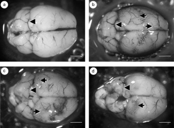
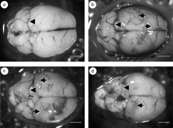
The brain of uninfected mice showed normal properties of blood vessels, whereas enlarged blood vessels and young adult worms were seen in infected-untreated mice. After treatment with 0.5% and 2% curcumin, enlarged blood vessels and young adult worms were still observed (fig. 1).
(a) Brain of uninfected mice, showing normal blood vessel (arrowhead); (b) brain surface of infected-untreated mice, showing enlarged blood vessel (arrowhead) and young adult worms of Angiostrongylus cantonensis (arrowed); (c) brain surface showing enlarged blood vessel (arrowhead) and young adult worms (arrowed) after treatment with 0.5% curcumin; (d) the blood vessel (arrowhead) could not reduce hyperaemia and young adult worms (arrowed) remain present after treatment with 2% curcumin. Scale bars = 2.5 mm.
Worm recovery
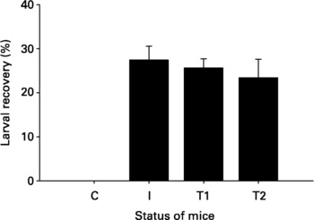
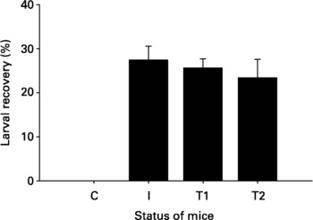
The efficacy of curcumin was assessed by recovery and counting of viable larvae from brain tissues. Recovered larvae in curcumin treated mice were not significantly decreased (P>0.05) compared with infected mice. There were no differences between 0.5% and 2% curcumin treatments (fig. 2).
Recovery of larvae of Angiostrongylus cantonensis in infected (I) and curcumin treated mice (T1, 0.5% curcumin; T2, 2% curcumin); larvae in curcumin treated mice were not significantly decreased (P>0.05) compared with infected mice. C, controls.
Influence of treatment on eosinophil counts
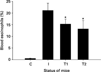
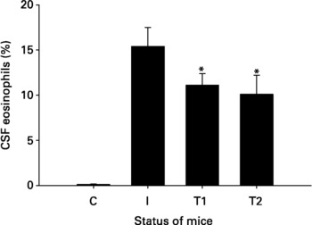
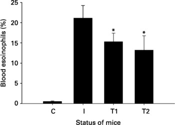
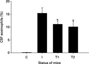
Eosinophilia of blood (fig. 3) and the cerebrospinal fluid (fig. 4) were significantly increased (P < 0.05) in mice infected with A. cantonensis compared with uninfected mice. Eosinophil percentages were mildly reduced by curcumin treatment and there were no significant differences between 0.5% and 2% treatments.
Changes in the percentage of blood eosinophils in mice infected with Angiostrongylus cantonensis (I) and in curcumin treated mice (T1, 0.5% curcumin; T2, 2% curcumin); eosinophils were significantly increased (P < 0.05) in mice infected with A. cantonensis compared with uninfected controls (C); eosinophils were mildly reduced (*P < 0.05) by curcumin.
Changes in the percentage of CSF eosinophils in mice infected with Angiostrongylus cantonensis (I) and in curcumin treated mice (T1, 0.5% curcumin; T2, 2% curcumin); eosinophils were significantly increased (P < 0.05) in mice infected with A. cantonensis compared with uninfected controls (C); eosinophils were mildly reduced (*P < 0.05) by curcumin.
Influence of treatment on MMP-9 activity
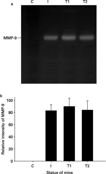
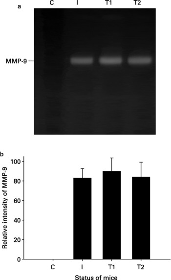
Gelatin zymography allows the activity of enzymatically active proteins such as MMP-9 to be measured. MMP-9 migrated electrophoretically as a 94 kDa species and the enzyme activity was significantly increased (P < 0.05) in the cerebrospinal fluid of BALB/c mice infected with A. cantonensis, as compared with controls. This enzyme activity remained unchanged after curcumin treatment (fig. 5).
(a) The influence of curcumin treatment (T1, 0.5% curcumin; T2, 2% curcumin) on MMP-9 activity; the activity was significantly increased in the CSF of BALB/c mice infected with Angiostrongylus cantonensis (I), as compared with controls (C). MMP-9 activity remained unchanged after curcumin treatment; (b) quantitative analysis of MMP-9 activity was performed using a computer-assisted imaging densitometer system and data shown are representative of five independent experiments with similar results.
Discussion
Curcumin has an extensive history as a food preservative and medicinal herb in India (Ammon & Wahl, Reference Ammon and Wahl1991). Studies have consistently shown that curcumin is relatively non-toxic and has few side effects at doses greater than the low dose tested in mice (Kelloff et al., Reference Kelloff, Crowell, Steele, Lubet, Malone, Boone, Kopelovich, Hawk, Lieberman, Lawrence, Ali, Viner and Sigman2000). Toxicity studies performed at 2000 mg kg− 1, which is 83-fold greater than a low-dose (24 mg kg− 1) curcumin treatment, revealed no mortalities in any group of mice tested (Srimal & Dhawan, Reference Srimal and Dhawan1973). Although side effects have been limited in chronic animal and short-term clinical studies, sustained clinical trials are needed to establish the safety of chronic curcumin at antioxidant and anti-inflammatory doses.
MMPs can promote the breakdown of the blood–brain barrier by directly degrading components of the vascular basement membrane (Romanic & Madri, Reference Romanic and Madri1994). Thus, degrading MMPs are implicated in the disruption of endothelial tight junctions and the blood–brain barrier during leukocyte diapedesis (Paul et al., Reference Paul, Lorenzl, Koedel, Sporer, Vogel, Frosch and Pfister1998; Leppert et al., Reference Leppert, Leib, Grygar, Miller, Schaad and Holländer2000). The anti-inflammatory property of curcumin suggests that it may improve meningitis through regulating MMP-9 activity. To test this possibility, the effects of curcumin in A. cantonensis-induced eosinophilic meningitis were examined by biochemical and histological methods. It was found that the cerebrospinal fluid MMP-9 activity remained unchanged and eosinophils were mildly decreased when mice were treated with curcumin. The fact that the enzyme cannot be inhibited in the cerebrospinal fluid probably indicates that cellular invasion of the central nervous system across the blood–brain barrier remains unchanged.
As a broad-spectrum anthelmintic, albendazole is an effective compound for the treatment of angiostrongyliasis through its larvicidal activity and facilitation of an improved inflammatory response via the reduction of MMP-9 activity (Lan et al., Reference Lan, Wang, Hsu, Chen, Lai and Lee2004a). In the present study, the inhibition of MMP-9 activity as measured by zymography remained unchanged by curcumin treatment alone. This may be because curcumin treatment alone could not kill the larvae, and live worms persisted to stimulate proteolytic enzymes. The important finding is that the anti-inflammatory curcumin must be combined with an anthelminthic drug for effective treatment, especially in parasitic meningitis.
In summary, the present results demonstrate the upregulation of MMP-9 in parasite-induced meningitis. It was found that curcumin treatment alone did not interfere with MMP-9 expression or action, and so may be not useful for larvicidal effect. The possible reasons include low level of curcumin across the blood–brain barrier and the larvae that survive stimulate MMP-9 production which in turn promotes blood–brain barrier damage, with leukocytes then crossing the blood–brain barrier to cause meningitis. Further studies will be required to test these possibilities.