Background
Systemic lupus erythematosus, systemic scleroderma, and mixed connective tissue disease are rheumatologic disorders characterised by systemic, multi-organ involvement with cardiac manifestations that can significantly contribute to morbidity and mortality.Reference Morello, Gholson and Huang1,Reference Quartier, Bonnet and Fournet2 Early and accurate detection of cardiac involvement is important to guide management and potentially improve outcomes in adults.Reference Mavrogeni, Pepe and Nijveldt3 While echocardiography remains the most accessible and widely utilised modality for assessing cardiac function in children, it has limitations, particularly in the qualitative assessment of the right ventricular function and geometric assumptions in calculating left ventricular ejection fraction, which can lead to variability.Reference Quartier, Bonnet and Fournet2,Reference Dorfman, Geva and Samyn4,Reference Lopez, Saurers and Barker5
Cardiac MRI offers advances in tissue characterisation and precise volumetric assessment, leading to unique advantages over echocardiography in detecting subclinical myocardial involvement, including oedema and fibrosis.Reference Quartier, Bonnet and Fournet2,Reference Mavrogeni, Smerla and Grigoriadou6,Reference Mavrogeni, Servos and Smerla7 In adults with systemic lupus and systemic scleroderma, cardiac MRI has proven valuable for identifying myocardial fibrosis and oedema, informing prognosis and therapy.Reference Habib, Romero and Huck8–Reference Panopoulos, Mavrogeni, Vlachopoulos and Sfikakis10 However, its clinical utility in paediatric patients with rheumatologic disorders remain largely unexplored, despite the potential for early detection of myocardial injury, which may influence treatment decision during the acute phases of disease.Reference Dorfman, Geva and Samyn4 This gap highlights the need for studies evaluating the role of cardiac MRI in the surveillance of children with systemic rheumatologic disorders.
Materials and methods
We performed a retrospective review of paediatric patients diagnosed with systemic lupus erythematosus, systemic scleroderma, and mixed connective tissue disease at Texas Children’s Hospital between 2014 and 2022. Among all patients with these diagnoses, we identified those who had both echocardiography and cardiac MRI within a three-month interval to minimise the potential impact of disease progression on imaging findings.
We collected baseline demographic characteristics, including age at diagnosis, sex, and disease at the time of cardiac MRI. We assessed right and left ventricular function by both modalities. For cardiac MRI studies, we documented the presence of myocardial oedema (based on increased signal intensity on standard Short Tau Inversion Recovery sequence and T2 mapping) and scarring or fibrosis (based on late gadolinium enhancement and extracellular volume—calculated based on T1 mapping and the patient’s haematocrit).
Finally, we reviewed medical records to identify any changes in clinical management that followed the cardiac MRI findings. This study was approved by the Institutional Review Board at the Baylor College of Medicine (protocol number: H-50497).
Results
Among 426 patients diagnosed with these three rheumatologic disorders (2014–2022), 309 patients (72.5%) received at least one echocardiogram, and eleven (2.5%) had cardiac MRI. The median age (at cardiac MRI) was 16 years old (IQR: 13–18), median weight 65.8 kilograms (IQR: 49.4–75.7), and median disease duration 18 months (IQR: 0–60). The median interval between echocardiography and MRI was 8 days (IQR: 4–15).
Most patients were female (10/11) and Hispanic (9/11); two were African American. Diagnoses included systemic lupus erythematosus (n = 4), systemic scleroderma (n = 4), mixed connective tissue disease (n = 2), and one patient with overlapping lupus and scleroderma features. Among five patients with systemic scleroderma, four had gastrointestinal manifestations, and all had pulmonary disease (three with pulmonary hypertension and two with interstitial lung disease).
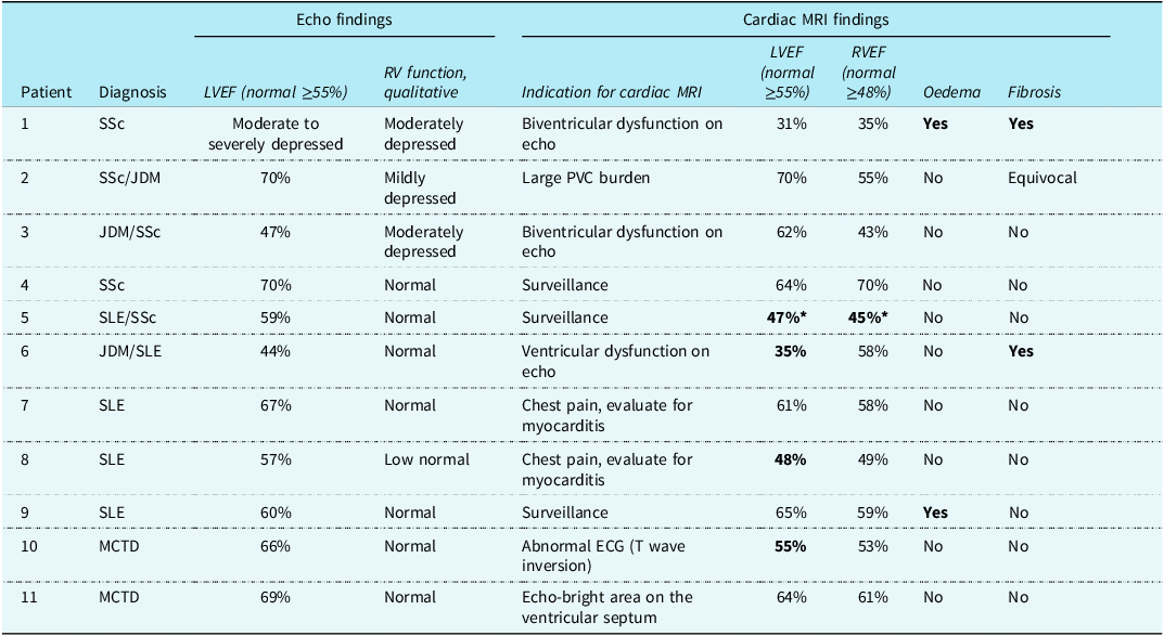
Echocardiography identified ventricular dysfunction in five patients (45%), all confirmed by cardiac MRI. Excluding one patient with transient myocardial dysfunction secondary to general anaesthesia (Table 1), cardiac MRI showed more severe ventricular dysfunction in three patients (27%). Cardiac MRI uniquely detected myocardial oedema in two patients (18%) and fibrosis in two patients (18%). One patient had both oedema and fibrosis. Notably, immunotherapy was subsequently escalated in all three patients with oedema and/or fibrosis identified on cardiac MRI (Figure 1).
Cardiac MRI findings in a 15-year-old female (patient #1) recently diagnosed with SSc: ( a ) arrowheads pointing to areas of myocardial scar (fibrosis; late gadolinium enhancement), ( b ) extracellular volume map of 16 myocardial segments demonstrating diffuse fibrosis of all segments (>32%), ( c ) T2 STIR sequence and ( d ) T2 mapping revealed borderline myocardial oedema, which can be considered a sign of active inflammation/myocarditis.

Echocardiographic findings and cardiac MRI findings in individual patients

JDM = juvenile dermatomyositis; LVEF = left ventricular ejection fraction; MCTD = mixed connective tissue disease, RVEF = right ventricular ejection fraction; SLE = systemic lupus erythematosus; SSc = systemic scleroderma.
Bold texts indicated additional findings or more significant abnormality by CMR compared to echocardiography.
* This patient underwent cardiac MRI under general anaesthesia, which showed decreased ventricular function. Non-sedated echocardiograms before and after cardiac MRI demonstrated normal function; therefore, ventricular dysfunction was deemed transient, secondary to general anaesthesia.
Discussion
This retrospective observational study provided preliminary insights into the role of cardiac MRI compared to echocardiography in paediatric patients with rheumatologic disorders. Cardiac MRI consistently confirmed ventricular dysfunction identified by echocardiography and revealed more severe impairment in 27% of patients. While this discrepancy may partly reflect differences in timing within individual disease courses, these findings highlight the potential of cardiac MRI to offer more precise ventricular function assessment. Additionally, cardiac MRI uniquely identified myocardial oedema and fibrosis in 27% (n = 3) of the paediatric patients, findings not detected by echocardiography.Reference Dorfman, Geva and Samyn4 While consensus guidelines support the use of cardiac MRI in symptomatic adults with newly diagnosed systemic lupus or scleroderma to evaluate cardiac involvement, monitor treatment response, and in select asymptomatic patients, our data showed that cardiac MRI was performed mostly in symptomatic children. Our study findings were consistent with prior reports in adults with rheumatologic conditions, demonstrating the utility of cardiac MRI for detecting myocardial inflammation and fibrosis.Reference Dorfman, Geva and Samyn4,Reference Mavrogeni, Smerla and Grigoriadou6 Although our data remained very limited, such information seemed to offer clinicians a better understanding of cardiac involvement and potentially informed decisions to alter immunosuppressive therapy in these few patients. Despite the potential benefit of cardiac MRI, our study results highlight that it remains underutilised in this patient population.
Our findings also reinforced earlier observations of severe cardiac involvement in paediatric systemic sclerosis, with cardiac MRI findings correlating with adverse outcomes.Reference Quartier, Bonnet and Fournet2 The detection of subclinical myocardial injury across multiple rheumatologic diagnoses highlighted the potential broader role of cardiac MRI in paediatric rheumatologic disorders. This study has limitations inherent to its retrospective design, including selection bias and the small sample size, which reflects the rarity of paediatric patients with systemic rheumatologic disorders undergoing both cardiac MRI and echocardiography within a defined period. Although we aimed to minimise variability by restricting inclusion to patients with imaging studies within three months, interim clinical changes could have contributed to some of the observed discrepancies between modalities.
In conclusion, our preliminary findings suggested that cardiac MRI provided additional diagnostic information beyond echocardiography in paediatric patients with rheumatologic disorders, particularly through its ability to detect subclinical myocardial involvement. While these observations supported the integration of cardiac MRI into the cardiac surveillance strategies for select paediatric patients, larger prospective studies are needed to better define the role of cardiac MRI and its therapeutic implication in the management of paediatric patients with rheumatologic disorders.
Acknowledgements
We thank the patients, families, and the cardiac imaging teams and rheumatology team at Texas Children’s Hospital for their support, and the Baylor College of Medicine IRB for facilitating this study. We thank Dr Marietta De Guzman for her assistance in compiling the list of all patients with rheumatologic disorders.
Financial support
None.
Competing interests
None.
Ethical standard
This study was approved by the Institutional Review Board at the Baylor College of Medicine (Protocol Number: H-50497).