I. INTRODUCTION
Palladium (Pd) complexes have been extensively studied concerning its antitumoral, antifungal, and antibacterial properties (Garoufis et al., Reference Garoufis, Hadjikakou and Hadjiliadis2009). In this context, our research group has contributed to the development of some biologically active Pd(II) complexes, mainly with amino acids and its derivatives. Carvalho et al. reported the synthesis of a Pd (II) complex with the amino acid L-tryptophan, [Pd(trp)2], which presents antibacterial activities against Gram-negative (Escherichia coli and Pseudomonas aeruginosa) and Gram-positive (Staphylococcus aureus) bacterial strains (Carvalho et al., Reference Carvalho, Souza, Paiva, Bergamini, Gomes, Gozzo, Lustri, Formiga, Rigatto and Corbi2012). In addition, a novel water-soluble Pd (II) complex with L-alliin (S-allyl-L-cysteine sulfoxide), with the coordination formula [Pd(alliin)2], was prepared and evaluated about its antitumor activity over HeLa cells. The compound [Pd(alliin)2] was shown to be active over HeLa tumorigenic cells, with an IC50 close to cisplatin, which is used in cancer treatment. Such compound has also shown to have antimicrobial activities against Gram-positive and Gram-negative microorganisms in the range 125–500 µg mL−1 (Abbehausen et al., Reference Abbehausen, Sucena, Lancellotti, Heinrich, Abrão, Costa-Neto, Formiga and Corbi2013).
Bergamini et al. also reported the synthesis and characterization of a dimeric Pd (II) complex with 2-mercaptothiazoline (mtz), with the formula [Pd2(mtz)4], where the ligand is coordinated to the metal by the sulfur and nitrogen atoms of the heterocyclic ring. However, this complex did not present antibacterial activities over the considered strains (Bergamini et al., Reference Bergamini, Abbehausen, Magalhães, Lustri, Gomes, Gozzo and Corbi2011).
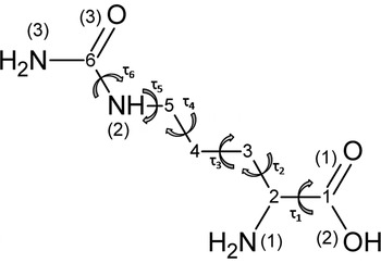
L-Citrulline (2-amino-5-ureidopentanoic acid, C6H13N3O3, Cit) is an endogenous mammal's amino acid intermediate in the urea cycle (Kaore et al., Reference Kaore, Amane and Kaore2013). Although endogenous, L-citrulline is also found in foods such as watermelons, cucumbers, pumpkins, muskmelons, and bitter melons, and it is known to act on body process of ammonia detoxification through its conversion to urea. This amino acid may also act as a potent hydroxyl scavenger (Kaore et al., Reference Kaore, Amane and Kaore2013). Sketch of L-citrulline is shown in Chart 1.
In this paper the synthesis, structural characterization, and ancillary analytical and spectroscopic data of a novel Pd(II) complex with the amino acid L-citrulline, [Pd(Cit)2], are reported.
II. EXPERIMENTAL
A. Materials and methods
L-Citrulline (98%), potassium hydroxide, and potassium tetrachloridopalladate(II) (98%) were purchased from Sigma-Aldrich Laboratories. Elemental analyses for carbon, hydrogen, and nitrogen were performed using a Perkin Elmer 2400 CHN Analyzer. IR spectra from 4000–400 cm−1 of citrulline and the Pd (II) complex were measured using a FT-IR spectrophotometer ABB Bomen MB Series; samples were prepared as KBr pellets.
1. Synthesis of [Pd(Cit)2]
The Pd (II) complex with citrulline was synthesized by the reaction of 5.0 ml of an aqueous solution of potassium tetrachloridopalladate(II), K2[PdCl4] (5.0 × 10⁻⁴ mol), with 10.0 ml of a freshly prepared aqueous solution of the alkaline salt of L-citrulline containing 1.0 × 10⁻³ mol of the ligand. The K2[PdCl4] aqueous solution was added dropwise to the alkaline solution of citrulline under magnetic stirring and at room temperature. After 2 h of constant stirring, the yellow solid obtained was vacuum-filtered, washed with cold water, and dried in a desiccator over P4O10. The [Pd(Cit)2] complex is insoluble in water and in common organic solvents such as methanol, ethanol, acetonitrile, dimethylsulfoxide, chloroform, and dichloromethane. Elemental analysis led to a 1:2 metal:ligand composition for the complex. Anal. Calcd. For PdC12H24N6O6 (%): C, 31.7; H, 5.32; N, 18.5. Found (%): C, 30.7; H, 5.56; N, 17.6; IR bands: ν asym (–(CO)NH2 ) 3446 cm−1, ν sym (–(CO)NH2 ) 3344 cm−1, ν asym (–NH2) 3217 cm−1, ν sym (–NH2) 3130 cm−1, ν asym (–COO−) 1639 cm−1, ν sym (–COO−) 1364 cm−1. The IR spectrum and additional discussions about the IR data are provided as supplementary material 1.
The alkaline salt of citrulline used in the synthesis of the Pd(II) complex was prepared by the reaction of equimolar quantities of the amino acid and potassium hydroxide under stirring and at room temperature.
B. X-ray powder diffraction data collection and structure determination
To perform the powder diffraction analysis, the polycrystalline [Pd(Cit)2] was gently grounded in an agate mortar and it was deposited in hollow of PMMA sample-holder plate. The diffraction data were collected by overnight scans in the 2θ range of 4°–105° with steps of 0.02° using a Bruker AXS D8 da Vinci diffractometer, equipped with Ni-filtered CuKα radiation (λ = 1.5418 Å), a Lynxeye linear position-sensitive detector and the following optics: primary beam Soller slits (2.94°), fixed divergence slit (0.3°) and receiving slit (8.73 mm). The generator was set at 40 kV and 40 mA. Approximate unit cell parameters were determined using 22 first standard peaks, followed by indexing through the single-value decomposition approach (Coelho, Reference Coelho2003) implemented in TOPAS (TOPAS, 2009), the Goodness-on-Fit value equal 53.13 (Wolff, Reference Wolff1968) was afforded for indexing stage. In the present case, space group P-1 was chosen and the cell parameters were refined using 4–55 2θ range by Pawley method (Pawley, Reference Pawley1981), giving a good R wp = 5.950. Then, the structure solution process was performed by the simulated annealing technique (Coelho, Reference Coelho2000) also implemented in TOPAS. No higher symmetric system was suggested by PLATON (Spek, Reference Spek2009). Since the P-1 space group has Z = 2 and density considerations suggested Z = 1 the Pd(II) ion was placed in fixed position at 0.0, 0.0, and 0.0. In addition, the citrullinate rigid body model, based on single-crystal data (Sridhar et al., Reference Sridhar, Srinivasan, Dalhus and Rajaram2002) was built using Z matrix formalism, with free rotations and translations as well torsion angles as described in Chart 1.
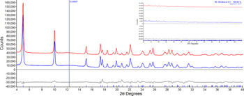
In the simulated annealing step only torsion angles, rotation, and translation parameters concerning the ligand were used. In the refinement stage, carried out by the Rietveld method (Young, Reference Young1981), 38 parameters were refined including 10 parameters for background modeled by a Chebyshev polynomial function and broad peak at 12.3° (2θ) was inserted to describe the presence of a small amorphous portion. The rigid body description introduced at the solution stage was maintained in the final refinement. An isotropic thermal parameter set up at 3.0 (0.2) Å2 was assigned to all atoms.
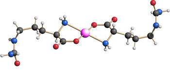
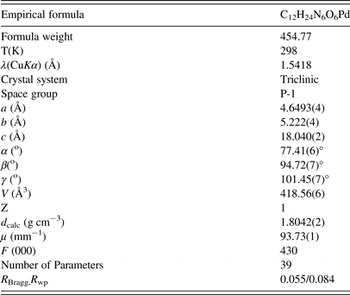
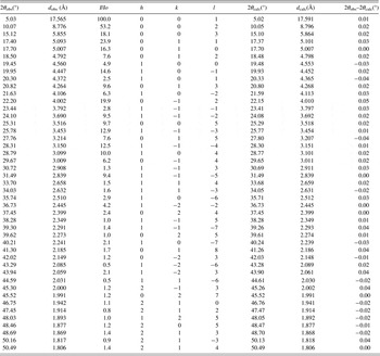
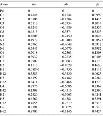
The final Rietveld refinement plot, the sketch of the crystal, and the molecular structure for [Pd(Cit)2] are shown in Fig. 1, Chart 1, and Fig. 2, respectively. Table I contains the relevant crystal data for [Pd(Cit)2] while Tables II and III contain relevant powder diffraction features and final fractional atomic coordinates for [Pd(Cit)2], respectively.
(Color online) Final Rietveld refinement plot in the range 4°–40° 2θ for [Pd(Cit)2] with difference plot as grey line and peak markers at the bottom. The experimental data is present in blue line while calculated data is in red. Horizontal axis is 2θ(°) and vertical axis is counts. A very broad peak at 12.35° was inserted to describe the presence of a small amorphous portion. The inset shows the high-angle region.

(Color online) Crystal structure of [Pd(Cit)2] drawn using SCHAKAL (Keller, Reference Keller1986). Color codes: Pd(II): pink; carbon: grey; hydrogen: white; nitrogen: blue and oxygen: red. The Pd(II) ion is lying on inversion center as pivot for citrullinate ligand.

Sketch of citrulline where numbers replaced carbon atoms and the nitrogen and oxygen atoms were numbered in parenthesis. The τ 1 to τ 6 symbols illustrate the torsion angles defining the conformation of the salt of citrulline present in the [Pd(Cit)2] complex.

Crystallographic data of [Pd(Cit)2].

List of the 50 first observed and calculated peaks for [Pd(Cit)2].

Fractional atomic coordinates for [Pd(Cit)2].

Isotropic thermal parameter was assigned to all atoms as 3.0(0.2) Å2.
III. DISCUSSION
In the [Pd(Cit)2] complex the Pd(II) ions were fixed at special position 0.0, 0.0, and 0.0 (see Table III). Since Pd(II) ions lie on inversion centers they act as pivot for the citrullinate ions where, as expected, the Pd(II) ions were surrounded by two ligands. Each ligand bonds to Pd(II) ions by one oxygen (O2) from the carboxylate group and by the nitrogen from alfa-amino group (N1) forming a square planar geometry around the metal center. The main distances and angles of [Pd(Cit)2] are listed on Table IV. The angle between N–Pd–O of [Pd(Cit)2] is 78.4(6)° and it falls in the range found in a complete Cambridge Structural Database (CSD) search, 82 ± 6° for 134 hits. The distance between O and Pd which is 2.07(1) Å is in good agreement than the average (2.05 ± 0.1 Å) found in the CSD database for Pd(II) complexes with N– and –O donor ligands. However, the Pd–N bond lengths 2.27(3) Å of [Pd(cit)2] is little longer compared with the CSD Pd–N average, 2.04 ± 0.16 Å. The intermolecular hydrogen-bond interactions are present between H atoms of N6 with O3 and H atoms of N1 with O1 and O2 (carboxylate group).
Main bond lengths (Å) and angles (°) of [Pd(Cit)2].

i code: 1 − x, 1 − y, 1 − z.
IV. CONCLUSION
A Pd(II) complex with citrulline was obtained and the molecular formula PdC12H24N6O6 was confirmed by elemental analysis. The IR data support that citrulline salt bonds to Pd(II) ions by oxygen and nitrogen atoms. In the absence of crystals of suitable size and quality amenable to conventional single-crystal characterization, the structure of the new Pd(II) complex was determined by standard laboratory X-ray powder diffraction techniques. The diffraction pattern of [Pd(Cit)2] was successfully indexed and the crystal structure was derived therefrom using state-of-the-art real-space structure solution methods: The [Pd(Cit)2] complex belongs to triclinic system (P-1) with cell parameters a = 4.6493(4) Å, b = 5.222(4) Å, c = 18.040(2) Å, α = 77.41(6)°, β = 94.72(7)°, and γ = 101.45(7)°. Its crystal structure consists in Pd(II) ions surrounded by N and O atoms from 2-citrulline ligands (as an ionic ligand) in a closed square-planar geometry. Once again, it has been shown that powder diffraction methods can supply relevant (otherwise inaccessible) structural information, though of lower quality than that can be obtained from single crystal analyses (Masciocchi and Sironi Reference Masciocchi and Sironi1997; Silva, et al., Reference Silva, Masciocchi and Cuin2014). Further studies about the antibacterial and antitumor activities of the complex are envisaged.
SUPPLEMENTARY MATERIAL
For supplementary material for this article, please visit http://dx.doi.org/10.1017/S0885715615000652
ACKNOWLEDGMENTS
This study was supported by grants from the Brazilian agencies CAPES and CNPq (grant # 442123/2014-0). The authors are thankful to anonymous referees for their valuable suggestions.