Introduction
Red panda (Ailurus fulgens) is an ambipolar animal and its evolutionary history plays a pivotal role in the higher level phylogeny of the ‘bear-like’ arctoides carnivore mammals (Flynn et al., Reference Flynn, Nedbal, Dragoo and Honeycutt2000). Its only known habitat exists in isolated mountain ranges in western China (Sichuan, Yunnan and Tibet) and the Himalaya mountain ranges of Nepal and India (Li et al., Reference Li, Wei, Goossens, Feng, Tamate, Bruford and Funk2005). As a result of environment pollution and the encroachment of humans, the red panda is now an endangered species. To date, the studies on red panda have focused on disease prevention, treatment and genetic classification (Slattery & O'Brien, Reference Slattery and O'Brien1995; Tian et al., Reference Tian, Nie, Wang, Yang and Yang2002). To date, the establishment of the fibroblast cell lines from red panda has not been reported. The establishment of a somatic cell line and its cryopreservation are basic but important steps in investigating the red panda's characteristics and storing valuable resources.
Since 1996, when the cloned sheep Dolly was produced from a differentiated cell line by somatic cell nuclear transfer (SCNT), many other species have been successfully cloned. Recently and increasingly, interspecies SCNT has been used not only in order to elucidate the embryo development mechanism, but also to potentially rescue the species. Unfortunately, these efforts have proved unsuccessful. In 1999, giant panda blastocysts were acquired by interspecies SCNT (Chen et al., Reference Chen, Sun, Liu, Li, Lian, Wang, Han, Song and Sun1999). The SCNT embryos were obtained from argali somatic cells and domestic sheep, but only one pregnancy using argali–sheep embryos was achieved and it terminated on day 59 (White et al., Reference White, Bunch, Mitalipov and Reed1999). Subsequently, reconstructed embryos were obtained from gaur bull somatic cells and domestic cow oocytes. The pregnancies from these embryos lasted until day 200 of gestation (Lanza et al., Reference Lanza, Cibelli, Diaz, Moraes, Farin, Farin, Hammer, West and Damiani2000). Recently, embryos were reconstructed with adult banteng fibroblasts and enucleated domestic bovine oocytes, but two pregnancies from these embryos were lost between days 30 and 90 of gestation (Sansinena et al., Reference Sansinena, Hylan, Hebert, Denniston and Godke2005). An ordinary cow gave birth to a baby bull by introducing a nucleus from the skin fibroblast of an adult male gaur into an enucleated oocyte of a cow and transferring it into the uterus of a surrogate cow after the reconstructed embryo developed to the blastocyst stage in vitro. Unfortunately, the calf succumbed to common dysentery within 48 hours after his birth (Wen et al., Reference Wen, Bi and Chen2004). Cross-species nuclear transfer embryos were produced from the postmortem somatic cells of a mouflon; the recipients were enucleated oocytes from sheep, a closely related domesticated species. Seven embryos were transferred into four surrogate sheep, which resulted in two pregnancies. One surrogate aborted between days 40 and 50 after the embryos were transferred; the other pregnancy went to term (Loi et al., Reference Loi, Ptak, Barboni, Fulka, Cappai and Clinton2001). This event was the first report of a viable offspring by subspecies SCNT. In a strict sense, however, the embryos involved in the reports noted above were not interspecies SCNT due to their heredity kindred. In the present study, we report for the first time that a fibroblast cell line from the red panda was established and cryopreserved and that interspecies embryos were reconstructed with enucleated rabbit oocytes as recipients.
Materials and Methods
Animals
The samples used in the present study were obtained from a red panda raised in the Hefei Wild Animal Park. It was a healthy 5-year-old female weighing 6.5 kg.
Chemicals and reagents
All the chemicals and reagents used were purchased from Sigma, unless otherwise noted.
Experiment 1. Establishment of fibroblast cell line
A biopsy from an ear skin piece was collected and fibroblast cells were cultured (Liu et al., Reference Liu, Zhang, Chen, Zhang, Zhang, Jin, Wang, Zhang, Li, Li, Zhao and Cheng2003). In brief, the skin biopsy was finely chopped into pieces measuring about 1 mm2 and seeded into 25 cm3 cell culture flasks. The pieces were dried in 5% CO2 at 37.5 °C for 4 h and cultured in Dulbecco's modified Eagle's medium/F12 (DMEM/F-12) supplemented with 20% fetal bovine serum (FBS, GIBCO, Life Technologies) in 5% CO2 at 37.5 °C with 100% humidity. In order to measure the growth curve and the logarithmic growth phase of fibroblast cells, the fibroblast cells, at third passage, were cryopreserved, thawed and then inoculated into a 24-well dish (Corning) at a density of 6 × 10⁴ cells/ml and cultured in DMEM/F12 and 20% FBS in the 5% CO2 at 37.5 °C. Over the next 7 days, the cells were digested and counted every day. Three replicates were taken.
Experiment 2. Cell cryopreservation and thawing
Red panda fibroblast cells were cryopreserved and thawed in accordance with our previous study with a minor modification (Liu, Y. et al., Reference Liu, Zhang, Zhang, Jin, Wang and Wu2004a). Briefly, after reaching 90% confluence, the cells were digested with 0.25% (w/v) trypsin for 90 s and resuspended in DMEM. A single cell suspension was prepared by pipetting; a portion of this suspension was to set up a culture in DMEM supplemented with 10% FBS and the remainder was resuspended in DMEM at 5 × 10⁵ cells/ml containing 20% FBS and 8% dimethyl sulphoxide (DMSO) and equilibrated in 4 °C for 30 min. For cryopreservation, the cells were kept at –80 °C for 12 h. Finally, the cells were put into liquid nitrogen for long-term preservation.
When necessary, the cells that were frozen in liquid nitrogen were thawed in a 37 °C water bath with gentle agitation. After three rinses with DPBS, the cells were resuspended in DMEM supplemented with 10% FBS and culture at 37.5 °C, 5% CO2 in humidified air.
Experiment 3. Interspecies embryo reconstruction
The cryopreserved fibroblasts, at passage 4–10, were used as somatic cell donors for embryo reconstruction. Sexually mature rabbits were superovulated by administering equine chorionic gonadotropin (eCG) and human chorionic gonadotropin (hCG). Each rabbit was injected with 120 IU eCG and with 180 IU hCG, 72 h after the eCG injection. Rabbits were euthanized 13–15 h after the hCG injection. Metaphase (MII)-arrested mature oocytes were collected and washed three times in DPBS. Micromanipulation was performed in accordance with our previously established protocol with minor modifications (Liu, Y. et al., 2003, Reference Liu, Zhang, Chen, Zhang, Zhang, Jin, Wang, Zhang, Li, Lin, Zhao and Cheng2004b; Zhang, X.R. et al., Reference Zhang, Liu, Zhang, Chen, Lian, Li, Jin, Zhang, Zhang and Wang2004; Zhang, Z.G. et al., Reference Zhang, Zhang, Liu, Jin, Zhang, Wang and Zhao2005; Zhang, Y.H. et al., Reference Zhang, Pan, Sun, Sun, Wang, Liu, Li, Dai and Li2006). After exposure to 0.5% hyaluronidase for 3–5 min, cumulus cells from oocytes were collected and mechanically removed by gently aspirating the oocytes with a fine pipette. Enucleation of the oocytes occurred within 1 h from collection to micromanipulation. The cumulus-free oocytes were incubated 30 min in M2 medium containing 7.5 μg/ml cytochalasin B (CB, Sigma). The first polar body and a small amount of the underlying cytoplasm were aspirated using an enucleation pipette. A single cell was selected and transferred into the perivitelline space of the enucleated oocytes. Electrofusion and activation were undertaken in accordance with our previously established method (Liu, Y. et al., Reference Liu, Zhang, Chen, Zhang, Zhang, Jin, Wang, Zhang, Li, Lin, Zhao and Cheng2004b). Thirty minutes after the operation, the reconstructed oocytes were electrofused by two direct currency pulses (140 V/mm, 80 μs) at a 1 s interval. The reconstructed oocytes were made by treating the cells with 5 μM ionomycin for 4–5 min and 2 mM 6-dimethylaminopurine (6-DMAP) for 4 h. Activated NT embryos were cultured in an embryo culture medium supplemented with 15% FBS (10 embryos/50 μl medium droplet). Embryonic development was observed daily. The control group was established using the rabbit oocytes, which were subjected to the same procedures as described above, including enucleation, nuclear transfer and activation. Electrofusion, however, was not performed prior to activation.
Statistical analysis
For the data, as analysed using the chi-squared test, p-values < 0.05 were deemed statistically significant.
Results
Fibroblast cell culture
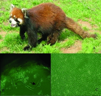
The fibroblasts from a red panda (Fig. 1a), which were dissociated by skin biopsy, were observed for 6 d after initial culturing. The cells showed spindle-shaped or uneven triangle morphology (Fig. 1b). Ninety per cent confluence was seen at 11 d (Fig. 1c).
Primary culture of red panda fibroblasts. (a) red panda sampled; (b) individual cells at day 6 after culture (×100), cells showed spindle-shaped (asterisk) or uneven triangle (arrow) morphology; (c) the cells 11 d after culture (×100).

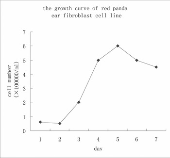
The S-like growth curve of the red panda fibroblasts appeared in the third passage culture (Fig. 2). A slightly descending phase was observed on the first day. For the next 3 days there was logarithmic growth. After fifth day, the density of fibroblasts decreased.
Growth curve of a red panda fibroblast cell line. Cells were inoculated in a 24-well dish (6 × 10⁴ cells/ml) and cultured in DMEM/F12 and 20% FBS in 5% CO2 at 37.5 °C. In the subsequent 7 days, the cells were digested and counted.

A great number of cells failed to attach when the fibroblast cells were cultured to the eleventh passage.
Cryopreservation and thawing
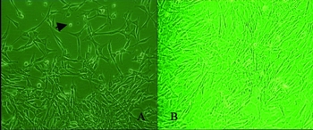
The fibroblasts were cryopreserved at –196 °C. The cells were thawed and then cultured. The results showed that the fibroblasts reached 90% confluence 48 h after culture. Typical spindle-shaped cells and some suspended single cells were observed in the medium (Fig. 3a) while 100% confluence was found 6 d after culture (Fig. 3b).
Fibroblasts from red panda after cryopreservation. (a) fibroblasts cultured for 48 h after cryopreservation and thawing. Some single cells were floating in the medium (arrow) while others attached to the wall (×100); (b) fibroblasts at 96 d culture after cryopreservation and thawing (×100).

The fibroblasts were cryopreserved, thawed and then cultured. They showed a rough appearance on the surface after trypsin digestion (Fig. 4).
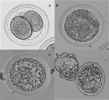
Red panda–rabbit interspecies reconstructed embryos at different stages. (a) at the 2-cell stage; (b) at the morula stage; (c) at the blastocyst stage; (d) a hatching blastocyst.

Reconstruction of interspecies embryos
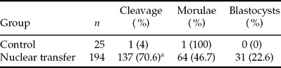
After micromanipulation, fusion, activation and in vitro culturing, the cleavage rate, morulae and blastocyst formation rate of reconstructed embryos were investigated. The results are shown in Table 1. The cleavage (Fig. 4a), morulae (Fig. 4b) and blastocyst (Fig. 4c, d), rates were 70.6, 46.7 and 22.6% respectively. In contrast, the control showed a 4% cleavage rate.
Development of interspecies reconstructed embryos.
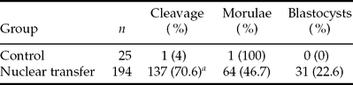
The control group was established as follows: rabbit oocytes were treated with the same procedures as noted above, including enucleating, nuclear transfer, activation, but fusion was not included before activation. Morulae or blastocyst formation rate was calculated by morula or blastocyst numbers divided by cleaved oocytes.
aPercentage indicates the most significant differences in the same column (p < 0.01) after t-test.
Discussion
Our study demonstrated, for the first time, that a fibroblast cell line from the red panda was successfully established and cryopreserved. Using this cell line, interspecies embryos were cloned with the fibroblasts as somatic cell donors and meiotically mature rabbit enucleated oocytes as recipients. The blastocyst rate reached 22.6%.
The evolutionary history of the red panda plays a pivotal role in the higher level phylogeny of ‘bear-like’ arctoides carnivore mammals (Flynn et al., Reference Flynn, Nedbal, Dragoo and Honeycutt2000). Since 1989, this species has become one of the endangered wild animals in China. The establishment and cryopreservation of the somatic cell line is essential not only to preserve the germ plasm resources of an important species, but also to provide valuable data for genome, postgenome and somacloning research. Moreover, the creation of this technological platform will provide the technology for storing the cell genetic resources of other endangered species. For the first time, the present study describes the successfully establishment of a fibroblast cell line from the red panda ear margin skin by direct tissue culture. The growth curve showed normal division and proliferation and no transformation tendencies. This indicated that the culture was suitable.
Our study established a cryopreservation procedure for red panda fibroblast cells using uncontrolled cooling. The preserved red panda genomic DNA material could be used for future SCNT to preserve fertility. The fibroblasts were transferred directly into a –80 °C freezer (freezing rate, 2 °C/min) after pretreatment and then to liquid nitrogen. The effect of controlled and uncontrolled cooling rate for freezing donor cells was investigated using reconstructed embryo development in cattle (Hayes et al., Reference Hayes, Rodriguez, Gonzalez, Falcon, Aguilar and Castro2005). It was found that freezing cell lines, when controlling the cooling rate at approximately 1 °C/min, reduced the fusion and blastocyst rate. Cells frozen with a non-controlled protocol were damaged more seriously. This suggested freezing donor cells for cloning is a critical step and should be monitored carefully. As for the mechanisms, it was speculated that the cryopreservation of donor cells might influence cell membranes and subsequently the fusion process in embryo reconstruction. Cryoprotectant can also influence the freezing outcome as well as the cooling rate (Kearney et al., 2005). It was reported that the combination of 10% DMSO and serum (2 or 10% albumin) resulted in better postcryopreservation growth than did a controlled-rate freezing with direct transfer into the –80 °C freezer on human corneal keratocytes (Borderie et al., Reference Borderie, Lopez, Lombet, Carvajal-Gonzalez, Cywiner and Laroche1998). In our study, we used 20% FBS and 8% DMSO as a cryoprotectant and this method proved feasible for cryopreservation.
It is known that the stage of the donor somatic cells affects the results of nuclear transfer and that the quiescent cell is able to support preimplantation development of the interspecies SCNT embryos. It was found that abducted cells were more suitable to be used in SCNT. Therefore, serum starvation and contact inhibition were widely used to induce donor cells to stay at the G0/G1 phase. The non-quiescent somatic cells were also used as donors in interspecies SCNT and the reconstructed embryos were harvested (Tani et al., Reference Tani, Kate and Tsunoda2000; Li et al., Reference Li, Han, Zhu, Wen, Liu, Wang, Lian, Du, Wang, Zhang and Chen2002). These studies indicated that non-quiescent cells could also be used as donors in interspecies SCNT. Our results also showed that the serum starvation of donor somatic cells was not absolutely necessary to produce the interspecies reconstructed embryos (data not shown).
We noticed that the fibroblasts had a rough surface after they were cryopreserved, thawed, in vitro cultured and treated with trypsin. Moreover, a great number of cells failed to survive the eleventh passage after trypsin digestion and serum starvation for 2 d. We speculated that the timing of trypsin treatment might be related to the fibroblast cells abnormality. According to previous reports, cell death and the appearance of surface roughness might be due to trypsin-induced DNA damage (Freshney, Reference Freshney and Freshney1994), or to exposure to a serum-deprived medium, which induced massive DNA fragmentation (Peura, Reference Peura2000; Peura et al., Reference Peura, Hartwich, Hamilton and Walker2003). Specific culture conditions and the time of enzymatic digestion should be optimized in further studies.
In many species, the technique of SCNT produces embryos, fetuses and offspring from embryonic, fetal and adult-derived cell types. Recently, interspecies SCNT has been an invaluable tool for studying nucleus–cytoplasm interactions and it may be a method for rescuing endangered species whose oocytes are difficult to obtain. Despite increasing interest in using cloning to rescue endangered species, reports of successful interspecies SCNT and in vitro embryo formation are sparse. These reports have shown that one species' oocytes could reprogramme the highly differentiated somatic cells of the other species. One of the first attempts of interspecies SCNT was reported by Dominko et al., who used the enucleated bovine oocyte as a recipient cytoplast and skin fibroblast cells from cows, sheep, pigs, monkeys and rats as sources of donor nuclei. Although interspecies embryos were obtained, no pregnancies were carried to term after the embryos were transferred into surrogate animals (Dominko et al., Reference Dominko, Mitalipova, Haley, Beyhan, Memili, McKusick and First1999).
Interspecies cloned embryos were produced between mountain bongo antelope and cattle (Lee et al., Reference Lee, Wirtu, Damiani, Pope, Dresser, Hwang and Bavister2003), buffaloes and cattle (Lu et al., Reference Lu, Shi, Wei, Yang and Wei2005), yak, dog and domestic cow (Murakami et al., Reference Murakami, Otoi, Wongsrikeao, Agung, Sambuu and Suzuki2005), whale and cattle or pig (Ikumi et al., Reference Ikumi, Sawai, Takeuchi, Iwayama, Ishikawa, Ohsumi and Fukui2004) and even mammal (rabbit) and chicken (Liu, S.Z. et al., Reference Liu, Zhou, Chen, Zhang, Wen, Kou, Li, Sun and Chen2004). Sun & Chen et al. have engaged in many interspecies SCNT studies (Wen et al., Reference Wen, Bi and Chen2004 review) and have obtained certain intraspecies and interspecies SCNT embryos such as cat–rabbit, cat–cat, rabbit–rabbit, panda–rabbit, mouse–mouse, rat–mouse and pig–mouse embryos (Wen et al., Reference Wen, Yang, Cheng, Li, Liu, Sun, Zhang, Lei, Wu, Kou and Chen2003, Reference Wen, Bi, Xu, Yang, Zhu, Sun and Chen2005; Zhong et al., Reference Zhong, Zhang, Meng, Zhang, Chen, Schatten and Sun2005).
In 2000, a cloned gaur (Bos gaurus) was created from gaur bull–domestic cow embryos. About 12% of cloned embryos developed to the blastocyst stage and 18% of these embryos developed to the fetal stage after they were transferred to surrogate mothers (Lanza et al., Reference Lanza, Cibelli, Diaz, Moraes, Farin, Farin, Hammer, West and Damiani2000). Unfortunately, the clones died within the first 48 h. Two births of cloned banteng were generated from male or female adult banteng fibroblasts (donors) and bovine oocytes (recipients). However, both pregnancies terminated prematurely, one between days 30 and 60 and the second between days 60 and 90 of gestation (Sansinena et al., Reference Sansinena, Hylan, Hebert, Denniston and Godke2005). In another study, two pregnancies were established from domestic sheep–argali embryos, but both terminated prematurely at day 59 of gestation (White et al., Reference White, Bunch, Mitalipov and Reed1999). A live offspring was obtained from domestic sheep oocytes (recipients) and mural granulose cells from deceased moufflons (donors) (Loi et al., Reference Loi, Ptak, Barboni, Fulka, Cappai and Clinton2001). These reports suggest that somatic cell cloning methods could be used to restore endangered, or even extinct, species.
Interspecies SCNT have been improved extensively. Direct nuclear injection using the Piezo drill proved to be an efficient method for interspecies SCNT in horse and cattle oocytes (Choi et al., Reference Choi, Love, Chung, Varner, Westhusin, Burghardt and Hinrichs2002). Whole cell intracytoplasmic injection combined with chemical enucleation was tried in a panda–rabbit SCNT and it resulted in an optimal enucleation. This method was efficient, less labour intensive and produced almost the same results (Jiang et al., Reference Jiang, Yang, Zhang, Zheng, Liu, Sun and Chen2004). It was found that the extrusion of a second polar body (PB2) of recipient oocytes influenced the developmental capacity of interspecies SCNT embryos. The karyotype of the panda–rabbit hybrid embryos consisted of chromosomes from both counterparts (Wen et al., Reference Wen, Bi, Xu, Yang, Zhu, Sun and Chen2005).
In our present study, we obtained red panda–rabbit interspecies embryos for the first time. In order to ensure that the embryos reconstructed by SCNT are cloned, chromosome analysis is often recommended. Instead, we set up a control, which was designed in accordance with SCNT procedures except for somatic cell introduction. The results showed that oocytes could not develop properly at the early stage; hardly any failed to cleave (1/25, 4% only), suggesting that the nucleus was entirely removed before SCNT was accomplished and that the embryos obtained in the study were from red panda genetically. The current study achieved a high blastocyst formation rate (23%), which was higher than reported previously (Li et al., Reference Li, Han, Zhu, Wen, Liu, Wang, Lian, Du, Wang, Zhang and Chen2002, Reference Li, Wei, Goossens, Feng, Tamate, Bruford and Funk2005; Wen et al., Reference Wen, Yang, Cheng, Li, Liu, Sun, Zhang, Lei, Wu, Kou and Chen2003; Murakami et al., Reference Murakami, Otoi, Wongsrikeao, Agung, Sambuu and Suzuki2005; Thongphakdee et al., Reference Thongphakdee, Numchaisrika, Omsongkram, Chatdarong, Kamolnorranath, Dumnui and Techakumphu2006). It is well known that the embryo transfer recipient selection is a substantial obstacle for interspecies SCNT. In order to investigate the development of these embryos, and eventually obtain viable births, a further study (probably utilizing cats) will be required to transfer reconstructed embryos successfully to surrogate recipients. The previous study found that the karyotype of red panda showed a high degree of homology with the domestic cat (Tian et al., Reference Tian, Nie, Wang, Yang and Yang2002).
In summery, this study marks the first report of a red panda fibroblast cell line and the cryopreservation and cloning of interspecies SCNT embryos with fibroblasts as somatic cell donors and rabbit oocytes as recipients. It demonstrates that the cytoplasm from a rabbit oocyte supports early development after SCNT and is capable of reprogramming the nucleus to generate blastocyst stage embryos.
Acknowledgements
The authors acknowledge the financial support of the Anhui Distinguished Youth Sci-tech Project (grant number 06041081), the Anhui ‘Eleventh Five-year’ Sci-tech Project (grant number 06013051A) and the International Cooperation Project (grant number 08080703034). The authors are also grateful to Dr Sun Qing-Yuan, Lei Lei, Fan Heng-Yu and Fang Changge for their invaluable assistance in preparing the manuscript.