The basic abnormality in congenitally corrected transposition is the combination of discordant atrioventricular and ventriculoarterial connections. Because of this, the systemic circulation is supported by the morphologically right ventricle and the tricuspid valve. Tricuspid regurgitation is a common concomitant problem in these patients. It is frequently associated with anatomical abnormalities, or is ascribed to increased pressure load on the valve. The cause-and-effect relationship between tricuspid regurgitation and morphologically right ventricular dysfunction remains to be determined. Patients without associated defects may be asymptomatic until late adulthood. Long-term follow-up of these patients has shown that cardiac failure occurs unpredictably, and is associated with increased morbidity and mortality.Reference Graham, Bernard and Mellen1 Most reports concerning neurohormonal activation in patients with congenitally malformed hearts have been confined to children, and limited to the assessment of a single neurohormonal system. The studies have often been undertaken in the setting of overt cardiac failure, and have not been focused on a specific type of anomaly.Reference Bolger, Sharma and Li2–Reference Tulevski, Groenink and van der Wall4 Neurohormonal activation characterises chronic cardiac failure, relates to the outcome, and constitutes an important therapeutic target in patients with this complication. The amino terminal pro brain natriuretic peptide has been included as a sensitive marker of cardiac dysfunction in a proposed algorithm for the diagnosis of cardiac failure.Reference Remme, Swedberg and Cleland5 Functional and morphological assessment of morphologically right ventricular function by conventional echocardiography is challenging because of the complex geometric shape of the ventricle. Cardiac magnetic resonance imaging and multislice computed tomography, therefore, have emerged as more appropriate quantitative methods for assessing right ventricular function.Reference Koch, Oellig and Oberholzer6, Reference Cluzel, Brochu and Izzillo7 Recently, nonetheless, tissue Doppler echocardiography has emerged as a promising technique for quantitative analysis of myocardial function.Reference Alam, Wardell, Andersson, Nordlander and Samad8, Reference Garcia, Thomas and Klein9 The management of patients with congenitally corrected transposition should include diagnostic tests for early identification of the syndrome of cardiac failure in the asymptomatic phase of the disease. The results may have implications for potential early medical or surgical therapeutic strategies. With this in mind, our aims in this study were, first to assess the level of neurohormonal activation of cardiac failure in both asymptomatic and symptomatic adults with congenitally corrected transposition, and second, to identify the most accurate markers of cardiac failure in these patients.
Methods
Patients
We included in our study all consecutive patients with congenitally corrected transposition collected in the database of adults with congenitally malformed hearts at University Medical Centre, Ljubljana, which is the only centre for adults with congenital cardiac diseases in Slovenia. We identified, and studied prospectively, 19 such adults, including ten women, aged from 15 to 68 years, with a mean age of 35 plus or minus 13.1 years. As a control group, we assembled 19 healthy volunteers matched for sex and age, without any history of cardiovascular disease, diabetes mellitus, or hypertension. Highly trained athletes were excluded. Their physical examinations, and resting 12-lead electrocardiograms, were all within normal limits. The study was approved by our local medical ethics committee, and conforms to the principles outlined in the Declaration of Helsinki. The patients and their control subjects gave written informed consent to participate in the study.
Clinical investigation
The patients underwent detailed clinical evaluation, including a review of medical history and physical examination. Historical data included the position of the patient within the functional classification of the New York Heart Association. The diagnosis of cardiac failure was made on the basis of symptoms and physical findings. We also analysed standard 12-lead electrocardiograms.
Echocardiography
Transthoracic cross-sectional echocardiography, echo-Doppler studies, and tissue Doppler echocardiography were carried out using a Hewlett-Packard Sonos 5500 echocardiographic machine. Transverse and longitudinal dimensions of the left atrium were measured in the apical 4-chamber view at the end of ventricular systole.Reference Weyman10, Reference Lang, Bierig and Devereux11 The degree of tricuspid regurgitation, assessed by colour-Doppler, was estimated as mild, moderate, or severe by the width and length of the jet, and by the pattern of Doppler flow in the pulmonary veins.Reference Pu, Griffin and Vandervoord12, Reference Miyatake, Okamoto and Kinoshita13 Diastolic systemic right ventricular function was assessed by measuring velocities of flow through the tricuspid valve and the right upper pulmonary vein. The following diastolic parameters were measured: E/A ratio, pulmonary vein S, D, Ar velocities, duration of A seen on the pattern of transtricuspid flow pattern (A dur) and on the pulmonary venous flow (Ar dur). The ratio of duration of A to Ar was calculated.Reference Alam, Wardell, Andersson, Samad and Nordlander14
Pulsed tissue Doppler echocardiographic images were recorded from the apical 4-chamber view. Maximal systolic (S), early (E), and late (A) diastolic peak velocities were measured. For assessing the velocity of longitudinal excursion of the ventricular wall, a sample volume was placed at the parietal part of the annulus of the morphologically tricuspid valve (St, Et, At), at the basal segment of the muscular ventricular septum (Sm, Em, Am), and at the parietal part of the mitral annulus in the morphologically left ventricle (Sml, Eml, Aml). For each subject, at least three consecutive cardiac cycles were analysed and averaged.Reference Alam, Wardell, Andersson, Samad and Nordlander14, Reference Suty-Selton, Mock, Piquemal, Vancon, Nippert and Juilliere15
Plasma neurohormones
Samples were obtained from the antecubital vein in all subjects. Blood was collected into chilled tubes containing preservative, promptly centrifuged, and stored at -70°C until final analysis.
Levels of amino terminal pro brain natriuretic peptide in the serum were measured by the electrochemiluminiscence immunoassay using a Roche Elecsys immunoassay analyzer (Roche Diagnostics GmbH D-68298 Mannheim). The measuring range of 5 to 35.000 ng/l was defined by the lower detection limit and the maximum of the master curve. The cut-off value for normality was 125 ng/l.Reference Gustafsson, Badskjaer, Stensgaard, Poulsen and Hildebrandt16 Plasma active renin was measured using a two-site immunoradiometric assay (Diagnostic Systems Laboratories, Inc., Webster, Texas USA). This is a non-competitive assay in which the analyte to be measured is »sandwiched« between two antibodies. The lowest detectable level was 0.05 pmol/l and the normal range from 0.57 to 3.67 pmol/l. Levels of epinephrine and norepinephrine in the plasma were measured by radioimmunoassay (TriCat TM RIA kit (IBL Immuno-Biological Laboratories, Hamburg, Germany). The lowest detectable levels for epinephrine and norepinephrine were 0.065 and 0.12 nmol/l; and normal values of epinephrine and norepinephrine were less than 0.55 nmol/l and less than 3.55 nmol/l.
Magnetic resonance imaging and multislice computed tomography
Magnetic resonance imaging was done in 10 patients, using a 1.5 Tesla scanner with high power gradients (Signa Magnetic resonance imaging 1.5 T Echospeed, General Electric medical systems). Imaging sessions were initiated with scout images, followed by electrocardiogram-triggered Tl-weighted spin echo series of axial images (minimum TE, slice thickness 7 mm, spacing 1.0 mm, nex 2, matrix 256 × 28).
Based on these images we performed breathold cine sequences in the short axis of the systemic ventricle from the base to the apex, in order to obtain a stack of 12 to 14 contiguous images, 8 mm thick, with no gap between slices. The repetition time was based on the R-R interval, the echo time was 4 miliseconds and the flip angle was 20 degrees.
Cardiac multislice computed tomography imaging was performed in 9 patients with a pacemaker. They were studied with multislice computed tomography using a 16-slice scanner Somatom Sensation 16 (Siemens, Erlangen Germany). The scan parameters were as follows: 120 Kv, 500 Eff mAs, tube rotation time was 0.42 s; slice width was 1.0 mm and collimation 12 × 0.75 mm; reconstruction increment was 0.6 mm and kernel B30 medium smooth. Scan was done over the whole cardiac cycle with retrospective electrocardiogram-gating. The heart was covered from the base to the apex, table feed/rotation was 2.8 seconds and scan time was 20–25 seconds. A bolus of 100 mililitres nonionic contrast media (300 mg I/ml) was injected through an 18-gauge catheter placed into the antecubital vein at a flow rate of 3.5/s, followed by a 30 millilitre saline chaser bolus at the same flow rate. Scanning started automatically after the density of contrast media in ascending aorta had reached 120 HU.
There followed post processing on the workstation. Short axis multiplaner reformatting images of the systemic ventricle from the base to the apex were reconstructed in end-diastole and end-systole (typically at 80 percent and 20 percent of RR interval) to get a stack of images 8 mm thick. MASS (Medis, Leiden, The Netherlands) image analysis software was used for processing multislice, multiphase images provided by cardiac magnetic resonance imaging and multislice computed tomography. Ejection fraction was determined by the manual outlining of the endocardial contours of end-diastolic and end-systolic frames. Papillary muscles and trabeculations were not included in the calculation of ventricular volumes. Ejection fraction was calculated as: 100 × (end-diastolic volume–end-systolic volume/end-diastolic volume. A right ventricular end-diastolic volume more than 58 millilitres for men and more than 48 millilitres for women, an ejection fraction more than 48 percent for men and more than 50 percent for women were defined as normal.Reference Lorenz17, Reference Alfakih, Plein, Thiele, Jones, Ridgway and Sivananthan18
Statistical analysis
Continuous variables are presented as mean plus or minus standard deviations, and discrete variables as relative frequencies. The Kolmogorov-Smirnov test was used to determine normal distributions of data. Differences between the parameters obtained in patients and their controls were analysed by the independent samples t-test or Mann-Whitney U-test. Pearson’s and Spearman’s rank correlation coefficients were used to determine bivariate correlation between the parameters in the two groups. Values for brain natriuretic peptide were natural log-transformed for the purpose of correlation analysis. A p value of less than 0.05 was considered statistically significant. Statistical analysis was performed using SPSS 13.0 for Windows (SPSS Inc., Chicago, Illinois).
Results
Demographic and clinical data
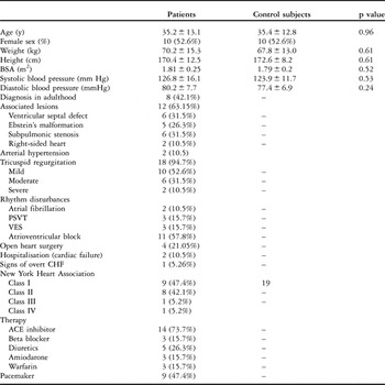
The demographics and clinical characteristics of the patients are listed in Table 1. In patients with atrioventricular block and sinus rhythm, atrioventricular synchrony was maintained.
Clinical characteristics of the patients and their controls.

NS = non significant, BSA = body surface area, PSVT = paroxysmal supraventricular tachycardia, VES = ventricular ectopic beats, CHF = congestive cardiac failure.
Neurohormonal levels
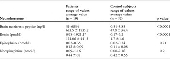
The patients had significantly higher circulating levels of brain natriuretic peptide and renin (both p less than 0.0001) than did their control subjects. Levels of epinephrine and norepinephrine were similar in the patients and their controls (Table 2).
Neurohormonal levels in patients and in control subjects.

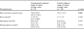
Levels of the brain natriuretic peptide were significantly higher in the subgroup of asymptomatic patients than in the controls (P less than 0.002). In 4 out of 9 asymptomatic patients, the levels of peptide were normal. There was no statistically significant difference between asymptomatic patients and controls as concerns the other three hormones assessed (Table 3).
Neurohormonal levels in asymptomatic patients and in control subjects.

Echocardiographic parameters
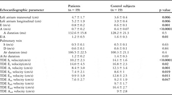
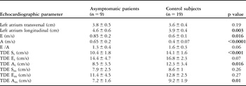
In Tables 4 and 5, we show the echocardiographic characteristics of the patients compared to their healthy controls. The patients had larger transverse and longitudinal left atrial dimensions and higher transtricuspid velocities. The E/A ratio of velocity of transtricuspid flow was lower in the patients. Statistically significant differences between the patients and their control subjects were found only for these conventional echocardiographic parameters. In contrast, there were statistical significant differences between the groups in all Doppler variables studied. The difference between tricuspid annular velocities at the lateral side of the systemic right ventricle, and mitral annular velocities at the lateral side of the left ventricle in controls, did not show any statistical significance.
Echocardiographic parameters of patients and control subjects.

E = early diastolic transtricuspid velocity, A = late diastolic transtricuspid velocity, TDE = tissue Doppler echocardiography, St = peak tricuspid systolic annular velocity (lateral side), Et = peak tricuspid early diastolic annular velocity (lateral side), At = peak tricuspid late diastolic annular velocity (lateral side), Sm = peak systolic velocity at the basal interventricular septum, Em = peak early diastolic velocity at the basal interventricular septum, Am = peak late diastolic velocity at the basal interventricular septum, Sml = peak mitral systolic annular velocity (lateral side) of systemic left ventricle in controls, Eml = peak mitral early diastolic annular velocity (lateral side) of systemic left ventricle in controls, Aml = peak mitral late diastolic annular velocity (lateral side) of systemic left ventricle in controls, Ar = atrial reversal wave.
Echocardiographic parameters of asymptomatic patients and control subjects.

E = early diastolic transtricuspid velocity, A = late diastolic transtricuspid velocity, TDE = tissue Doppler echocardiography, St = peak tricuspid systolic annular velocity (lateral side), Et = peak tricuspid early diastolic annular velocity (lateral side), At = peak tricuspid late diastolic annular velocity (lateral side). Sm = peak systolic velocity at the basal interventricular septum, Em = peak early diastolic velocity at the basal interventricular septum, Am = peak late diastolic velocity at the basal interventricular septum.
The severity of tricuspid regurgitation in our patients is indicated in Table 1. Out of 18 patients studied, 5 had Ebstein’s malformation of the morphologically tricuspid valve. We took note of 3 proposed risk factors for tricuspid regurgitation in such patients, namely previous open-heart surgery, anatomic valvar abnormalities, and atrioventricular block.Reference Prieto, Hordof, Secic, Rosembaum and Gersony19 In our series, only one patient was without tricuspid regurgitation, and he had no predisposing factors. In those with mild tricuspid regurgitation, 3 of 10 patients were free of the predisposing factors studied, while 3 had more than one predisposing factor. All patients in the group with moderate tricuspid regurgitation had one or more predisposing factors. In those with severe tricuspid regurgitation, one patient had no predisposing factors, and one presented with all of them, as well as Ebstein’s malformation.
Global right ventricular systolic function
The mean end-diastolic right ventricular volume was 163.63 millilitres, with a range from 115 to 230 millilitres, and mean end-systolic right ventricular volume was 88.38 millilitres, with a range from 39 to 136 millilitres, as assessed by magnetic resonance imaging or multislice computed tomography. Mean right ventricular ejection fraction was 47.86 plus or minus 15.3%, ranging from 28.3 to 66.1%. In the group of asymptomatic patients, mean right ventricular ejection fraction was 49.2 plus or minus 14.9%, ranging from 28.3 to 66.1%, and mean end-diastolic right ventricular volume was 144 plus or minus 37.8 millilitres. No statistical significant differences were found in comparison between asymptomatic and symptomatic patients with regards to right ventricular ejection fractions, or end diastolic and endsystolic volumes.
Comparison of neurohormonal levels, echocardiographic findings, and magnetic resonance imaging and multislice computed tomographic scans
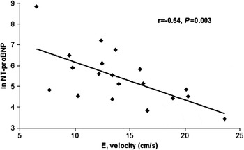
The levels of brain natriuretic peptide showed significant positive correlation with the clinical functional class (r equal to 0.49, p equal to 0.032), the age of patients (r equal to 0.46, p equal to 0.048), and the transverse (r equal to 0.85, P < 0.0001) and longitudinal left atrial dimensions (r equal to 0.80, p less than 0.0001). Among tissue Doppler echocardiographic velocities, we found a significant negative correlation between Et velocity and levels of the peptide (r equal to – 0.64, p equal to 0.003 – Fig. 1).
Correlation between levels of amino terminal pro brain natriuretic peptide in the plasma and tissue Doppler tricuspidal annular early diastolic velocity (Et).

Levels of the peptide also showed significant negative correlation with morphologically right ventricular ejection fraction assessed by magnetic resonance imaging or multislice computed tomography (r equal to – 0.53, p equal to 0,02). They correlated positively with the end-diastolic and end-systolic morphologically right ventricular volumes assessed by magnetic resonance imaging or multislice computed tomography (r equal to 0.50, p equal to 0.026; r equal to 0.61, p equal to 0.006).
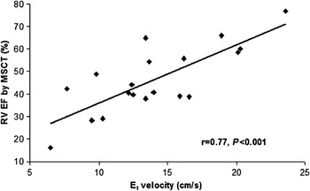
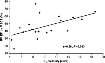
The severity of tricuspid regurgitation did not correlate significantly with either the levels of brain natriuretic peptide or the age of the patient. Neither did tricuspid regurgitation correlate with the systolic function of the systemic ventricle as assessed by magnetic resonance imaging or multislice computed tomography. A negative correlation was found between right ventricular ejection fraction determined by magnetic resonance imaging or multislice computed tomography and the transverse and longitudinal left atrial dimensions (r equal to – 0.453, p equal to 0.05 vs. r equal to – 0.485, p equal to 0.03). A significant positive correlation was established between right ventricular ejection fraction assessed by magnetic resonance imaging or multislice computed tomography and Et velocity (r equal to 0.77, p less than 0.0001) and Em velocity (r equal to 0.56, p equal to 0.013) (Figs. 2, 3).
Correlation between right ventricular ejection fraction determined by magnetic resonance imaging and multislice computed tomography and tissue Doppler tricuspid annular early diastolic velocity (Et).

Correlation between right ventricular ejection fraction determined by magnetic resonance imaging and multislice computed tomography and tissue Doppler peak early diastolic velocity measured at the basal muscular ventricular septum (Em).

Discussion
In only 11 of our patients had the correct diagnosis been established in childhood. This proves that congenitally corrected transposition may remain asymptomatic and undetected for many years, and contribute to the consequences arising from the delay of preventive measures and commencement of therapy. Nearly half of our patients were asymptomatic, with only two presenting with severe symptoms and with cardiac failure requiring frequent hospitalisation. The main haemodynamic problems in these two patients included systemic ventricular dysfunction and severe tricuspid regurgitation, yet they did not fulfil the criterions for cardiac transplantation because of high pulmonary vascular resistance in one case, and advanced age in the other. As expected, the asymptomatic patients were younger than those who were symptomatic.
Our studies show that neurohormonal activation indicative of cardiac failure is present as early as the asymptomatic period in patients with congenitally corrected transposition. This is especially true for amino terminal pro brain natriuretic peptide. The levels of circulating epinephrine and norepinephrine were also elevated, but the differences did not reach statistical significance, probably because of the small number of patients. The neurohormonal system is known to be activated in many patients with different types of congenitally malfomed hearts. Studies focusing on specific types of anomaly, however, are scarce.Reference Bolger, Sharma and Li2 Our current findings, therefore, may promote further studies on early introduction of medical treatment directed at incipient cardiac failure in asymptomatic patients with congenitally corrected transposition.
As expected, left atrial dimensions were larger in the patients, regardless of the severity of tricuspid regurgitation. Longitudinal dimensions were significantly larger even in those who were asymptomatic. The increased left atrial dimensions, and a lower E/A ratio, were most likely due to diastolic dysfunction of the systemic ventricle, as levels of the brain natriuretic peptide were found to increase with left atrial dimensions. These dimensions, therefore, may serve as a simple and accurate conventional echocardiographic marker of cardiac failure in the setting of congenitally corrected transposition.
When using tissue Doppler echocardiographic parameters, we compared values for the systemic morphologically right ventricle in the patients with congenitally corrected transposition with those derived from the pulmonary right ventricle in the normal subjects. It is not appropriate to compare the myocardial velocities of the systemic right ventricular free wall in the patients, and left ventricular myocardial velocities in controls, because of the different architecture of the aggregated myocytes, the main bulk of the pulmonary right ventricular myocardium being composed of myocytes aggregated together in longitudinal fashion.Reference Rushmer, Crystal and Wagner20 On the other hand, we deemed it logical to measure myocardial velocities of the muscular ventricular septum, since both ventricles share the septal myocardium.Reference Sanchez-Quintana, Anderson and Ho21 Systolic and diastolic myocardial velocities measured at the lateral free wall of the systemic right ventricle and at the basal segment of the ventricular septum were significantly lower in patients than in their controls. Furthermore, the subgroup of asymptomatic patients had lower tissue Doppler echocradiographic velocities, yet only St, Et and Em velocities reached statistical significance. These results clearly indicate that systolic and, above all, diastolic function of the systemic ventricle is impaired in the setting of congenitally corrected transposition. These results agree with the results of tissue Doppler echocardiographic studies in patients with regular transposition after an atrial switch operation.Reference Rentzsch, Abd El Rahman and Hui22 A significant negative correlation between Et and levels of brain natriuretic peptide confirms that early diastolic myocardial velocities are sensitive markers of systemic right ventricular failure.
More than 10 years ago, a large study was performed on a group of 40 patients with congenitally corrected transposition.Reference Prieto, Hordof, Secic, Rosembaum and Gersony19 These results indicate that right ventricular failure appears almost always to be secondary to long standing tricuspid regurgitation. In our study, in contrast, the velocities of pulmonary venous flow suggested that tricuspid regurgitation was not a major haemodynamic problem. No correlation was found between the severity of tricuspid regurgitation and levels of brain natriuretic peptide, or systolic function of the systemic ventricle assessed by magnetic resonance imaging or multislice computed tomography. We cannot confirm, therefore, the notion that failure of the morphologically right ventricle in patients with congenitally corrected transposition is secondary to tricuspid regurgitation, albeit that the number of our patients was too small to draw a firm conclusion.
The ejection fraction and volumes of the morphologically right ventricle as determined using magnetic resonance imaging and multislice computed tomography correlated significantly with the levels of brain natriuretic peptide, and also with some easily derived non-invasive parameters measured by tissue Doppler echocardiography. The significant correlation between the results from tissue Doppler echocardiography and magnetic resonance or computed tomography parameters of right ventricular function confirms the reliability of tissue Doppler echocardiography for the assessment of global ventricular function in the setting of congenitally corrected transposition. Magnetic resonance imaging and multislice computed tomography have previously been shown to be valuable tools for measuring right ventricular size and quantitatively assessing right ventricular ejection fraction.Reference Koch, Oellig and Oberholzer6, Reference Cluzel, Brochu and Izzillo7
Our study was limited in that our group of 19 patients may seem small, but it represents a sizable group for this rare entity. Some of our patients were medicated with inhibitors of angiotensin-converting-enzyme and beta blockade due to symptoms of cardiac failure, which could have had an unpredictable effect on our results. The subgroups of patients on different therapeutic regimes were too small to analyse statistical differences between them.
When considering brain natriuretic peptide as a marker of cardiac failure, therefore, we can conclude that neurohormonal activation in the syndrome of cardiac failure is present at an early stage of the disease in the setting of congenitally corrected transposition, and is noticeable even in asymptomatic patients. Neurohormonal activation is strongly related to the functional class, morphologically right ventricular function, and to some easily derived conventional echocardiographic and tissue Doppler echocardiographic parameters. Levels of the hormone, and early diastolic myocardial velocity at the lateral border of the tricuspid annulus, therefore, are the earliest and most sensitive markers of systemic right ventricular dysfunction in patients with congenitally corrected transposition. We believe that these findings could have important clinical benefits for management, in particular with regard to optimal timing of medical treatment of cardiac failure or surgical management of tricuspid regurgitation. Use of either levels of brain natriuretic peptide or tissue Doppler parameters may also facilitate quantitative follow-up of right ventricular function in these patients. The combination of echocardiography, magnetic resonance imaging and multislice computed tomography data opens the door not only to a thorough investigation of anatomical changes, but provides a marked benefit to research.
Acknowledgments
The study was supported by Roche Diagnostics GmbH, Mannheim, Germany and Krka d.d., Novo Mesto, Slovenia. Tomaž Podnar was supported in part by the Slovenian Agency for Research grant # J3-9663.