Introduction
In vitro devices are playing a key part in modern neuroscience as they represent an efficient, easy to use and reliable way to record the electrical activity of neurons in cell cultures[ Reference Pine 1 , Reference Nam and Wheeler 2 ] and brain slices.[ Reference Steidl, Neveu, Bertrand and Buisson 3 , Reference Sessolo, Khodagholy, Rivnay, Maddalena, Gleyzes, Steidl, Buisson and Malliaras 4 ] In vitro experiments allow a more profound understanding of the neural signaling mechanisms, and represent an important step for testing new technologies before applying them in vivo.[ Reference Arnold, Hofmann, Bengtson, Wittmann, Vanhoutte and Bading 5 , Reference Kanagasabapathi, Massobrio, Barone, Tedesco, Martinoia, Wadman and Decre 6 ] Moreover, in vitro models are of great importance in modern drug discovery as they promote a biologically-driven approach in drug development, in accordance with the new trend in pharmacological research that dictates the restriction of in vivo experiments due to ethical reasons.[ Reference Soldatow, LeCluyse, Griffith and Rusyn 7 ] More precisely, the development of alternative approaches to animal testing, consistent with the 3Rs (Replacement, Reduction, Refinement) principle is a necessity. Finally, the REACH regulation is an extra driving force towards in vitro testing as it necessitates the acquisition of information regarding the toxicity of chemicals manufactured or imported in Europe at significant volumes.[ Reference Rivnay, Owens and Malliaras 8 ] Consequently, the design and validation of high performance in vitro screening platforms is very important.
Hippocampal cell cultures represent a good example of a vehicle for in vitro testing, as these cells show a great variety of neural activity. Local field potentials and action potentials (APs or spikes) are the two that gather the greatest interest of the scientific community. The former are created by the contribution of multiple active sources.[ Reference Buzsaki, Anastassiou and Koch 9 ] Those are slow events (typically 1–200 Hz) and their amplitude varies from a few hundred μV up to a few mV. The latter are faster events (typically 1 kHz) with extracellular amplitude around 100 µV since they are generated from single neurons.[ Reference Nam and Wheeler 2 , Reference Sessolo, Khodagholy, Rivnay, Maddalena, Gleyzes, Steidl, Buisson and Malliaras 4 , Reference Buzsaki, Anastassiou and Koch 9 , Reference Ludwig, Langhals, Joseph, Richardson-Burns, Hendricks and Kipke 10 ] These particular characteristics of APs (high frequency, low amplitude) make their extracellular recording a challenging task due to the fact that they can be easily lost inside the background noise collected by the sensing device. The most common device for recording the activity of hippocampal cultures (and of electrogenic cell cultures in general) is the microelectrode array (MEA). MEAs consist of microfabricated electrodes on a glass substrate, on top of which cells are cultured.[ Reference Pine 1 , Reference Nam and Wheeler 2 ] Commercially available MEAs, with electrodes made of conductors such as Pt, TiN, Ir and Au are being used extensively in conjunction with hippocampal cultures to study the influence of various compounds on neural activities.[ Reference Nam and Wheeler 2 , Reference Arnold, Hofmann, Bengtson, Wittmann, Vanhoutte and Bading 5 , Reference Nam, Wheeler and Heuschkel 11 , Reference Geissler and Faissner 12 ] The quality of the recordings depends on both the quality of the cell culture (ion channel expression, ion cell density, cell size, cell density) and on the quality of the electrode (lower impedance is firmly translated into better quality recordings).[ Reference Yang, Zhao, Keefer, Liu, Bengio, Schuurmans, Lafferty, Williams and Culotta 13 ] A fundamental trade-off in MEAs involves an increase in impedance as the electrodes are made smaller in order to gain spatial resolution.[ Reference Ludwig, Uram, Yang, Martin and Kipke 14 , Reference Kovacs, Stenger and McKenna 15 ] This increase in impedance, which arises from the decrease of the capacitance of electrode/media interface, makes recording small neural signatures challenging.[ Reference Ludwig, Langhals, Joseph, Richardson-Burns, Hendricks and Kipke 10 ]
Conducting polymers have emerged as a promising class of materials for improving the electrical contact between metals and cells, both in vitro and in vivo.[ Reference Berggren and Richter-Dahlfors 16 , Reference Green and Abidian 17 ] This is mainly because of their mixed electronic/ionic conductivity, which increases capacitance, hence decreases impedance at the biotic/abiotic interface.[ Reference Martin and Malliaras 18 , Reference Proctor, Rivnay and Malliaras 19 ] As a result, coating MEA electrodes with conducting polymer films was shown to lead to higher quality recordings from cell cultures[ Reference Nyberg, Shimada and Torimitsu 20 ] and brain slices.[ Reference Sessolo, Khodagholy, Rivnay, Maddalena, Gleyzes, Steidl, Buisson and Malliaras 4 ] The use of conducting polymers enables novel form factors such as mechanically-flexible MEAs,[ Reference Blau, Murr, Wolff, Sernagor, Medini, Iurilli, Ziegler and Benfenati 21 ] and these materials can even be deposited around living neural cells (SH-SY5Y) to create cell-mimetic surfaces.[ Reference Richardson-Burns, Hendricks, Foster, Povlich, Kim and Martin 22 ] Biomolecules can be incorporated in or on conducting polymer electrodes to promote cell growth and control morphology.[ Reference Cui, Lee, Raphael, Wiler, Hetke, Anderson and Martin 23 – Reference Green, Hassarati, Bouchinet, Lee, Cheong, Yu, Dodds, Suaning, Poole-Warren and Lovell 25 ] Cell morphology can also be controlled by modifying the structure of the conducting polymer film.[ Reference Abidian, Corey, Kipke and Martin 26 ]
In this paper, we show that conducting polymer MEAs can be used as in vitro platforms for assessing the influence of different drugs on cell cultures. We fabricate MEAs with electrodes consisting of the conducting polymer poly(3,4-ethylenedioxythiophene) doped with poly(styrene sulfonate) (PEDOT:PSS), coated with the polypeptide poly-L-lysine (PLL).[ Reference Nam and Wheeler 2 ] The use of the polypeptide constitutes a biofunctionalization treatment, essential for establishing a cell compatible interface on our recording sites as it facilitates cell attachment and neurite outgrowth. Overall, it allows for activity measurements to be performed rendering our platform suitable for in vitro neuron culture electrophysiology with the use of conducting polymer electrodes. Hippocampal cell cultures were maintained for 3–6 weeks on these MEAs, showing mature synaptic network formation and neural activity. Their viability was confirmed with fluorescence microscopy and patch clamp recordings. The low impedance of the electrodes led to high-quality recordings of neural activity, enabling the observation of drug-induced changes. These results validate the use of conducting polymers in MEAs for in vitro drug screening and toxicology.
Experimental section
MEA fabrication
The MEAs consisted of 32 electrodes, each with a 10 × 10 µm2 (nominal) recording area (Figs. S1, S2, Supplementary material). They were fabricated as previously reported.[ Reference Sessolo, Khodagholy, Rivnay, Maddalena, Gleyzes, Steidl, Buisson and Malliaras 4 ] The process involved the deposition and patterning of metal (gold), parylene-C and PEDOT:PSS on a glass substrate. The substrate (a 26 × 76 mm2 glass slide) was thoroughly cleaned by sonication in a soap bath for 15 min followed by a 15 min sonication in a mixture of acetone/isopropanol (1:1). S1813 (Shipley) photoresist was spun cast on the substrate before exposed to UV light with the use of a SUSS MBJ4 contact aligner and a chromium mask (1st photolithography step). The exposed parts of the photoresist were then developed in a MF-26 developer bath and a 10 nm of chromium/100 nm of gold deposition step in a metal evaporator followed. Lift-off in a 1:1 mixture of solvents (acetone/isopropanol), removed the excess gold creating the desired electrode pattern on the substrate. Two layers of parylene, 2 µm each, were deposited with the help of a SCS Labcoater. Between the two layers commercially available soap solution (1%, Micro-90) was deposited in order to act as an anti-adhesive layer. At the same time, 3-(trimethoxysilyl)propyl methacrylate (A-174 Silane) was used as an adhesion promoter between the first layer and the substrate. A second photoresist (AZ 9260) was then spun cast and a second photolithography step took place (development in AZ Developer). Reactive ion etching with O2 plasma (Oxford 80 Plasmalab plus) resulted in a window opening on top of each electrode. A peeling-off step of the sacrificial second parylene layer defined the active area of the recording site just after PEDOT:PSS dispersion [Clevios PH 1000 from Heraeus Holding GmbH, with 5 wt.% ethylene glycol, 0.1 wt.% dodecyl benzene sulfonic acid and 1 wt.% of (3-glycidyloxypropyl)trimethoxysilane)] was spun cast on the device. The thickness of the PEDOT:PSS layer was 350 nm, as this value minimizes impedance at 1 kHz.[ Reference Sessolo, Khodagholy, Rivnay, Maddalena, Gleyzes, Steidl, Buisson and Malliaras 4 ] The devices were then hard baked at 140 °C for 1 h and were immersed in deionized water over night so that any excess of low molecular weight compounds inside the PEDOT:PSS dispersion would be removed. After fabrication, the devices were sterilized in 70% ethanol for 2 h, rinsed with water and incubated in a solution of 0.5 mg/ml PLL diluted in water for 2 h, before being rinsed again with water and dried.
Preparation of the rat hippocampal cell cultures
Primary hippocampal cell cultures were performed from embryonic day 18-Wistar rats. Hippocampi were collected in Hanks’ balanced salt solution, dissociated with trypsin and plated at a density of 12 × 105 cells/cm2 on PLL coated PEDOT:PSS MEAs. The hippocampal neurons were cultured in Neurobasal supplemented with 2% B-27, 1% penicillin-streptomycin and 0.3% glutamine in a humidified atmosphere containing 5% CO2 at 37° C. All animal experiments were carried out according to the animal care and experimentation committee rules approved by CNRS, France. A vital fluorescent dye (4-Di-2-ASP, 3 µM, Sigma-Aldrich) was used to stain living neurons and assert the viability of the culturing process on the electronic devices. Cultures were immersed for 15 min in a warm (37 °C) solution of the dye and then abundantly rinsed with the recording extracellular solution.
Statistical analysis
Data are reported in the text as mean values ± standard error of the mean. For the reported impedance values, 60 electrodes from four different devices were characterized.
Electrical characterization
Impedance measurements were performed with a potentiostat (Autolab PGSTAT128N) in a three electrode configuration. A Ag/AgCl electrode was used as the reference electrode, a Pt electrode was the counter electrode while the recoding electrode of the MEA was the working electrode. The characterization was performed in 0.1 M NaCl solution.
Extracellular recordings
During the electrophysiological recordings a three-dimensional (3D) printed holder with pogo pins was used in order to establish contact to the MEA. The holder was printed with the use of a 3D printer (Model EDEN 260 V from Stratasys). All data were recorded with a multichannel amplifier chip RHD 2000 (INTAN technologies, Los Angeles, CA, USA), which was connected to the MEA through the 3D printed holder and to a PC through an interface board. The sampling rate was set to 20 kS/s and the recordings were analyzed in Matlab (Mathworks) with custom-written tools. A Morlet wavelet analysis was used for the frequency content determination of the recordings after the signal was high passed filtered (f c = 100 Hz). The recordings were performed in extracellular medium (in mM: NaCl, 140; KCl, 3; HEPES, 10; glucose, 10; CaCl2, 2.5; MgCl2, 1; adjusted to pH 7.4 with NaOH).
Patch clamp recordings
Whole-cell recordings were performed using an Axopatch200B amplifier (Axon Instruments, Axon Digidata 1550) under visual control (InfraRed Differential Interference Contrast, Microscope, Zeiss Examiner A1; camera, Jenoptik ProgRes MF) and patch microelectrodes (1.5 mm OD, borosilicate filament glass, BF150 from WPI); PP-830 electrode puller, Narishige) filled with (in mM): potassium chloride, 140; N-2-hydroxyethylpiperazine-N-2-ethanesulphonic acid (HEPES), 10; ethylene glycol-bis(b-aminoethylether)-N,N,N’, N-tetraacetic acid (EGTA), 10; MgCl2, 1; CaCl2, 1; and Mg-ATP, 4/Na2-GTP, 0.4 added the day of the experiment (adjusted to pH 7.4 with KOH). Pipettes (4–6 MΩ) were directed onto neurons using a motorized Sutter microdrive (ROE200, Sutter Instrument Co). The offset between the reference electrode and the patch pipette was zeroed when touching the recording chamber extracellular medium (in mM: NaCl, 140; KCl, 3; HEPES, 10; glucose, 10; CaCl2, 2.5; MgCl2, 1; pH 7.4 with NaOH). The reference electrode was an Ag/AgCl wire connected to the extracellular solution. The resting membrane potential values ranged from −53 to −68 mV and were not corrected for junction potential. Selected neurons had gigaohm seals (typically 1–5 GΩ) and a stable resting membrane potential. In current-clamp mode output bandwidth was set at 10 kHz and the series resistance was not adjusted. After membrane rupture with a negative pressure the input resistance ranged 340 MΩ to 1.5 GΩ. Selected neurons had an access resistance <15 MΩ that was not compensated for.
Results and analysis
Biocompatibility assessment and device characterization
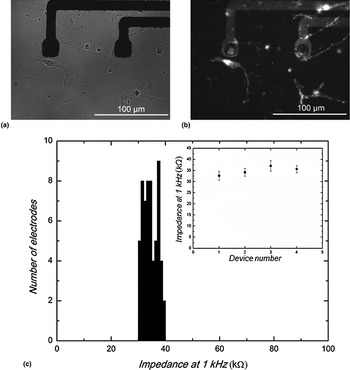
The MEAs were coated with PLL, and hippocampal cells were cultured on them for at least 21 days. In fact, maintaining the culture 3 weeks in vitro allows the neural network to become spontaneously active. Figure 1(a) shows a close-up of a cell culture after 21 days in vitro (DIV) on the MEA, with cells near the electrodes. Figure 1(b) shows a fluorescence image of the same frame. The cells were stained with vital fluorescent dye 4-Di-2-ASP, which stains the mitochondria of living neurons. This image, hence, shows that cells remain viable on the MEA. Figure 1(c) presents a histogram of impedance at 1 kHz (a frequency value used to benchmark neural electrodes) of 60 electrodes randomly selected from four devices. The value of impedance (34.6 ± 0.3 kΩ) is about two orders of magnitude smaller than that of Au electrodes of the same size, in agreement with previous work on conducting polymer electrodes.[ Reference Martin and Malliaras 18 ] This results in a background noise that is 9 µV peak-to-peak [3.2 µV root mean square (RMS)], when measured in the presence of cells, during a period when spiking activity was blocked by Tetrodotoxin (TTX) (see below). For comparison, in a 0.1 M NaCl solution, the background noise was 3.8 µV peak-to-peak (1.4 µV RMS). It should be noted that noise is considerably reduced compared with that measured in Au electrodes [Fig. S1(d), Supplementary material].
Biocompatibility assessment and device characterization. (a) Optical micrograph of a hippocampal cell culture on PEDOT:PSS electrodes and (b) a fluorescence image of the same frame. The fluorescent dye used is 4-Di-2-ASP (c) Histogram showing the variation in impedance at 1 kHz of 60 PEDOT:PSS-coated electrodes, randomly selected out of four different MEA devices. The inset shows the device-to-device reproducibility.

Electrophysiological recordings
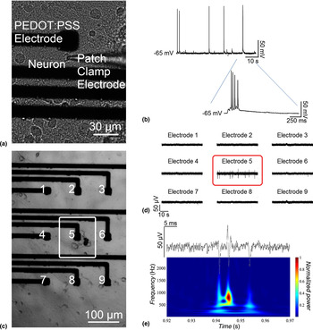
The cell cultures were tested for their excitability through a standard, well-established technique shown in Fig. 2(a). Patch clamp recordings were performed on 21 DIV cell cultures and on multiple hippocampal cells. Bursts of APs of 150 mV in amplitude were successfully recorded validating the ability of our devices to sustain a healthy neural culture (Fig. 2(b)). Extracellular recordings were subsequently performed with our conducting polymer MEAs. The cell-covered electrode [marked as electrode 5 in Fig. 2(c)] recorded extracellular spontaneous activity in contrast with the uncovered electrodes surrounding it [Figs. 2(c) and (d)]. Time-frequency analysis reveals the frequency content of the high-pass filtered (f c = 100 Hz) signal as the recordings show 50 µV of amplitude peaks with a frequency around 1 kHz, values that are characteristic of hippocampal cell APs. The time trace of the same recordings presents the extracellular APs in the time domain, displaying spikes originated, putatively, from multiple cells [Fig. 2(e)].
Electrophysiological recordings from cell cultures with different recording methods. (a) Infrared differential interference contrast (DIC) micrograph of a patch clamp electrode sealed at the surface of the membrane of a hippocampal neuron (21 DIV) and (b) the subsequent recording. The expanded trace below depicts a burst of action potentials. (c) DIC micrograph of a cell culture on the recording device and (d) extracellular recordings from the same culture. The micrograph shows the coverage of electrode 5 with cells. The covered electrode records extracellular activity while the non-covered surrounding electrodes do not. (e) Time-frequency analysis during a short period of the previous extracellular recordings showing peaks of activity around 1 kHz.

Chemical modulation of cell activity
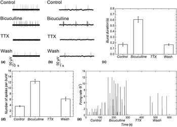
To demonstrate the potential of PEDOT:PSS MEAs in pharmacological applications, we tested different compounds on a different cell culture [Figs. S2(f,e)], with time duration of 2.5 min each. Figs. 3(a) and (b) depict the recorded activity under these conditions and for different time scales. The spontaneous activity (referred to as control) shows the initial firing behavior of the cells. Bicuculline (4 µM), a blocker of the ionotropic GABAA receptor, was consequently perfused in the recording chamber. Bicuculline induces hyperexcitability by blocking the inhibitory action of GABAA receptors resulting in an increased cell activity in the form of bursts of spikes as reported before.[ Reference Arnold, Hofmann, Bengtson, Wittmann, Vanhoutte and Bading 5 ] Indeed, the initial number of recorded cell spikes increased after drug application. TTX (300 nM), which inhibits AP firing by binding to the voltage gated sodium channels and blocking the passage of sodium ions, was subsequently administered. Its presence in the recording chamber resulted in the cell activity extinction as shown in Figs. 3(a) and (b). TTX sodium channel inhibition is reversible, so washing both drugs (bicuculline and TTX) and restoring the cell environment to a normal recording solution resulted in the partial recovery of the cell activity. The above qualitative analysis is followed by a quantitative one in Figs. 3(c)–(e) with the study of the burst duration, the number of spikes per burst and the firing rate (FR) for each condition. Figure 3(c) presents the mean burst duration for the different drug conditions. The initial duration of 0.17 ± 0.03 s for the spontaneous activity (which is referred to as control) increased to 0.61 ± 0.05 s with the use of bicuculline and dropped to zero after the perfusion of TTX. Interestingly, drug removal restored the duration to 0.17 ± 0.02 s. Figure 3(d) presents the change in the mean number of spikes per burst. As expected, the initial average number of 3.67 ± 0.21 spikes per burst increased to 12.42 ± 0.67 spikes per burst after the use of bicuculline, before returning to 6.20 ± 0.66 spikes per burst after the washing step. The use of TTX in between had set this value to zero.
Drug modulation of the extracellular activity. (a) Electrophysiological recordings for different chemical conditions. The initial spontaneous cell activity was modified by the application of bicuculline. TTX suppressed any activity while the activity recovers after washing with recording solution. (b) The smaller time scale depicts better the changes in the firing rate (number of spikes per second) during each condition. (c–d) Quantification of the experimental results. (c) The recorded burst duration and (d) the number of spike per burst increase and decrease corresponding to chemical application. (e) Firing rate profile evaluated over 600 s of activity under different conditions (bin size = 500 ms).

Another way to characterize neural activity is the cell FR and how it changes throughout the experiment. The FR is defined as the number of spikes recorded by an electrode in a time interval T divided by T. The smaller this interval is, the more accurately the FR simulates the instantaneous FR.[ Reference Kanagasabapathi, Massobrio, Barone, Tedesco, Martinoia, Wadman and Decre 6 ] In our case, the time interval was set to T = 500 ms (a typical value for this type of analysis) and it results in the histogram presented in Fig. 3(e). As expected, the initial FR, in the control condition, was increased (tripled) after the use of bicuculline. TTX set the FR to zero, as the cell activity was blocked, while the removal of both drugs increased it again. Similar experiments were performed with the use of a different drug and other chemical agents (4-Aminopyridine and KCl solutions), which also resulted in changing the firing behavior of the cells (Fig. S3, Supplementary material).
Conclusions
In conclusion, high-quality measurements of neural activity from hippocampal neuron cell cultures were performed using conducting polymer MEAs. A variety of drugs were used to modify the firing behavior of cells maintained in culture for 3–6 weeks. The results show that these MEAs are suitable for in vitro drug screening and toxicology studies.
Supplementary Material
The supplementary material for this article can be found at https://doi.org/10.1557/mrc.2017.34
Acknowledgements
The authors would like to thank the Marie Curie initial training network OLIMPIA for the financial support. The MEAs were fabricated at the Centre Microélectronique de Provence (Gardanne, France) clean room facilities while the cell culturing (B.H. and C.F.) and the electrophysiology (E.D.) measurements were performed at MyEnterix laboratories in Aix-Marseille University, CNRS, CRN2M, Marseille, France. The authors declare no competing financial interest.