Introduction
Congenital nasolacrimal duct obstruction is a common problem in infancy, affecting up to 20 per cent of newborns.Reference Wallace, Cox, White and MacEwen1–Reference Ram, Barras, White, MacEwen and Young5 The usual cause is a membranous obstruction at the distal end of the nasolacrimal duct resulting from incomplete canalisation. The clinical presentation varies from mild epiphora to sticky mucopurulent discharge.
In up to 96 per cent of children affected, the symptoms resolve spontaneously before the age of 1 year.Reference Wallace, Cox, White and MacEwen1, Reference MacEwen4–Reference Hanovar, Prakash and Rao6 After 12 months of age, the likelihood of spontaneous resolution decreases. For children who continue to suffer from epiphora, the surgical treatment of choice is probing and irrigation: the nasolacrimal drainage system is probed to open the blockage mechanically, and irrigation is performed with dilute fluorescein solution to confirm patency. Other surgical options include nasolacrimal silicone intubation and dacryocystorhinostomy.
However, there is still some controversy in the literature regarding the optimal timing and efficiency of probing and irrigation for the treatment of congenital nasolacrimal duct obstruction. Some studies report high success rates (90–97 per cent) when probing is performed within the first year of life, with success rates subsequently decreasing with increasing age.Reference Katowitz and Welsh7–Reference Gardiner, Forte, Pashby and Levin10 Some other studies claim that success is dictated by the type of obstruction.Reference Wallace, Cox, White and MacEwen1, Reference Kashkouli, Beigi, Parvaresh, Kassaee and Tabatabaee11, Reference Zilelioglu and Hosal12
In the majority of previous studies, probing was performed in a conventional manner; the probing was a blind procedure with recognised complications. In recent years, the development and use of rigid and flexible endoscopes in the nasal cavity has enabled the visualisation of the inferior meatus and the distal end of the duct. This has led to a better understanding of rhinological disease and the nature of obstruction in the lacrimal system.
The aim of the present study was to evaluate the results of lacrimal probing assisted with nasal endoscopy performed in our hospital, and to determine whether age at probing or site of obstruction had any prognostic significance for the outcome of probing.
Materials and methods
The current study comprised a retrospective, comparative case series of 50 eyes of 38 consecutive children with congenital nasolacrimal duct obstruction. The children were aged 12 months or older. They underwent nasolacrimal duct probing for the first time (performed by two ophthalmologists) in conjunction with nasal endoscopy (conducted by one otolaryngologist) at Victoria Hospital in Kirkcaldy (NHS Fife) between May 2007 and December 2011 (a study period of 56 months).
The diagnosis of presumed congenital nasolacrimal duct obstruction was based on a history of epiphora and discharge, and an abnormal fluorescein dye disappearance test result on examination.
Only those cases in which probing and irrigation alone were the initial treatments were included in the study. Children who had undergone previous probing treatments, and those for whom the epiphora had other causes such as eyelid malposition, were excluded from the study.
In all cases of presumed nasolacrimal duct obstruction, probing with irrigation was performed as day-case surgery under general anaesthesia. The technique of probing and irrigation has been described previously.Reference MacEwen, Young, Barras, Ram and White2, Reference Ram, Barras, White, MacEwen and Young5 Briefly, five drops of xylometazoline hydrochloride 0.05 per cent (paediatric Otrivine (Novartis AG, Basel, Switzerland)) were placed in the nasal cavity immediately after the induction of anaesthesia. This was followed by the precise placement of two neurosurgical pledgets soaked in paediatric Otrivine: one was placed under the inferior turbinate, and the other was placed between the inferior turbinate and nasal septum in order to constrict the vascular nasal mucosa and improve visualisation. Thereafter, the pledgets were removed and a careful intranasal examination was performed. This examination was conducted to identify any pre-existing nasal pathology, for example, an impacted inferior turbinate. The lacrimal punctae were inspected, and punctal stenosis, if present, was treated by dilatation.
The patency of the system was tested by irrigation with diluted fluorescein dye. A cannula was introduced via the upper canaliculus as far as the lacrimal sac, and the dye was injected through the system using a syringe. Nasal endoscopy and irrigation were performed simultaneously, and free flow of fluorescein from the lacrimal sac to the nose confirmed anatomical patency of the system.
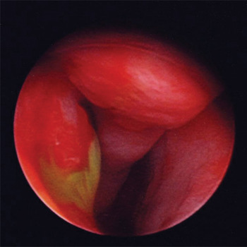
If no anatomical obstruction was present, and dye was seen to enter the nose in a smooth, steady flow, the child was diagnosed with ‘functional’ epiphora; that is, tearing secondary to physiological dysfunction. If no dye was seen passing into the inferior meatus, a diagnosis of atresia was made. If fluorescein had to be injected through the system by force with resultant ballooning of the nasal mucosa and poor flow of fluorescein (Figure 1), a diagnosis of stenosis was made. Probing was performed in those patients diagnosed with either stenosis or atresia.
Endoscopic view of fluorescein emerging from the stenosed Hasner's valve at the distal end of the nasolacrimal duct (right nostril).

Inferior turbinate infracture was performed as a prelude to probing. This not only improved visualisation of Hasner's valve at the distal end of the nasolacrimal duct, but it may also have established patency in some cases by stretching open the mucosal exit of the duct.
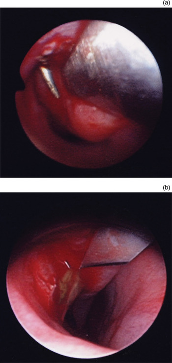
The lacrimal probe was passed through the upper punctum and canaliculus, into the sac and onwards into the duct. The probe was observed endoscopically as it entered the inferior meatus (Figure 2a), and cut down was performed with a phaco knife (a 3.2 mm keratome) (Figure 2b). Re-establishment of patency was confirmed by repeating the fluorescein injection.
Endoscopic views showing (a) a Freer elevator, with the probe emerging from Hasner's valve, entering the inferior meatus, and (b) cut down with a phaco knife.

Patients were evaluated post-operatively at three months. The primary outcome measure was treatment success or failure (three months after the probing surgery) based on an assessment of clinical signs. Success was defined as the absence of epiphora and mucoid or mucopurulent discharge. Success was confirmed by a normal fluorescein dye disappearance test result or near-complete resolution, with significant improvement of signs and minimal symptoms brought on by respiratory tract infections or exposure to wind or cold. If obstructive symptoms persisted beyond three months after an attempted probing, patients were referred to a larger centre for a repeat procedure or other treatment (silicone intubation). Such cases were considered probing and irrigation treatment failures.
Results and analysis
Thirty-eight children were included in the study and 50 lacrimal systems underwent primary probing with nasal endoscopy (Table I). The mean age of the children was 31.21 months (standard deviation ±18.19), with an age range of 17–109 months. The median age was 25 months. Of the 38 children, 19 were boys and 19 were girls. Twelve of the patients were affected bilaterally (five boys and seven girls).
Patient demographic and clinical characteristics

Data represent numbers of children (total n=38). *At time of procedure. Mth = months
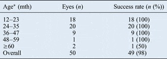
For the entire group, probing resulted in the successful resolution of symptoms in 98 per cent of cases. Forty-nine out of 50 eyes had complete resolution of symptoms. There was only one case in which probing failed to treat the nasolacrimal obstruction. The success rates according to the various age groups are shown in Table II.
Probing success according to age group

*At time of procedure. Mth = months
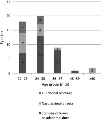
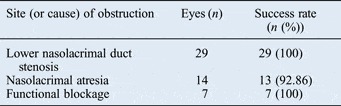
The anatomical sites of blockage within the drainage system were identified for each age group; these are summarised in Figure 3 and Table III. Overall, 50 eyes (of 38 children) were diagnosed with nasolacrimal duct obstruction: 29 eyes had lower nasolacrimal stenosis and 14 had nasolacrimal atresia. In 7 eyes (14 per cent; 4 children), the flow of the dye was free; these were considered to be anatomically patent and to have a functional blockage. Probing was successful in all cases of stenosis at the distal end of the nasolacrimal duct and in those with a functional blockage. However, the symptoms persisted for one child (one eye), but these were resolved at a later follow-up examination with histamines. The one probing treatment that failed was in a case with atresia. Nevertheless, 13 eyes of those (11 children) with atresia had resolution of symptoms after probing, giving a success rate of 92.86 per cent for this subgroup.
Site (or cause) of obstruction for each age group. Mth = months

Probing success according to site of obstruction

The one case of treatment failure occurred in a nine-year-old child with Down's syndrome. During the operation, complete ductal atresia was reported; the fluorescence dye failed to appear in the nose, and the probe could not penetrate the nasal mucosa in front of the inferior meatus. This suggested a more complex developmental abnormality, rather than the simple failure of Hasner's membrane to open. In particular, there was difficulty probing through what might have been scar tissue, and cut down was attempted twice. Although some minor improvement in fluorescein flow was reported, the flow was not satisfactory. The patient subsequently underwent dacryocystorhinostomy and her symptoms resolved.
Discussion
Epiphora is common in infants. It is most frequently the result of a congenital nasolacrimal duct obstruction.Reference Cassady13 This is an obstructive membrane at Hasner's valve, which opens spontaneously in approximately 95 per cent of affected cases by 1 year of age.Reference Paul14, Reference Nelson, Calhoun and Menduke15 Probing is the surgical treatment of choice for children with congenital nasolacrimal duct obstruction. However, there is some debate in the literature regarding the effectiveness of primary probing in children relating to the timing of this intervention.Reference Katowitz and Welsh7, Reference Mannor, Rose, Frimpong-Ansah and Ezra16 Some studies suggest that increasing age is associated with a lower cure rate of probing, while others have proposed that age may not be the major factor and that the type of obstruction may be crucial. The symptoms of patients with simple membranous obstruction at the nasal end of the lacrimal duct resolve spontaneously at a younger age, while the symptoms of more complex and resistant cases persist and worsen with time.Reference Wallace, Cox, White and MacEwen1, Reference Zilelioglu and Hosal12
In the majority of studies, probing was performed in a blind manner, without the use of nasal endoscopy to accurately identify the site of obstruction. In those studies, the success rate of primary probing in children older than 1 year fluctuated between 55 per cent and 90 per cent.Reference Katowitz and Welsh7, Reference Robb8, Reference Zilelioglu and Hosal12 Very few other studies within the last 10 years have achieved a success rate of 90 per cent or higher.Reference El-Mansoury, Calhoun, Nelson and Harley9, Reference Gardiner, Forte, Pashby and Levin10 In our study, probing was assisted with nasal endoscopy and cut down. The overall success rate was 98 per cent (49 out of 50 eyes). This is slightly higher than the success rate reported in two recent works, in which the authors also used endoscopy for congenital nasolacrimal duct obstruction in older children.Reference Wallace, Cox, White and MacEwen1, Reference Kouri, Tsakanikos, Linardos, Nikolaidou and Psarommatis17 The advantages of endoscopy are well-known.Reference Gardiner, Forte, Pashby and Levin10, Reference Sener and Onerci18 Visualisation of the inferior meatus can help in the identification and treatment of intranasal anomalies that obstruct the distal end of the nasolacrimal duct, and can facilitate guidance of the probe thereby avoiding the formation of false passages.
• Congenital nasolacrimal duct obstruction in children older than one year warrants probing as a first-line therapy
• The optimal timing for probing remains controversial
• The success rate of duct probing in children of different ages, using nasal endoscopy, was 98 per cent
• Age and site of obstruction did not affect the outcome of probing
• Lacrimal probing should be the primary treatment for congenital nasolacrimal duct obstruction in children of all ages
• Nasal endoscopy and cut down allow accurate localisation of the obstruction site, and can increase success
Seven eyes of four patients had anatomically patent lacrimal systems. These children, who had symptomatic epiphora and delayed fluorescein dye disappearance test results, seemed to have a functionally inadequate drainage system and were diagnosed with a physiological obstruction. This condition may have been caused by a number of factors, including an inadequate pumping mechanism, nasal mucosal oedema (if present with colds or allergy) or a tight inferior meatus. All four children in this group achieved a successful outcome after the operation, although one required treatment with histamines. The authors recognise that this successful outcome may have been partly attributable to the infracture of the inferior turbinate, which has been shown to improve the cure rates of probing due to stretching of the inferior meatus.Reference Wallace, Cox, White and MacEwen1 However, there were no signs of trauma around the valve and so a diagnosis of functional block was made.
The one patient who did not show any improvement in her symptoms had different nasolacrimal duct pathology and the child had Down's syndrome. Down's syndrome is thought to be a risk factor for failed probing.Reference Arora, Koushan and Harvey19, Reference Coats, McCreery, Plager, Bohra, Kim and Paysse20 Nasolacrimal system abnormalities, particularly punctual agenesis and canalicular atresia, are common in patients with Down's syndrome. The presence of these abnormalities makes standard bicanalicular probing difficult or even impossible, and more than one procedure may be required.Reference Coats, McCreery, Plager, Bohra, Kim and Paysse20
We have shown that anatomical anomalies within the lacrimal system are quite common in older children. Lacrimal probing remains the primary treatment for congenital nasolacrimal duct obstruction. Nasal endoscopy is a useful adjunct to probing, which significantly increases the success rate of the procedure. Our data indicate that the important factor for the success of probing is not age, but rather the presence of more complex obstructions. The findings suggest that lacrimal probing with nasal endoscopy and cut down should be the first-line treatment for congenital nasolacrimal duct obstruction. Endoscopy and cut down allow accurate localisation of the obstruction site, and significantly increase the success rate of the procedure.