Case report
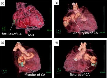
A 9-month-old female infant weighing 7.8 kg underwent a routine clinical work-up. Tachypnea and a continuous murmur were noted during the physical examination. On chest X-ray, the heart was enlarged and the pulmonary vasculature was engorged. An electrocardiogram demonstrated the presence of a normal sinus rhythm with right ventricular hypertrophy and a right axis deviation. Echocardiography revealed a dilated right atrium, a giant coronary artery with a diameter of 13 mm, and abnormal flow from the coronary artery into the right atrial cavity. Heart catheterisation revealed pulmonary hypertension. The pulmonary artery pressure was 56/32 mmHg, and the mean pressure was 30 mmHg. A giant right coronary artery and shunt flow from the coronary artery to the right atrium were also detected. However, these preoperative tests could not clearly identify the site and route of the coronary artery fistulas. To clearly visualise the fistulas and surrounding materials, we performed three-dimensional computed tomography, and the images were transferred as novel visualised three-dimensional images (Supplementary Figures S1 and S2). In this reported case, β-blockers or nitro-glycerine were not used before the computed tomography exam. The scanning protocol included a collimation of 64 sections with an individual thickness of 0.5 mm. The study data were transferred to a workstation for further reconstruction with maximum intensity projection, curved planar reconstruction, and volume-rendering techniques. The diagnosis was established by two cardiologists and an experienced radiologist, with the transverse images combining various reconstructions. A consensus of the specialists detected the coronary artery origin and the route of the anomalous vessel. The images successfully revealed three holes on the lateral side of the right atrium. The relationship between the coronary artery fistulas and the surrounding structures was easily identified (Fig 1).
( a ) Fistulas are present on the lateral side of the inner right atrium (green). ( b , c , d ) The series of still frames of a supplemental file 2. The fistulas drain into the right lateral wall of the right atrium (the drainage sites of the fistulas are green). ASD=atrial septal defect; CA=coronary artery.

The surgical repair was performed through median sternotomy. Cardiopulmonary bypass was established after heparinisation, and the right atrium was opened under hypothermic circulatory arrest. The fistulas of the coronary artery had three drains as illustrated by three-dimensional computed tomography. The fistula drains were directly closed using a 5–0 prolene suture. After rewarming, the patient was easily weaned from cardiopulmonary bypass without arrhythmias, and the postoperative hemodynamic state of the patient was stable. On postoperative day 1, cardiac angiography revealed that the fistulas were completely closed and blood flow in the aneurysm of the right coronary artery remained slow. We started warfarin and aspirin therapy to prevent postoperative complications such as thromboembolism and myocardial infarction. Echocardiography performed on postoperative day 3 confirmed the disappearance of blood flow through the fistulas and good cardiac function. The patient was discharged from our hospital on postoperative day 4 in a good condition.
Comment
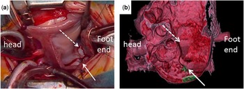
Coronary artery fistula is a rare anomaly that accounts for 0.2–0.4% of all cases of congenital heart disease. Affected patients may experience complications including heart failure, endocarditis, myocardial infarction, or coronary aneurysms. Over 90% of these fistulas drain into the venous structures of circulation, including the right heart chambers, pulmonary artery, coronary sinus, and superior vena cava. Drainage into the left heart chambers is less frequent. Fistulous drainage into the right ventricle, right atrium, pulmonary artery, left ventricle, coronary sinus, and superior vena cava occurs in 40%, 26%, 17%, 3%, 7%, and 1% of cases, respectively.Reference Dodge-Khatami, Mavroudis and Backer 2 – Reference Levin, Fellows and Abrams 4 Although the natural history of coronary artery fistulas varies and spontaneous closure is sometimes reported, treatment is generally recommended for symptomatic coronary artery fistulas. Treatment of asymptomatic patients may be considered to prevent related complications such as heart failure, endocarditis, and myocardial ischemia, the risk for which increases with age. Because the patient presented in our study had cardiac failure, surgical treatment was preferred.Reference Darwazah, Eida, Batrawy, Isleem and Hanbali 5 Echocardiography and angiography are effective examination techniques, but they cannot always accurately indicate the course and location of drainage of the coronary artery. Coronary angiography was reliable in identifying the size and anatomical features of the fistulous tract. Coronary angiography remains the gold standard for imaging the coronary arteries, but the relationship with other structures as well as their origin and route are not always apparent. In our institution, although coronary angiography is routinely performed, chest three-dimensional computed tomography is performed for selected patients whose diagnosis is inadequate from other exams or whose spatial structure of the cardiac anomaly is considered important for surgical treatment. Because medical radiation exposure at a young age is considered a risk for the future development of cancer,Reference Han, Lindberg, Overman, Schwartz, Grant and Lesser 1 and because the contrast medium may induce adverse effects, we selected a minimum number of cases for three-dimensional computed tomography. We used a 64-section multi-detector computed tomography scanner. The actual operative findings were similar to the preoperative imaging data (Fig 2). The preoperative three-dimensional computed tomography images may have allowed the surgeons to minimise the aortic cross clamp time. As a result, three-dimensional computed tomography might improve the safety of the operation and reduce the risk for residual shunting. The effective use of preoperative three-dimensional computed tomography could possibly facilitate the conduct of non-operative innovative therapeutic methods for the closure of coronary artery fistulas, such as catheter embolisation techniques using coils, a detachable balloon polyvinyl foam, and double-umbrella devices. Many articles described the surgical management of coronary artery fistula as safe and effective, resulting in high survival and closure rates.Reference Kamiya, Yasuda and Watanabe 6 – Reference Balanescu, Sangiorgi, Castelvecchio, Medda and Inglese 8 Accurate detection of the location of fistulous drainage is the most crucial point in the surgical treatment of coronary artery fistulas. In the surgical management of coronary artery fistulas, three-dimensional computed tomography may potentially be the most useful method. The three-dimensional computed tomography images may also provide diagnostic information for patients with coronary artery fistula who have coexistent congenital cardiac anomalies. In conclusion, this case report suggests that reconstructed three-dimensional computed tomography images may be useful to facilitate planning for therapeutic interventions in patients with coronary artery fistulas.
( a ) An image illustrating the opening of the RA and suturing of the ASD and coronary artery fistula. ( b ) The 3D-CT image of the surgical view. Both figures ( a and b ) show the inner structures of the RA. Fistulas of the coronary artery are seen on the lateral wall of the RA (solid arrow), and an ASD is seen under the swelling of the coronary aneurysm (dotted arrow). ASD=atrial septal defect; RA=right atrium; 3D-CT=three-dimensional computed tomography.

Acknowledgement
The authors would like to thank Mr Sakamoto, whose technical support and suggestions were of inestimable value for this study.
Financial Support
This research received no specific grant from any funding agency, commercial, or not-for-profit sectors.
Conflicts of Interest
None.
Supplementary material
To view supplementary material for this article, please visit http://dx.doi.org/10.1017/S1047951114000286.