Atrial septal defect is one of the most common forms of congenital heart diseases, defined as a defect in the interatrial septum.Reference Carlgren 1 , Reference Hijazi and Hellenbrand 2 On the basis of the anatomic position of the defect, it is divided into four subtypes including ostium secundum or fossa ovalis type (75%), ostium primum type (15%), sinus venosus type (10%), and coronary sinus defects.Reference Russell, Rouine-Rapp and Stratmann 3 Due to the atrial remodelling that occurs in patients with septal defects that confer a higher risk to arrhythmias. Therefore, closure of the defect is important because of the increased risk of arrhythmias in addition to haemodynamic consequences and thromboembolic events.Reference Roberts-Thomson, John and Worthley 4 Both surgical and percutaneous transvenous device techniques have been utilised to correct the interatrial septal defect.Reference Kharouf, Luxenberg and Khalid 5 , Reference Konstantinides, Geibel and Olschewski 6 Percutaneous transvenous device closure is more commonly used for the ostium secundum-type defects.Reference Hijazi, Cao and Patel 7 However, some patients with ostium secundum-type atrial septal defects undergo open-heart surgery for the repair of their defects.Reference Donti, Bonvicini and Placci 8
P-wave duration is prolonged in patients with atrial septal defects compared with healthy people.Reference Ho, Chia and Yip 9 Post-operative changes in P-wave duration after surgical and percutaneous device closure of atrial septal defects indicate an ongoing atrial remodelling in these patients.Reference Yavuz, Nisli and Oner 10 , Reference Guray, Guray and Mecit 11 Comparing two different techniques of atrial septal defect closure and their effect on P-wave, it can be noted that PR segment duration and PR interval is of great importance.
Owing to a lack of standard guidelines for evaluation of internodal conduction pattern and atrial electrical conductance in patients with atrial septal defect,Reference Kharouf, Luxenberg and Khalid 5 our study was designed to compare specific electrocardiographic changes following either surgical or percutaneous closure of atrial septal defects. We hypothesised that there was a decrease in P-wave, PR segment duration, and PR interval before the procedure and 6 months after the procedure among the patients with percutaneous and surgical closure. To test this hypothesis, we compared the duration of P-wave, PR segment, and PR interval in adults with atrial septal defect.
Methods
The study design was reviewed and approved by the Institutional Review Board and Ethics Committee at Shahid Madani Heart Center, a major teaching hospital of Tabriz University of Medical Sciences. The study design was a prospective non-randomised clinical observation.
Patient selection and preparation
The patients were examined pre-operatively and then were assigned to either surgical or percutaneous transvenous closure of their isolated ostium secundum-type atrial septal defect between 2004 and 2010. Assignment to either treatment group was not randomised, and it was based on size and rim margins of the defects at the discretion of the treating cardiologist, cardiac surgeon, and patient preference. The patients were approached and informed consent was obtained. The surgical group underwent an open-heart surgical procedure with cardiopulmonary bypass where a median sternotomy was performed with a right atriotomy and application of a CardioSEAL patch (NMT Medical Incorporation, Boston, Massachusetts, United States of America) under direct visualisation. The patients in the percutaneous group underwent a closure approach that consisted of percutaneous access to the defect via the femoral vein with subsequent application of the Amplatzer occlusion device (AGA Medical Corporation, Plymouth, Minnesota, United States of America) under fluoroscopic and echocardiographic guidance.
The following patients were excluded: coexisting congenital, ischaemic and valvular heart disease, cardiac failure, chronic pericardial effusion, thyroid disease, diabetes mellitus, chronic obstructive pulmonary disease, smoking history, use of antiarrhythmics, any amount of residual shunt by transthoracic echocardiography at the time of follow-up, less than 6 months’ time interval from treatment, history of persistent paroxysmal atrial fibrillation, and missing follow-up data within 1 year after therapeutic intervention. In all, 15 patients in the surgical group were excluded from our study because of either an incomplete hospital database or lack of interest in participation in the study; nine patients in the percutaneous group were excluded from the study because of either a time interval of less than 6 months between treatment and evaluation or they had a residual shunt.
Electrocardiographic studies
Two 12-lead electrocardiograms were obtained. The first electrocardiogram was obtained 24 hours before therapeutic intervention. The second electrocardiogram was obtained at an outpatient hospital clinic after treatment. A paper speed of 25 millimetres per second, 1 millivolt per centimetre standardisation was used. Post-treatment electrocardiograms were obtained in the same manner that the pre-treatment electrocardiograms had been obtained. Patients were asked to lie down for at least 15 minutes and be completely calm before electrocardiogram recordings. The P-waves, PR intervals, and PR segments were measured by two physicians blinded to each other's measurements and to the type of atrial septal defect treatment. The point where the first visible upward or downward movement was taken as the P-wave beginning and the junction between the P-wave and the isoelectric line was taken as the P-wave end point. PR interval was defined as the distance between the onset of the P-wave and the beginning of the QRS complex. PR segment was defined as the distance between the end point of the P-wave and the beginning of the QRS complex. Average P-wave and PR segment duration and PR interval for each patient, before and after treatment, was calculated using the sum of the measurable P-waves, PR segments, and PR intervals in each of the 12 leads divided into the number of used leads and then the two physicians’ measurements were averaged to obtain the final data. More than a 20-millisecond difference between the two physicians’ measurements was defined as the measurement of repeating criterion. Measurement repeating occurred in 37 waves. In each lead, one wave was measured. Undetectable waves were omitted from the study.
Ultrasound examinations
All patients were examined using transthoracic echocardiography (Vivid7, GE-Healthcare, Milwaukee, Wisconsin, United States of America) and 3.0 MHZ probe. Four standard views were obtained for all patients. Apical four-chamber view was used to measure the size of the right and left atria. After tracing the area, the results were reported in square centimetre. Diameters of the atria were also measured and the averages of three measurements were reported. Transoesophageal echocardiographic examinations were performed with using a multiplane 7.0 MHZ probe. Transeosophageal echocardiography was performed 2 days before therapeutic intervention was used for evaluation of the atrial septum and to confirm the size – the largest diameter, definite location, morphology, and visualisation of the rims of the defect in all patients. Mid-oesophageal four-chamber and bicaval views in two-dimensional imaging and color Doppler exams were obtained. One experienced cardiologist performed all transesophageal echocardiography studies during both surgery and percutaneous procedures. Right ventricular diastolic diameter, right atrium size, atrial septal defect size, right ventricular systolic pressure, and pulmonary flow-to-systemic flow ratio were evaluated only before therapeutic intervention. Transthoracic echocardiography was performed 1 hour before transoesophageal echocardiography evaluation.
Patient follow-up
All patients were seen in outpatient Cardiology Clinic between 6 months and 12 months after their initial corrective treatment. During the follow-up visit, a transthoracic echocardiographic examination was performed and a 12-lead electrocardiogram was obtained, and the results were recorded and analysed.
Statistical analyses
Data are shown as mean plus or minus standard deviation. Continuous variables were compared using Mann–Whitney U-test, and Kendall's tau-b rank correlation coefficients were used to evaluate the correlation between atrial septal defect size and P-wave, PR segment, and PR interval changes in surgical and percutaneous groups. The null hypothesis was rejected when alpha was less than 0.05. The SPSS 13.0 statistical software (SPSS Incorporation, Chicago, Illinois, United States of America) was applied for all statistical calculations.
Results
A total of 32 patients were included in the study of whom 16 patients (8 women, 8 men; mean age, 31.3 plus or minus 11.5 years) underwent surgical closure (SURG group) and 16 patients (10 women, 6 men; mean age, 30.8 plus or minus 10.8 years) underwent percutaneous transvenous closure (PCTV group).
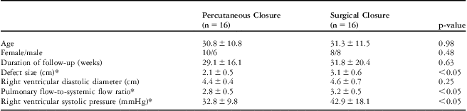
Demographic characteristics and transthoracic echocardiographic data of our studied population are shown in Table 1. In the echocardiographic evaluation, pulmonary flow-to-systemic flow ratio was similar between the surgical group and the percutaneous group (3.2 plus or minus 0.5 versus 2.8 plus or minus 0.5, p-value equal to 0.09). Patients who underwent surgery also had larger atrial septal defect sizes compared with patients who were treated with percutaneous transvenous technique (3.1 plus or minus 0.6 versus 2.1 plus or minus 0.5 centimetres, p-value less than 0.05). The surgical group patients also showed higher right ventricular systolic pressure compared with the percutaneous group (42.9 plus or minus 18.1 versus 32.1 plus or minus 9.8 millimetres of mercury, p-value less than 0.05). In our study, right ventricular diastolic diameter measurement results before treatment were not statistically different between percutaneous and surgical groups (4.5 plus or minus 0.6 versus 4.6 plus or minus 0.7 centimetres, p-value equal to 0.66) (Table 1).
Demographic and Transoesophageal echocardiographic data of studied population.

*p-value < 0.05 is considered statistically meaningful
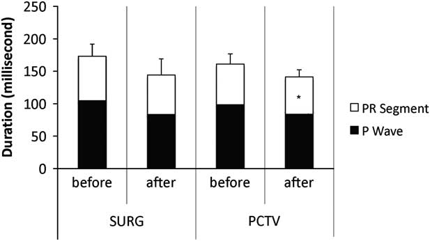
Baseline pre-operative data for the percutaneous group included P-wave duration of 98.5 plus or minus 15.4 milliseconds, PR interval of 162.9 plus or minus 18.5 milliseconds, and PR segment duration of 63.4 plus or minus 14.5 milliseconds. Baseline pre-operative data for the surgical group included P-wave duration of 104.5 plus or minus 24.7 milliseconds, PR interval of 173.2 plus or minus 38.7 milliseconds, and PR segment of 68.6 plus or minus 18.7 milliseconds. Baseline pre-operative data were similar in both groups. Most patients had decreases in the duration of P-wave, PR segment, and PR interval within 6 months of closure of their defects. There were a few patients in both groups who either had increases or had no changes in the duration of these parameters (Table 2). The patients who had either an increase or no change in duration also had a baseline right atrial size that was considered to be within normal limits and not enlarged. P-wave duration reduced from 98.5 plus or minus 15.4 to 86.4 plus or minus 13.2 milliseconds (p-value less than 0.05) in the percutaneous group. Similarly, P-wave duration reduced from 104.5 plus or minus 24.7 to 83.2 plus or minus 13.3 milliseconds (p-value less than 0.05) in the surgical group. A similar trend was observed in the duration of PR interval. PR interval decreased from 162.9 plus or minus 18.5 to 140.6 plus or minus 15.2 milliseconds (p-value less than 0.05), in the percutaneous group. A similar reduction in PR interval (173.2 plus or minus 38.7 versus 144.3 plus or minus 32.0 milliseconds, p-value less than 0.05) was observed in the surgical group (Fig 1). P-wave duration changes after treatment was not significantly different in the percutaneous group compared with the surgical group (−12.3 plus or minus 14.1 versus −21.3 plus or minus 22.6 milliseconds, p-value equal to 0.22). Difference in the duration of PR interval changes was also not significant between the two groups (−18.3 plus or minus 12.7 versus −28.9 plus or minus 24.1 milliseconds, p-value equal to 0.47).
Changes in P-wave duration, PR segment, and PR interval between two treatment groups.

Any changes less than 1 millisecond in duration was considered as “No Change”
*p-value < 0.05 is considered statistically meaningful
PR segment (□) and P-wave (▪) duration is shown before closure and six months after either percutaneous closure (PCTV) or surgical closure (SURG). Asterisks indicate a significant difference comparing before to after measurements.
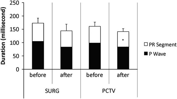
PR segment duration after percutaneous transvenous device closure was reduced significantly (63.4 plus or minus 14.5 to 52.1 plus or minus 10.8 milliseconds, p-value less than 0.05). PR segment duration did not significantly change after surgical closure (68.6 plus or minus 18.7 versus 61.1 plus or minus 24.7 milliseconds, p-value equal to 0.16). Although the mean difference of PR segment duration did not significantly decrease in the surgical group, the majority of surgical group patients did see a decrease in PR segment. In addition, the difference in PR segment changes after treatment between the two study groups was not significant (−11.3 plus or minus 15.0 versus −7.6 plus or minus 20.5 milliseconds, p-value equal to 0.98).
Right atrium was measured 25.8 plus or minus 10.3 square centimetres in the surgical group and 23.8 plus or minus 9.7 square centimetres in the percutaneous group. There was a direct correlation between baseline duration of P-wave and the size of the right atrium (R2 equal to 0.68, p-value less than 0.001). During the follow-up electrocardiogram (6–12 months), the size of the right atrium was 16.2 plus or minus 9.4 square centimetres in the surgical group and 18.2 plus or minus 9.6 square centimetres. There was no correlation between changes of P-wave, PR segment, and PR intervals with the atrial septal defect size in either group. There were no signs of failed repair in the form of recurrent shunt in either group.
Discussion
Atrial dysrhythmias add to post-cardiotomy morbidity. Studies have shown associations between increased conduction markers, such as P-wave duration and subsequent development of arrhythmias.Reference Dixen, Larsen and Ravn 12 Conduction markers, such as P-wave durations and P-wave dispersion, are increased in patients with atrial septal defects.Reference Donti, Bonvicini and Placci 8 , Reference Ho, Chia and Yip 9 There are several studies regarding P-wave duration and dispersion changes after surgical correction and percutaneous techniques.Reference Donti, Bonvicini and Placci 8 , Reference Yavuz, Nisli and Oner 10
Our study compared the effects of surgical and percutaneous transvenous closure of atrial septal defect on P-wave, PR interval, and PR segment duration. In our study, we used mean P-wave, PR interval, and PR segment duration instead of dispersion in adults. There were significant reductions in P-wave duration and related PR interval after treatment in surgical and percutaneous groups. Decreases in PR interval were related to concurrent reductions in P-wave duration and PR segment duration following both treatments.
Veldtman et alReference Veldtman, Razack and Siu 13 also showed electrophysiologic improvement after percutaneous transvenous device closure of atrial septal defects. They demonstrated a significant reduction in PR interval, right atrium and ventricle sizes after device treatment of isolated atrial septal defects in adult patients supporting the idea of improvement in right atrium and right ventricle physiology after closure of atrial septal defects. However, their study did not examine the effects after surgical closure. Guray et alReference Guray, Guray and Mecit 11 also studied the effects of surgical closure on the electrophysiology of the atrium. Their study specifically looked at P-wave duration and P-wave dispersion. Their study did not examine the effects after percutaneous transvenous device closure. Considering the findings from our study and the other studies preceding it, it is clear that there are electrophysiologic improvements after both surgical and percutaneous transvenous device closure of the atrial septal defects. The interatrial pathway is located in the interatrial septum that is prone to iatrogenic damage. Regional conduction disturbances during surgery can potentially occur through manipulation of the interatrial septum.Reference Morton, Sanders and Vohra 14 , Reference Thilén, Carlson and Platonov 15 It is noteworthy to mention that Guray et alReference Guray, Guray and Mecit 11 have examined the impact of P-wave duration on arrhythmias after surgical closure, and they have found a decrease in P-wave duration and P-wave dispersion without associated arrhythmias. The causes of decreased arrhythmias observed after closure are yet to be known.
In conclusion, our analysis showed that percutaneous transvenous device closure was equivalent to open-heart surgical closure of atrial septal defects in terms of improving the electrophysiology of the heart, specifically in reducing the P-wave duration, PR segment, and PR interval. A limitation of our study was that our patients were not randomised. The characteristics of each treatment group were different in terms of baseline atrial size, pulmonary flow–to-systemic flow ratio, and right ventricular systolic pressure. This was mainly due to the limitations of each closure technique. Therefore, future studies, once technical advancements can allow for randomisation, are warranted.