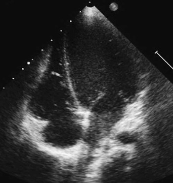
Simple transposition of great arteries had been diagnosed at birth in a 3.4 kilograms baby who underwent a successful arterial switch operation with an uneventful post-operative course. At the age of 6 years, the child was asymptomatic but echocardiography showed an isolated left ventricular volume overload. The end diastolic left ventricular diameter was 5.3 centimetres, ejection fraction 65%, and shortening fraction 36% (Fig 1). No other abnormalities or signs of ischaemia were found at echocardiography. The electrocardiogram was normal.
A four-chamber view showing left ventricular dilatation. Systolic function was normal.

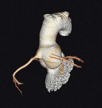
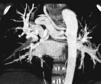
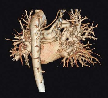
The patient was scheduled to undergo cardiac computed tomography, which clearly assessed the patency of both coronary arteries (Fig 2) but revealed three anomalous aorto-pulmonary vessels originating from the thoracic aorta, mimicking enlarged and abnormal bronchial arteries directed to the right lung (Figs 3 and 4).
Volume rendering reconstructions by cardiac computed tomography showing patent coronary arteries late after arterial switch operation.

Maximal intensity projection reconstructions revealed three enlarged and tortuous aorto-pulmonary collaterals from the thoracic aorta to the right lung.

Volume rendering reconstruction of the aorto-pulmonary collaterals.

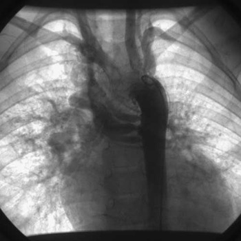
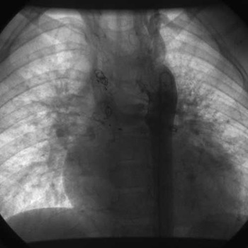
The patient underwent cardiac catheterisation, which confirmed the findings of cardiac computed tomography (Fig 5); during the same exam, he underwent percutaneous embolisation of multiple collaterals by using vascular plugs and spring coils (Fig 6).
Aortic angiography confirmed the number and morphology of the aorto-pulmonary collaterals as detected by computed tomography.

Aortic angiography after percutaneous embolisation.

During a mid-term follow-up, serial echocardiographic evaluations confirmed the complete regression of left ventricular volume overload.
These images underline the clinical usefulness of cardiac computed tomography to exclude coronary complications after arterial switch operation, to detect misdiagnosed aorto-pulmonary collaterals, and to plan percutaneous interventions.