Introduction
Congenital tracheal stenosis is defined as tracheal stenosis caused by a complete tracheal ring forming the posterior wall of the stiff trachea and a membranous trachea lacking normal structure. The occurrence of airway obstruction can be caused by the formation of lumen with the proliferation of tracheal submucosal glands and connective tissue, or by other reasons such as tracheal intubation injury, vascular ring compression, and tracheomalacia. Reference Cantrell and Guild1 It was first reported by Wolman in 1941, who described the disease as tracheal stenosis of different lengths and degrees caused by the presence of complete tracheal cartilaginous ring, with an incidence rate between 0.3 and 1% of all laryngobronchial stenosis. Reference Du, Jinlong and Jinfen2 Congenital tracheal stenosis is rarely isolated, accounting for only 10–25%. Reference Benjamin, Pitkin and Cohen3 It is often combined with other congenital malformations, the most common is cardiovascular and cerebrovascular abnormalities, and the incidence is as high as 50% in foreign countries, Reference Loeff, Filler and Vinograd4 including cardiovascular abnormalities (pulmonary artery sling, patent ductus arteriosus, ventricular septal defect, double aortic arch, and subclavian artery abnormalities), respiratory abnormalities (absence or hypoplasia of pulmonary arteries, tracheobronchus), and others. It is widely believed that the abnormal development of the respiratory system during embryonic development leads to congenital tracheal stenosis and related diseases. Researchers speculate that the occurrence of congenital tracheal stenosis may be mainly related to the abnormal development of the embryo in the fourth week. This may be highly related to the faster longitudinal elongation rate of the stenotic segment of the trachea than normal growth in fetal patients. Reference Chen, Wu, Wang, Chou, Chen and Huang5 With the in-depth understanding of the embryonic development of trachea, molecular studies have found that some molecular channels and transcription factors play an important role in the development of trachea. Herrera et al. Reference Herrera, Caldarone and Forte6 summarized the transcription factors and gene locations related to the formation of bronchial tree reported in the relevant literature in recent years and believed that the formation of congenital tracheal stenosis was closely related to Shh gene at 7q36 locus, Gli2 gene at 2q14 locus, Gli3 gene at 7p13 locus, etc.
In the early stage, due to the lack of understanding of the disease, the blind use of conservative treatment resulted in long-term dependence on ventilator after surgery, which was prone to cause respiratory infection, leading to respiratory failure and death. It has been a great challenge for paediatric cardiac surgeons for a long time. Although a variety of methods have been used to correct congenital tracheal stenosis, including costal cartilage tracheoplasty, pericardial patch tracheoplasty, tracheectomy, autologous tracheal transplantation, and slide tracheoplasty. However, in the past 20 years, slide tracheoplasty has gradually become the mainstream method for the correction of congenital tracheal stenosis, especially long-segment congenital tracheal stenosis, in many medical centres around the world due to the advantages of the tracheal diameter expanding to twice of the original and the length of the tracheal stenosis segment only shortening by half of the original stenosis segment. Reference Maltezeanu, Aldriweesh and Chan7 This article reviews the classification, past and current situation, advantages, surgical indications, surgical techniques, prognosis, related risk factors, and prospects of slide tracheoplasty in the treatment of congenital tracheal stenosis in children.
Classification of congenital tracheal stenosis
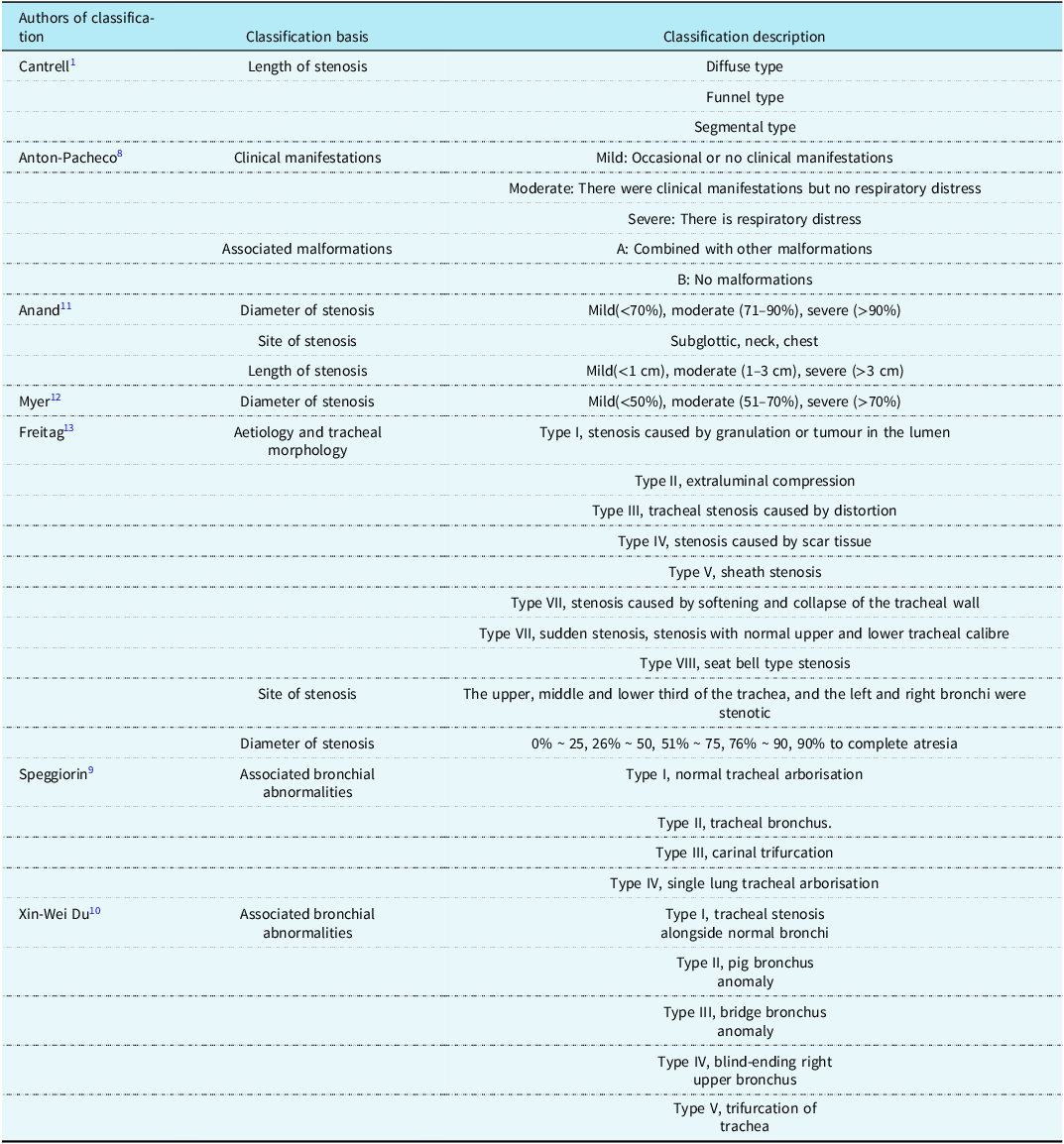
In the past 60 years, clinicians and scholars have proposed more than a dozen different classifications of the disease based on its anatomical characteristics and clinical manifestations (Table 1). Cantrell and Anton-Pacheco classification is still the most widely used classification. According to the anatomical structure of congenital tracheal stenosis, the classical classification of congenital tracheal stenosis was first proposed by Cantrell and Guild. In each classification, complete tracheal rings are present, while the main bronchus has normal morphology and is not involved. Reference Cantrell and Guild1 In 2003, Anton-Pacheco proposed a clinical classification based on patients’ clinical manifestations and airway function. Reference Anton-Pacheco, Cano, Garcia, Martinez, Cuadros and Berchi8 Anton Pacheco classification and Cantrell classification have been widely used for a long time to determine the surgical indications and provide important clinical evidence for the selection of surgical methods. The Great Ormond Street Hospital for Children team Reference Speggiorin, Torre, Roebuck, Mclaren and Elliott9 believes that Cantrell classification and Anton-Pacheco classification, which are widely used nowadays, cannot clearly describe more than 30% of airway morphology, especially the trachea with abnormalities. So, in 2008, their group proposed a new classification describing the association between different tracheobronchial arborisations and the site and length of the stenosis. All the morphologic variants were divided into 4 main types according to tracheobronchial arborisation, and each of these types included congenital tracheal stenosis or congenital tracheobronchial stenosis, depending on the distal extent of the stenotic segment. In their system, type I includes normal tracheobronchial arborisation, where the trachea divides at the carina into the left and right main bronchi. Type II includes the presence of a tracheal right upper lobe bronchus with the tracheal right upper lobe bronchus origin at any point on the lateral aspect of the trachea and at least 5 mm above the carina. Type III includes carinal trifurcation, where the trachea divides into 3 bronchi. Type IV includes a single right or left lung where the trachea continues mainly into 1 right or left main bronchus and the contralateral bronchus is absent or severely hypoplastic. In 2025, the team of Shanghai Children’s Medical Center Reference Du, Wang and Wang10 also proposed a new classification. Type I is tracheal stenosis alongside normal bronchi, type II is a pig bronchus anomaly, type III is a bridge bronchus anomaly, type IV is a blind-ending right upper bronchus (resembling a diverticulum-like anomaly), and type V is a trifurcation of the trachea.
Description and basis of the classification of congenital tracheal stenosis

Past and current status of slide tracheoplasty in the treatment of congenital tracheal stenosis
Cantrell Reference Cantrell and Guild1 in 1964, Carcassone Reference Harrison, Heldt, Brasch, de Lorimier and Gregory14 in 1973, and other surgical teams Reference Grillo and Zannini15,Reference Mansfield16 in the early 1980s successfully performed anastomosis after resection of segmental tracheal stenosis, which is suitable for children with Cantrell type III. In general, the stenotic trachea with less than one-third of the total length of the trachea can be safely resected, while the resection of more than one-third of the stenotic trachea will be accompanied by significantly increased complications, such as postoperative restenosis and anastomotic leakage. Reference Lobe, Hayden, Nicolas and Richardson17 Until 1989, Tsang et al. Reference Tsang, Murday, Gillbe and Goldstraw18 proposed a surgical technique named “slide tracheoplasty” for the first time. In 1994, Grillo Reference Grillo19 improved this method and popularised it. Slide tracheoplasty was defined by determining the length and midpoint of the tracheal stenosis under bronchoscopy. The trachea was cut at the midpoint, the middle of the anterior wall of the upper trachea was cut beyond the stenosis, and the middle of the posterior wall of the lower trachea was also cut to the distal end of the stenosis opening, and the posterior wall of the upper trachea was anastomosed with the anterior wall of the lower trachea. However, Grillo Reference Grillo19 cut the posterior wall of the upper trachea and the anterior wall of the lower trachea, and then anastomosed the upper and lower trachea. In 2008, Brazdil et al. Reference Brazdil, Tlaskal, Vojtovic, Pohunek and Cihar20 successfully used sliding anastomosis between the trachea and the right bronchus to correct children with tracheal stenosis above carina and right bronchial opening stenosis. Therefore, for children with carina or bronchial stenosis, it is often necessary to extend the longitudinal incision of the lower trachea to one main bronchus (Figure 1a). In 2009, Tokyo Municipal Pediatric General Medical Center Reference Toma, Kamagata and Hirobe21 described a modified slide tracheoplasty in which the anterior wall of the lower segment was cut into an inverted “Y” shape and extended to the main bronchi on both sides, solving the problem of long-segment congenital tracheal stenosis above the carina combined with stenosis of the left and right bronchial openings (Figure 1b). In 2010, Huang et al. Reference Huang, Wu and Wang22 reported the case of treating congenital tracheal stenosis with bridging bronchus by sliding combined with autologous tracheal patch (Figure 1c). Specifically, the bridging bronchus was cut-off between the carina and the opening of the right upper bronchus, the right side of the upper end was cut to the opening of the right upper bronchus, and the lower segment of the bridging bronchus was removed above the carina. Then, the carina was incised to the lateral side of the left main bronchus and the enlarged opening of the right bronchus, and the upper and lower segments were gliding anastomosed to form the anterior and posterior walls of the new carina, respectively. The excised tracheal bridge was trimmed into an autologous tracheal patch, which was added to the anterior wall of the new tracheal carinal to enlarge the right bronchial opening. In 2015, Wang et al. Reference Wang, Zhang, Zhu, Zhen, Liu and Xu23 from Shanghai Children’s Medical Center used side-to-side slide tracheoplasty for the first time to solve the problem of supracarinal tracheal stenosis combined with bronchial bridge stenosis (Figure 1d). They severed the trachea at the upper edge of the first carinal process, the proximal end was cut-off along the right side wall of the trachea, and the distal bridge bronchus was cut-off along the left side wall to the secondary carinal process. The upper and lower ends were then anastomosed side to side. Subsequently, in 2020, Chen Hao et al. Reference Chen, Shi, Zhu, Wang, Lu and Xu24 from Shanghai Children’s Medical Center reported a clinical study of modified slide tracheoplasty in the treatment of sucarinal tracheal stenosis combined with bridging bronchus and distal secondary bronchus (Figure 2).
Clinical application of different types of slide tracheoplasty. (a) Sliding anastomosis was performed between the trachea and the right bronchial opening. (b) Sliding anastomosis was performed between the trachea and the left and right main bronchi. (c) Autologous tracheal patch combined with sliding anastomosis. (d) Sliding anastomosis of side-to-side anastomosis was performed.

The extending distal incision made in the presence of right lower bronchial stenosis.

Advantages of slide tracheoplasty in the treatment of congenital tracheal stenosis in children
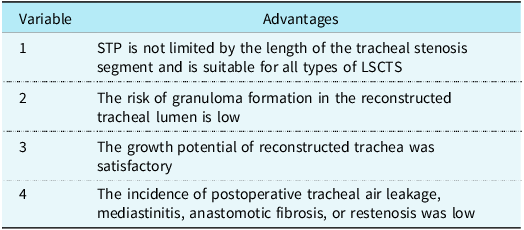
Slide tracheoplasty has greatly improved the prognosis of long segment and diffuse tracheal stenosis, because it has a variety of advantages that are irreplaceable by other surgical procedures (Table 2) : (1) slide tracheoplasty is not limited by the length of the tracheal stenosis segment. The diameter of the stenotic trachea was enlarged by 2 times, and the length of the stenotic trachea was shortened by only 1/2. Therefore, even if the whole tracheal stenosis or long tracheal stenosis sliding anastomosis will not produce intolerable anastomotic tension. Through modification, it can be successfully applied to all types of long-segment congenital tracheal stenosis, including bronchial stenosis and tracheal bypass. Therefore, we considered that for congenital tracheal stenosis with a stenosis length of more than 30% of the main tracheal length, slide tracheoplasty was recommended. (2) The risk of granuloma formation in the reconstructed tracheal lumen is low. The upper and lower segments of the trachea glide and anastomose with each other, and the continuous eversion suture makes the mucosa of the trachea smooth and continuous, and no foreign patch is introduced. Therefore, there are normal airway cilia and epithelial cells in the wall of the trachea after plasty, which reduces the risk of postoperative granulation formation and ultimately reduces the probability of postoperative restenosis. After weaning from ventilator, spontaneous breathing can be established earlier. (3) The growth of reconstructed trachea was satisfactory. Speggiorin et al. Reference Speggiorin, Gilbert, Broadhead, Roebuck, Mclaren and Elliott25 reviewed the follow-up data of 14 children 2 years after surgery and showed that the tracheal growth in children with congenital tracheal stenosis was not inhibited after slide tracheoplasty, and the tracheal growth was the fastest in the first 6 months after surgery. Wilcox et al. Reference Wilcox, Schweiger, Hart, de Alarcon, Peddireddy and Rutter26 conducted a follow-up study on 40 patients after slide tracheoplasty, and they found that the average growth rate of tracheal diameter after slide tracheoplasty was 0. 42 mm/ year. Liqin Chen et al. Reference Chen, Zhu and Wang27 reported the experience of slide tracheoplasty in the treatment of congenital tracheal stenosis in children at their institution. The median follow-up time was 4.44 (0.26–66.73) months. Ninety-two patients underwent chest CT reexamination, and the trachea diameter had increased significantly from 2.32 ± 0.72 mm to 5.46 ± 1.24 mm. Therefore, after slide tracheoplasty, the growth of the reconstructed trachea is not inhibited and full growth can be obtained. (4) The incidence of anastomotic problems is low, and the incidence of postoperative tracheal air leakage, mediastinitis, anastomotic fibrosis, or restenosis is low. Reference Maltezeanu, Aldriweesh and Chan7
Advantages of slide tracheoplasty in the treatment of congenital tracheal stenosis in children

Indications of slide tracheoplasty in the treatment of congenital tracheal stenosis in children
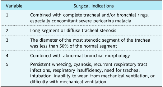
The poor outcome of early surgical repair has led to the consensus that the severity of congenital tracheal stenosis should not only focus on the diameter of the stenotic segment. Contemporary surgical treatment of congenital tracheal stenosis is essentially based on the overall clinical presentation of the child. Based on our clinical experience in surgical treatment of congenital tracheal stenosis and literature reports, we have summarised 5 surgical indications (Table 3) :
Surgical indications for slide tracheoplasty in the treatment of congenital tracheal stenosis in children

-
1. Pathological features: combined tracheal and/or bronchial rings, especially concomitant severe pericarinal malacia. Wilcox et al. Reference Wilcox, Hart and de Alarcon28 suggested that most children with complete tracheal rings have biphasic respiratory murmurs, laboured breathing, and cyanosis in infancy, which require early surgical repair. Rutter et al. Reference Rutter, Cotton, Azizkhan and Manning29 believed that the clinical manifestations of congenital tracheal stenosis are usually related to complete tracheal rings, which is a rare disease with a high mortality rate. Slide tracheoplasty is currently the intervention of choice for this condition. Studies have reported that preoperative tracheobronchomalacia is a risk factor for poor prognosis in patients with congenital tracheal stenosis. Butler et al. Reference Butler, Speggiorin and Rijnberg30 believed that slide tracheoplasty is a universal and reliable treatment method for congenital tracheal stenosis with tracheobronchomalacia.
-
2. Stenosis length: slide tracheoplasty is preferred for long segment or diffuse tracheal stenosis. Short-segment tracheal stenosis is often treated by resection of the stenotic segment followed by end-to-end anastomosis. Long segment tracheal stenosis requires slide tracheoplasty, and the definition of long segment tracheal stenosis is different in different centres. Wang et al. Reference Wang, Zhang, Zhu, Zhen, Liu and Xu23 considered that the length of the stenosis segment > 30% of the total length of the trachea was defined as long segmental tracheal stenosis. Similarly, studies have shown that resection of the length of the trachea < 30% of the total length of the trachea will not cause postoperative high tension of end-to-end anastomosis. Reference Maeda and Grillo31 Dayan et al. Reference Dayan, Dunham, Backer, Mavroudis and Holinger32 reported that end-to-end anastomosis after resection of the stenosed segment is not suitable for cases in which the length of the stenosed segment is more than 50% of the total length of the trachea, but such cases can be successfully corrected by slide tracheoplasty. Elliott et al. Reference Elliott, Roebuck and Noctor33 considered that slide tracheoplasty should be the first choice of surgical treatment when the length of the stenosed segment of the trachea was more than 2/3 of the total length of the trachea.
-
3. The diameter of the stenotic segment: generally, children can tolerate at least 50% of the normal tracheal diameter stenosis before showing severe respiratory symptoms. Children with obvious respiratory symptoms such as persistent wheezing, dyspnoea or even tracheal intubation, recurrent respiratory infections, and unable to wean from mechanical ventilation or hypoxaemia after mechanical ventilation are important surgical indications. Therefore, surgical treatment is often performed in children with the diameter of the most stenotic segment of the trachea less than 50% of the normal segment. Reference Cheng, Manson and Forte34 Huang et al. Reference Huang, Wu and Wang35 considered that tracheal diameter < 3 mm required surgical treatment. Mei Dongyu Reference Huang, Wu and Wang35 reviewed the medical records of 129 consecutive children with CHD complicated with tracheal stenosis admitted to Shanghai Children’s Medical Center, studied the relationship between the degree of tracheal stenosis and surgical treatment in children with CHD complicated with tracheal stenosis, and concluded that children with tracheal stenosis greater than 55% should receive slide tracheoplasty. Harada et al. 37 evaluated 42 children with congenital tracheal stenosis combined with pulmonary artery sling and used the receiver operating characteristic curve for diagnostic analysis. At the same time, diameter/length ratio, the definition reported by Hong et al.37, was used. Diameter/length ratio was defined as the ratio of the narrowestern diameter/length of the stenosis segment to the total length of the trachea, and it was finally concluded that diameter/length ratio < 5.9 could be used as a new surgical indication. Xin-Wei Du et al. Reference Du, Wang and Wang10 retrospectively analysed the clinical data of 304 children with congenital ring sling syndrome who underwent surgical treatment in three domestic centres from January 2010 to June 2023. They concluded that concurrent sling and tracheal surgeries for children with the ringsling complex are safe and effective and are especially preferable for those with narrow-width ratio < 0.6. N is the diameter of the narrowest part of the trachea, W is the diameter of the widest part of the trachea, and the narrow-width ratio is calculated as N/W.
-
4. Combined abnormal bronchial morphology: Song et al. Reference Harada, Shimojima and Shimotakahara38 analysed the morphological characteristics of CHD combined with abnormal bronchial branches, and found that 76.9% of congenital disease patients with abnormal bronchial branches required surgical correction. Wang et al. Reference Wang, Zhang, Zhu, Zhen, Liu and Xu23 also showed that congenital tracheal stenosis combined with abnormal bronchial branches should also consider slide tracheoplasty to correct stenosis.
-
5. Symptoms and signs: persistent wheezing, cyanosis, recurrent respiratory tract infection, respiratory insufficiency, need for tracheal intubation, inability to leave mechanical ventilation, or difficulty with mechanical ventilation. Patients with cardiovascular abnormalities should be considered for simultaneous repair. Yokoi Reference Hong, Zhou, Liu, Liu, Wang and Feng39 indicated that respiratory failure, extubation failure, and acute life-threatening events are clear indications for surgery in children with congenital tracheal stenosis. Rutter et al. Reference Rutter, Cotton, Azizkhan and Manning29 reported that children with respiratory failure or continuous deterioration of respiratory symptoms required surgical treatment. And the younger the age at initial diagnosis, the more likely slide tracheoplasty is to be required. The need for ventilator support in children with tracheal stenosis implies a series of serious pathological basis such as high degree of tracheal stenosis, long stenosis segment, and obvious tracheomalacia. Timely and effective surgical correction of tracheal stenosis and treatment of tracheomalacia are effective methods for clinical management of ventilator-dependent children.
Surgical procedures
The child was placed in the supine position with the neck hyperextended back to better expose the trachea. For tracheal intubation under general anaesthesia, the tube usually cannot pass through the stenosis and is suspended directly above the stenosis. Before tracheal intubation, fiberoptic bronchoscopy was performed to verify the tracheobronchial branches under spontaneous breathing for further preparation for tracheoplasty. Subtotal thymectomy was performed through a median sternotomy, and autologous pericardium was reserved for intracardiac repair, with preservation of the pericardial pedicle at the right pleural reflex. During cardiopulmonary bypass, the blood supply of trachea and recurrent laryngeal nerve should be paid close attention to. Cardiac vascular malformation is usually completed before tracheoplasty. The proximal trachea was exposed by release of the thyroid isthmus or, rarely, hyoid bone release. The entire stenotic segment of the trachea and the proximal ends of the left and right bronchial branches were exposed by separation in the space surrounded by the superior vena cava, ascending aorta, right pulmonary artery, and innamed vein. The lymph nodes below the carina of the trachea were generally removed, but the tissue on both sides of the trachea and posterior carina wall was preserved. Special attention should be paid to the protection of the recurrent laryngeal nerve (in which the left recurrent laryngeal nerve is close to the trachea). Under the guidance of fibre bronchoscope, and through the appearance of the visual and lens tracheal side on the position of the light indicator to determine the narrow section of the rim (the outer wall of trachea intubation top lien tag line, to determine the top the narrow section). The lower edge of the stenotic segment is generally considered to be at the site of the carina. The length of the stenotic segment was thus determined, and the trachea was transected at its midpoint. The proximal segment of the trachea was cut from the anterior wall and the distal segment from the posterior wall, and two pieces of triangular tissue were removed on each side of the top of the broken end to create a tongue shape. Slide tracheoplasty anastomoses generally extend from the carina to the head end on both sides, respectively, to the uppermost end. The continuous eversion of 5–0 or 6–0 (< 1 year old) Polydioxanone suture was used for sliding oblique anastomosis to ensure tight alignment of the inner membrane of the tracheal anastomosis. Intermittent reinforcement given at about 5 mm intervals after continuous suturing is considered to be helpful in relieving tension at the tracheal anastomosis in long segments of tracheal stenosis. Stop after extracorporeal circulation line immediately before bronchoscopy, evaluate anastomotic and bronchial and secretion of airway lumen.
Prognosis and related risk factors of slide tracheoplasty in the treatment of congenital tracheal stenosis in children
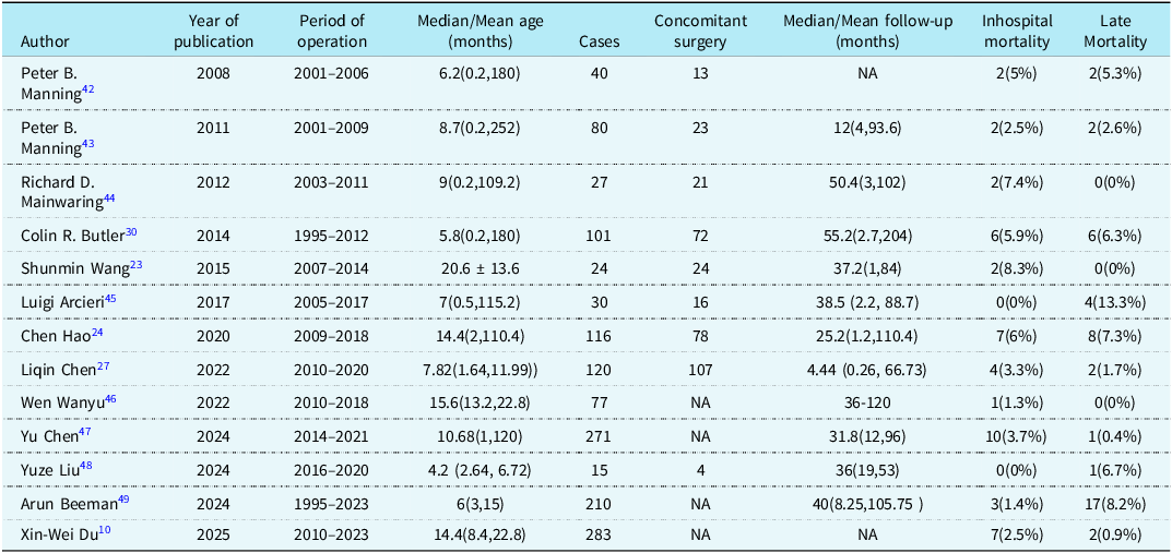
Because of the rarity of the cases, we do not have global epidemiological statistics and population-based incidence surveys, and the literature on congenital tracheal stenosis has mainly focused on a single clinical centre with many years of experience in the diagnosis and treatment of congenital tracheal stenosis. In the early years, foreign reports reported that the 20-year survival rate ranged from 25 to 77%, Reference Song, Lu, Zhu, Du, Wang and Xu40 among which the mortality rate of long-segment stenosis was the highest, but with the development of surgical techniques and other standardised treatments, the survival rate could reach more than 95%. In 2008, Peter B. Manning et al. Reference Benjamin, Pitkin and Cohen3 reported the patient characteristics, hospital course, and outcome of 40 children who received slide tracheoplasty in their institution. There were 2 early and 2 late deaths, none related to the tracheoplasty. One patient required repair of a recurrent tracheal stenosis, 4 patients required tracheotomy, and 3 required temporary stent placement. Twenty-one patients (52.5%) were extubated within 48 hours after treatment tracheoplasty. Univariate and multivariate analysis revealed only preoperative mechanical ventilatory support and duration of cardiopulmonary bypass support to be significant predictors of the need for prolonged intubation. And then in 2011, they also collected the preoperative characteristics, operation and outcome data of 80 children after slide tracheoplasty. The results showed that age at surgery, cardiopulmonary bypass time, and mechanical ventilation time were associated with operative mortality, and postoperative mechanical ventilation time was associated with the need for airway reintervention. Preoperative mechanical ventilation support, cardiopulmonary bypass time, previous airway surgery history, and need for airway reintervention are predictive factors for prolonged hospital stay. Reference Yokoi41 In 2012, Richard D. Mainwaring et al.42 summarised the surgical treatment experience of 27 patients with CHD complicated with airway obstruction. There were 2 postoperative deaths, both in children with single ventricle. The median duration of follow-up for the entire cohort of 25 surviving patients was 4 years. None of the patients have required reoperations on the trachea; 5 have had minor reinterventions. Studies have shown that tracheal obstruction is often associated with CHD, and nearly one-third of patients present with delayed manifestations of airway obstruction after previous repair of CHD. Regardless of the cause of airway obstruction, tracheal reconstruction techniques are effective. In 2014, Butler et al. Reference Butler, Speggiorin and Rijnberg30 collected the demographic characteristics, preoperative conditions, surgical data, and prognostic indicators of 101 children who received slide tracheoplasty. Multivariate analysis showed that preoperative use of extracorporeal membrane oxygenation, preoperative tracheomalacia, and bronchial stenosis were risk factors for poor prognosis, and preoperative tracheomalacia was an important risk factor for stent implantation. In 2015, Shunmin Wang et al. Reference Wang, Zhang, Zhu, Zhen, Liu and Xu23 reported the results of 24 children with congenital tracheal stenosis who underwent slide tracheoplasty. There were 2 operative deaths, due to postoperative tracheomalacia or residual main bronchial stenosis. The follow-up duration was from 1 month to 7 years. Tracheal granulation occurred in 1 case. Clinical symptoms were significantly improved in the remaining patients. In 2017, Luigi Arcieri et al. Reference Manning, Rutter and Border43 retrospectively analysed the surgical results of slide tracheoplasty in 30 patients. No in-hospital deaths occurred. Overall mortality was 4 of 30 cases (13.3%). Twenty-four endoscopic reinterventions were performed required in 19 children (63%) and consisted of stent positioning in 13 (43.3%), balloon dilatation in 5 (16.7%), granulation removal in 4 (13.3%), and tracheostomy in 2 (6.7%). Of the survivors (26 of 30, 86.7%), 11 children (42.3%) did not require further examination on adequate tracheal diameter for age and absence of symptoms after a median follow-up period of 3.5 years. In 2020, Chen Hao et al. Reference Chen, Shi, Zhu, Wang, Lu and Xu24 reviewed the clinical data and surgical outcome indicators of 116 children who underwent slide tracheoplasty, and the results of multivariate analysis showed that cardiovascular abnormalities were significantly associated with postoperative tracheomalacia. Preoperative carina stenosis is significantly associated with postoperative restenosis. Bronchial stenosis with carina stenosis and tracheobronchial compression are significantly associated with postoperative tracheomalacia, restenosis, and mortality. In 2022, Liqin Chen et al. Reference Chen, Zhu and Wang27 retrospectively analysed the clinical data of children under 1 year old with congenital tracheal stenosis admitted to a single centre from April 2010 to September 2020. Of 120 infants, 71.7% (86/120) had a pulmonary artery sling and 37.5% (45/120) had simultaneous intracardiac repairs. Additionally, 52.5% (63/120) of the patients had anomalous tracheobronchial arborisation, and 17.5% (21/120) had diffuse tracheal stenosis. Six airway reoperations (5%) and 6 deaths (5%) occurred, and the mortality decreased annually. Multivariate analysis revealed that a low body weight, cardiovascular anomalies, and normal tracheobronchial arborisation predicted a longer intubation duration. Univariate analysis revealed that a low body weight, preoperative invasive ventilation, a long cardiopulmonary bypass time, and granulation tissue were associated with death. Nineteen and 29 patients underwent spirometry before and after surgery, respectively, and showed improvements in ventilation function, with the ratio of time to peak tidal expiratory flow to total expiratory time and ratio of volume to peak tidal expiratory flow to total expiratory volume values significantly improved from 19.80% and 23.10% to 26.80% and 30.20%, respectively (P < .05). In the same year, Wen Wanyu et al. Reference Manning, Rutter, Lisec, Gupta and Marino44 included 77 children with congenital tracheal stenosis who underwent slide tracheoplasty in their institution with complete clinical data as the observation group. The diameter and length of the tracheal stenosis, the total length of the tracheal stenosis, and the carina angle were measured by CT images before and after operation, and the ratio of the length of the stenosis to the total length of the trachea was calculated. A group of children with normal tracheal morphology with the same number and no significant difference in age, height, and weight were enrolled as the control group. The study found that the diameter of the reconstructed trachea and trachea increased significantly, the length and carinal angle of the reconstructed trachea decreased significantly, and the shape of the reconstructed trachea was close to that of the control group. In 2024, Yu Chen et al. Reference Mainwaring, Shillingford and Davies45 analysed the prognosis of 271 congenital tracheal stenosis patients treated with slide tracheoplasty. Multivariate analysis showed that unilateral lung hypoplasia and bilateral bronchomalacia contributed to postoperative airway complications. Full-length stenosis, bilateral bronchomalacia, and secondary surgery accounted for increased mechanical ventilation time. Using 99 hours as a benchmark, weight and bilateral bronchomalacia were factors for prolonged mechanical ventilation time. In addition, full-length stenosis, unilateral lung hypoplasia, unilateral bronchomalacia, and secondary surgery were positively associated with the duration of hospitalisation, whereas weight was negatively correlated. Taking 21 days as a benchmark, weight, full-length stenosis, unilateral lung hypoplasia, and reoperation associated with prolonged hospital stay. Weight and tracheal diameter were positively correlated with survival. Subsequently, Yuze Liu et al. Reference Arcieri, Pak and Poli46 reported the results of 15 children with congenital tracheal stenosis who underwent slide tracheoplasty in Fuwai Hospital. There was no in-hospital death, and one late death occurred during a median follow-up of 36 months. In the same year, Arun Beeman et al. Reference Wen, Du, Wang, Xu and Lu47 retrospectively analysed all patients who underwent slide tracheoplasty for long-segment congenital tracheal and tracheobronchial stenosis at Great Ormond Street Hospital for Children, London, UK, from March 1995 to February 2023. A total of 210 patients underwent slide tracheoplasty for long segmental congenital tracheal and tracheobronchial stenosis. The median age at surgery was 6 months (interquartile range, 3–15 months), and the median weight was 6.4 kg (interquartile range, 4.2–8.7 kg). Of these patients, 40% (n = 85) had abnormal arborisation, and tracheobronchial morphology was the most common. A total of 24% (n = 50) patients had stenosis extending to 1 or more bronchi. Patients with carinal trifurcation presented early with critical airway stenosis, requiring ventilation support (60%) and extracorporeal membrane oxygenation support (35%) as bridge therapy to slide tracheoplasty. The duration of postoperative ventilation was higher in the patients with congenital tracheobronchial stenosis (P = .006). Patients with a morphology with trifurcation arborisation had higher mortality (23%), and stent requirement (35%). Slide tracheoplasty remains the standard surgery for long segmental congenital tracheal and tracheobronchial stenosis even with different arborisations. Preoperative identification of tracheal arborisations and of the extent of stenosis aids surgical strategies for better outcomes. In 2025, Xin-Wei Du et al. Reference Du, Wang and Wang10 reported the results of 304 children with ring-sling complex who underwent surgical treatment. Studies have shown that concurrent sling and tracheal surgeries for children with the ring-sling complex are safe and effective and are especially preferable for those with narrow-width ratio < 0.6, long-segment or diffuse tracheal stenosis, anomalous tracheobronchial arborisation, and pronounced respiratory symptoms. In summary, slide tracheoplasty has been widely accepted, and in recent years, many clinical centres around the world have adopted it as the standard procedure for long-segment congenital tracheal stenosis. Combined with accurate intraoperative and perioperative bronchoscopy evaluation, slide tracheoplasty has greatly improved the success rate of surgery for long-segment congenital tracheal stenosis. Table 4 presents the results of slide tracheoplasty procedures in different centres.
Previous clinical studies of slide tracheoplasty for the treatment of congenital tracheal stenosis in children during the past 15 years

Perspectives
At present, intraoperative extracorporeal circulation brings convenience to the simultaneous correction of cardiovascular abnormalities and greatly improves the efficiency of operation. The comprehensive multidisciplinary team cooperation of cardiothoracic surgery, otorhinolaryngology surgery, respiratory medicine, ICU, and fiberoptic bronchoscopy lays the foundation for the good prognosis of children with congenital tracheal stenosis.
Biodegradable stent implantation is a promising new technique for anastomotic restenosis after slide tracheoplasty. Anton-Pacheco JL et al. Reference Chen, Wen and Du48 reported 14 cases of biodegradable endotracheal stent implantation after slide tracheoplasty, with an average follow-up of 6 years, and all cases had good clinical symptoms. Juan L et al. Reference Liu, He, Dou, Ma, Chen and Li49 reported 13 cases of severe congenital tracheal stenosis treated with biodegradable stents, which confirmed that biodegradable stents were safe and effective. The symptoms of respiratory obstruction in children were relieved rapidly, and only slight granulation tissue grew in the trachea during the follow-up of 5–40 months. Compared with traditional metal and plastic stents, biodegradable stents can significantly reduce the incidence of airway restenosis and airway malacia. The promotion of this technique requires more clinical cases to be reported, so that a set of clinical criteria can be established for children.
It has been more than 30 years since the rise of tissue engineering trachea research, but it is still in the stage of animal experiments, and only a few cases have been applied in clinical practice. Tissue-engineered trachea is a method of using tissue-engineered trachea seed cells and biological scaffold materials to construct a suitable tissue-engineered trachea and then transplanting it into the body of children. The selection of seed cells and scaffold materials and the culture conditions of tracheal prosthesis are the key points of tissue engineering trachea and also the research hotspots and difficulties in this field. According to the principle of in vivo bioreactor, Elliott et al. Reference Beeman, Ramaswamy and Thiruchelvam50 injected tracheal epithelial cells and bone marrow stromal cells into the scaffold to obtain tissue-engineered trachea to replace the tracheal cartilage ring of children with congenital tracheal stenosis. During the follow-up of 12 weeks after operation, it was found that the trachea had completed angiogenesis and physiological changes such as epithelial coating. Compared with other kinds of tracheal substitutes, tissue-engineered trachea has many advantages: no immunogenicity, no rejection, biological activity, solving the problem of insufficient donors, and maintaining good tracheal morphology. However, there are still many problems to be solved, such as how to effectively grow the blood vessels, coat the endothelium, control the infection after implantation, and how to make the implants survive for a long time, which still need to be further studied and discussed.
In recent years, the application of 3D printing technology in the medical field has been growing rapidly, including in vitro medical models and customised medical devices. Bioprinting of artificial organs and tissues, or bioprinting, is at the forefront of 3D printing technology. Using stem cells as printing materials, the printed tissues and organs will automatically form blood supply and other internal structures. In 2012, a boy with congenital left bronchomalacia in the United States received the transplantation of a 3D synthetic bronchus. Reference Anton-Pacheco51 This is the first time in the world that a 3D printed model has been successfully implanted into a patient’s body. 3D printing technology has shown great potential in the field of tracheal stenosis. With the development of 3D printing technology, it may be possible to implant stem cell-based 3D synthetic tracheal with normal physiological structure into the body to replace the stenosis or dysplasia of the trachea in the near future.
Conclusions
In summary, there have been encouraging achievements in the outcomes of surgical correction of children with congenital tracheal stenosis over the past two decades. Slide tracheoplasty has become the standard procedure for the correction of long-segment congenital tracheal stenosis with abnormal bronchial morphology in many medical centres around the world. Identification of the shape and degree of tracheal stenosis before operation is helpful to develop a better surgical strategy. Flexible application of slide tracheoplasty can effectively correct different types of tracheal stenosis. Cardiopulmonary bypass and intraoperative fiberoptic bronchoscopy are helpful to improve the efficiency of surgery. Postoperative multidisciplinary cooperative management can improve the prognosis of children. Biodegradable scaffolds, tissue-engineered trachea, and 3D printing technology are based on a completely different perspective from traditional medicine. The initial attempts in the biomedical field provide a new idea for the treatment of congenital tracheal stenosis. These new diagnosis and treatment tools are still a long way from direct clinical application, but we believe that with the development of technology, they will open up a new direction of diagnosis and treatment in the near future.
Data availability statement
All data are incorporated into the article, and the data underlying this article are available in the article.
Acknowledgements
We acknowledge the roles of our colleagues, perfusionists, nurses, and others involved in the care of the study participants.
Author contributions
Zhangwei Wang: Conceptualisation; data curation; formal analysis; investigation; methodology; software; validation; visualisation; writing – original draft. Honghao Fu: Writing – review & editing. Shoujun Li: Writing – review & editing.
Financial support
No funding was received for this work.
Competing interests
None declared.