Introduction
The Megaloptera, which was most diverse in the late Permian period, contains ~350 extant species in the world (Wang et al. Reference Wang, Liu, Winterton and Yang2012; Yang et al. Reference Yang, Makarkin, Winterton, Khramov and Ren2012). Currently, there are 35 species belonging to eight genera (Corydalidae: Acanthacorydalis Weele, Eochauliodes Liu, Wang, Shih, Ren, and Yang, Jurochauliodes Wang and Zhang, Neochauliodes Weele, Parachauliodes Weele, Protohermes Weele; Sialidae: Nipponosialis Kuwayama, Sialis Latreille) from the eastern Palaearctic region, with seven fossil species (Oswald Reference Oswald2013). The larval stage is exclusively aquatic inhabiting diverse lotic and lentic freshwater habitats, whereas the pupal and adult stages are terrestrial. The larvae prey on small aquatic invertebrates but the adults normally do not feed while they live for a week or two in their one-year to three-year life span (Takeuchi and Hoshiba Reference Takeuchi and Hoshiba2012, Reference Takeuchi and Hoshiba2013). The larvae are frequently used in freshwater biomonitoring programmes (Yang and Yang Reference Yang and Yang1995; Liu and Yang Reference Liu and Yang2004; Flint et al. Reference Flint, Evans and Neunzig2008).
The order contains two families, Corydalidae and Sialidae, each of which includes two subfamilies, Corydalinae and Chauliodinae in Corydalidae and Sialidinae and Sharasialinae in Sialidae (Liu et al. Reference Liu, Hayashi and Yang2014). The order has been classified as either monophyletic or paraphyletic based on adult morphology or DNA sequences (Cameron et al. Reference Cameron, Sullivan, Song, Miller and Whiting2009; Beutel et al. Reference Beutel, Friedrich, Hörnschemeyer, Pohl, Hünefeld and Beckmann2010; Aspöck et al. Reference Aspöck, Haring and Aspöck2012; Liu et al. Reference Liu, Wang, Shih, Ren and Yang2012). Megaloptera larvae can be easily distinguished by morphological characters. Species of Corydalidae can be characterised by the colour pattern of the head and pronotum, respiratory tubes on the abdominal segment VIII, spiracle size and arrangement, macrosetae on the abdomen, and shape of the submentum of the labium (Neunzig and Baker Reference Neunzig and Baker1991; Contreras-Ramos and Harris Reference Contreras-Ramos and Harris1998). Species of Sialidae can be characterised by the colour pattern on the head and pronotum, postoccipital sutures, and colour pattern on the abdominal tergum (Elliott Reference Elliott1996; Meinander Reference Meinander1996).
Despite their large body size and importance in aquatic ecosystems, larval stages are poorly known since Megaloptera taxonomy is largely based on adult morphology. Six species of Korean Megaloptera belonging to four genera in two families were recorded based on male adults (Jung and Bae Reference Jung and Bae2012). The larvae, however, have not been formally recorded in South Korea, except for one species, Sialis koreana Jung and Bae, which was described recently in both the adult and larval stages.
Mitochondrial DNA (mtDNA) sequences such as partial mitochondrial cytochrome c oxidase subunit I (COI) gene sequences have been widely used for species level identification in different life stages with cryptic species. Previous studies showed that the COI gene sequences can be effectively used as a DNA barcode to identify or associate immature stages with identified adult stages (Miller et al. Reference Miller, Alarie and Whiting2007; Ruiter et al. Reference Ruiter, Boyle and Zhou2013) and for resolving evolutionary relationships among closely related species of insects (Lunt et al. Reference Lunt, Zhang, Szymura and Hewitt1996; Hwang et al. Reference Hwang, Yoon, Suh and Bae2013).
The purposes of this study were to apply DNA barcoding for Megaloptera and associate unknown larvae of South Korean Megaloptera with taxonomically identified adults using COI gene sequences; and to provide keys to the known larvae of South Korean Megaloptera.
Materials and methods
Taxonomy. All larval materials were collected using a hand net (mesh size 1.0 mm) or a Surber net (30×30 cm or 50×50 cm, mesh size 0.2 mm) in streams and wetlands, whereas adults were collected with a sweeping net or a light trap (particularly for Corydalidae) near streams, wetlands, and mountains. Dorsal larvae whole bodies were photographed with a digital camera (Nikon D90, Tokyo, Japan). Male genitalia and larvae were illustrated using a dissecting microscope with an image analyser (Carl Zeiss Discovery V12 with AxioCam I Cc 1, Oberkochen, Germany). The tip of the abdomen and larvae was dissected and cleared with 10% KOH for two days for examination, and then preserved in microtube with glycerin. The morphological terminology for larvae generally follows Neunzig and Baker (Reference Neunzig and Baker1991). Province abbreviations used in this study are as follows: CB, Chungcheongbuk-do; CN, Chungcheongnam-do; GB, Gyeongsangbuk-do; GG, Gyeonggi-do; GN, Gyeongsangnam-do; GW, Gangwon-do; JB, Jeollabuk-do; JN, Jeollanam-do. All materials are preserved in 80–90% ethanol and deposited in the Entomological Museum of Korea University, Seoul, South Korea (KU). One adult male of Parachauliodes asahinai Liu, Hayashi, and Yang is deposited in the Entomological Museum of China Agricultural University, Beijing, China (CAU).
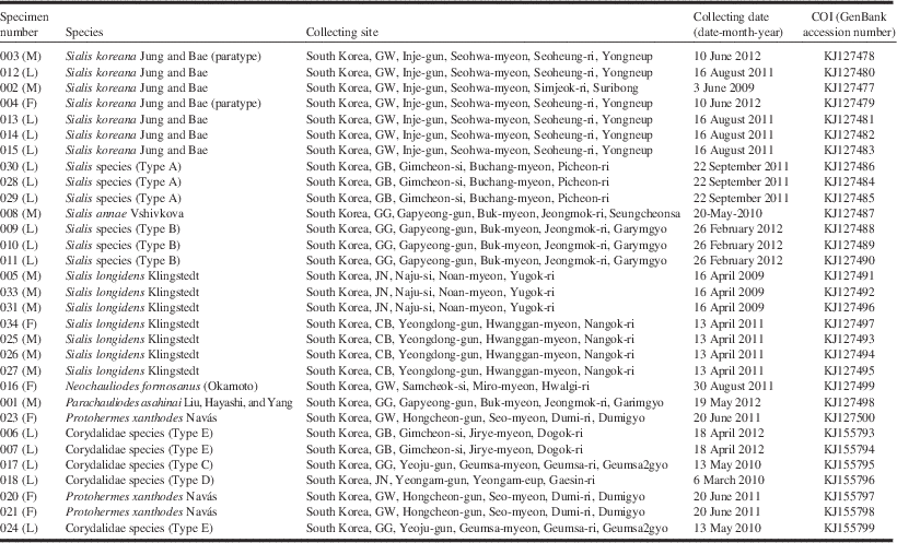
DNA sequencing. A piece of tissue from the thorax (for larvae) or femur (for adults) was taken from 31 specimens (16 adults and 15 larvae) and preserved in 80% ethanol (Table 1). Total DNA extraction was performed using the DNeasy Tissue Kit (Qiagen, Valencia, California, United States of America) extraction protocol and the DNA was stored at −20 °C. The DNA barcoding region of ~660 base pairs (bp) of the COI gene was amplified using the primer pair LCO1490 (5′-GGTCAACAAATCATAAAGATATTGG-3′) and HCO2198 (5′-TAAACTTCAGGGTGACCAAAAAATCA-3′) described by Folmer et al. (Reference Folmer, Black, Hoeh, Lutz and Vrijenhoek1994). Each 50 μL polymerase chain reaction (PCR) contained ~40–100 ng of template DNA, 0.5 U of Taq DNA polymerase with 1X Taq buffer containing 20 mM Tris-HCL (pH 8.2) and 50 mM KCl, 4 mM MgCl2, 200 μM total dNTP, and each primer at 1 μM. Thermal cycling started with incubation at 94 °C for one minute, followed by 35 cycles at 94 °C for one minute, annealing at 47 °C for 30 seconds, and extension at 72 °C for one minute, with a final extension step at 72 °C for five minutes. All PCR products were visualised on 1.5% agarose gels using GelRed (Biotium Inc, Hayward, California, United States of America). Gel purified PCR products were acquired using a Gel Extraction Kit (Koma Biotech Inc, Seoul, South Korea) and sequenced on an ABI 3730 Automated Sequencer (Macrogen Inc., Seoul, South Korea). All sequences were submitted to GenBank (accession numbers: KJ127477-KJ127500, KJ155793-KJ155799) (Table 1) and BOLD systems (http://www.boldsystems.org) wherein voucher data can be accessed in the public dataset “Megaloptera in Korea” (KMDS001-14–KMDS031-14).
Specimen number, species used in analysis, collecting site and date, and GenBank accession numbers for COI sequences.

Note: L, larva; M, male adult; F, female adult; CB, Chungcheongbuk-do; GB, Gyeongsangbuk-do; GG, Gyeonggi-do; GW, Gangwon-do; JN, Jeollanam-do.
Sequences (660 bp) were aligned using CLUSTAL X version 1.81 (Thompson et al. Reference Thompson, Gibson, Plewniak, Jeanmougin and Higgins1997). Maximum parsimony analyses of 31 nucleotide sequences were conducted in MEGA 6.0 (Tamura et al. Reference Tamura, Stecher, Peterson, Filipski and Kumar2013) and sequence divergence was calculated using the Kimura 2-parameter (K2P) model (Kimura Reference Kimura1980). The percentage of replicate trees in which the associated taxa clustered together in the bootstrap test (1000 replicates) is shown next to the branches (Felsenstein Reference Felsenstein1985). The maximum parsimony (MP) tree was obtained using the tree-bisection-regrafting (Nei and Kumar Reference Nei and Kumar2000) algorithm with random addition of sequences (10 replicates). The species of Parainocellia bicolor (Costa) (Raphidioptera: Inocelliidae) was used to root the trees as an outgroup species (sequence available in GenBank (http://www.ncbi.nlm.nih.gov/genbank/) under accession number EU839733).
Results and discussion
Morphological types of larvae and their DNA-based identification
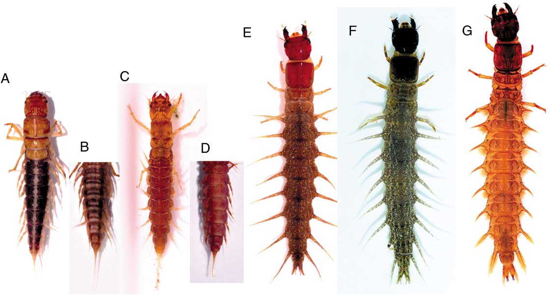
In total, 15 larval specimens for DNA extraction were examined based on diagnostic external morphology in this study. Megalopteran larvae were separated by family levels: Sialidae (Figs. 1A–1D) has small body size (less 20 mm), sickle-shaped mandibles, and abdominal segment X with caudal filament; whereas Corydalidae (Figs. 1E–1G) has large body size (above 40 mm), robust mandibles, and abdominal segment X with a pair of anal prolegs. Within two families, all materials were classified into the following five types (Fig. 1) morphologically.
(A–G) Larval habitus: (A) Sialis koreana Jung and Bae, (B) abdomen of S. koreana, ventral view, (C) Sialis annae Vshivkova, (D) abdomen of S. annae, ventral view, (E) Neochauliodes formosanus (Okamoto), (F) Parachauliodes asahinai Liu, Hayashi, and Yang, (G) Protohermes xanthodes Navás.

Type A (Fig. 1A). Family Sialidae. Body elongate; labrum subtriangular; prothorax wider than mesothorax and metathorax. Abdomen dark brown to black, with white longitudinal stripe dorsally and transverse dark stripes ventrally (Fig. 1B). The specimens belonging to this type were the larvae from the type locality of S. koreana Jung and Bae (Reference Jung and Bae2012) and additional larvae from other localities that were morphologically identical to the larvae of S. koreana (Table 1).
Type B (Fig. 1C). Family Sialidae. Body elongate; labrum distinctly protruding; prothorax as wide as mesothorax and metathorax. Abdomen dorsally pink, without white longitudinal stripe.
Type C (Fig. 1E). Family Corydalidae. Body grey, flattened, with long respiratory tubes on abdominal segment VIII.
Type D (Fig. 1F). Family Corydalidae. Body green, flattened, with short and stout respiratory tubes on abdominal segment VIII.
Type E (Fig. 1G). Family Corydalidae. Body brown, flattened, without respiratory tubes on abdominal segment VIII (Fig. 2).
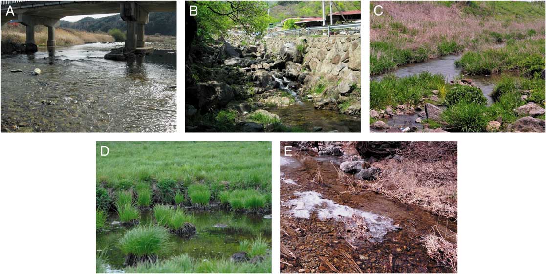
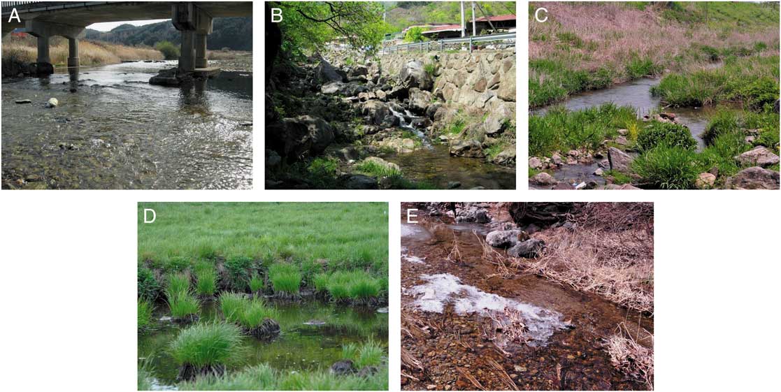
(A–E) Larval habitats of Megaloptera in South Korea: (A) Seomjin river (Protohermes xanthodes), (B) Ganmun stream (Neochauliodes formosanus), (C) Geumsa stream (Parachauliodes asahinai), (D) Yongnup swamp (Sialis koreana), (E) Garim stream (Sialis annae).
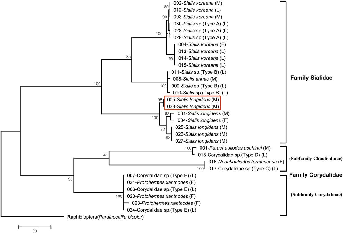
Thirty-one specimens (16 adults and 15 larvae) of South Korean Megaloptera were sequenced successfully, resulting in identification of larvae with adults through the high bootstrap support (⩾90%) by MP tree (Fig. 3), and there was a low genetic distance (K2P model) between larvae and adults (0.50±0.21%). A MP tree using the COI sequence also supported the monophyly of Megaloptera (family Sialidae+family Corydalidae (Chauliodinae+Corydalinae)) (Wang et al. Reference Wang, Liu, Winterton and Yang2012). The average intraspecific K2P distances of the 31 specimens belonging to the families Sialidae and Corydalidae were 0.6% (range, 0.1–1.0%) and 0.3% (range, 0–0.5%), respectively, whereas the average interspecific K2P distances were 10.1% (7.3–11.6%) and 16.6% (15.0–18.5%), respectively. Furthermore, the average maximum intraspecific K2P distance of the compared species was 1.5% within the species of Sialis longidens Klingstedt, whereas the minimum interspecies distance was 7.3% between S. koreana and Sialis annae Vshivkova. High intraspecific divergence was revealed for S. longidens and S. koreana, which had the COI gene sequence variations ranging from 4–11 and 2–7 bp, respectively.
Maximum-parsimony tree of 31 cytochrome c oxidase I sequences from six species (adults and larvae) of Corydalidae and Sialidae (length=425, consistency index=0.715, retention index=0.926). The red-boxed specimens represent a genital variation of Sialis longidens Klingstedt. Nodal support values are bootstrap values (percentage of 1000 replicate). The tree is drawn to scale with branch lengths calculated using the average pathway method and represents the number of changes over the whole sequence. Scale bar indicates 20 nucleotide substitutions. L, larva; M, male adult; F, female adult.
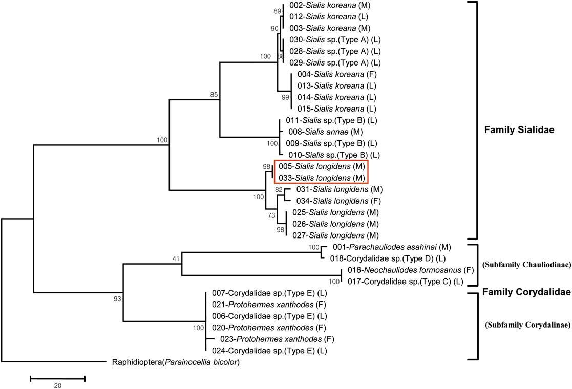
As a result, the larval types were associated with the known species as follows: Type A (S. koreana Jung and Bae), Type B (S. annae Vshivkova), Type C (Neochauliodes formosanus (Okamoto), Type D (P. asahinai Liu, Hayashi, and Yang), and Type E (Protohermes xanthodes Navás). The larva of S. longidens was not sequenced in this study (see the species account below).
Taxonomic accounts: Corydalidae
Neochauliodes formosanus (Okamoto, Reference Okamoto1910)
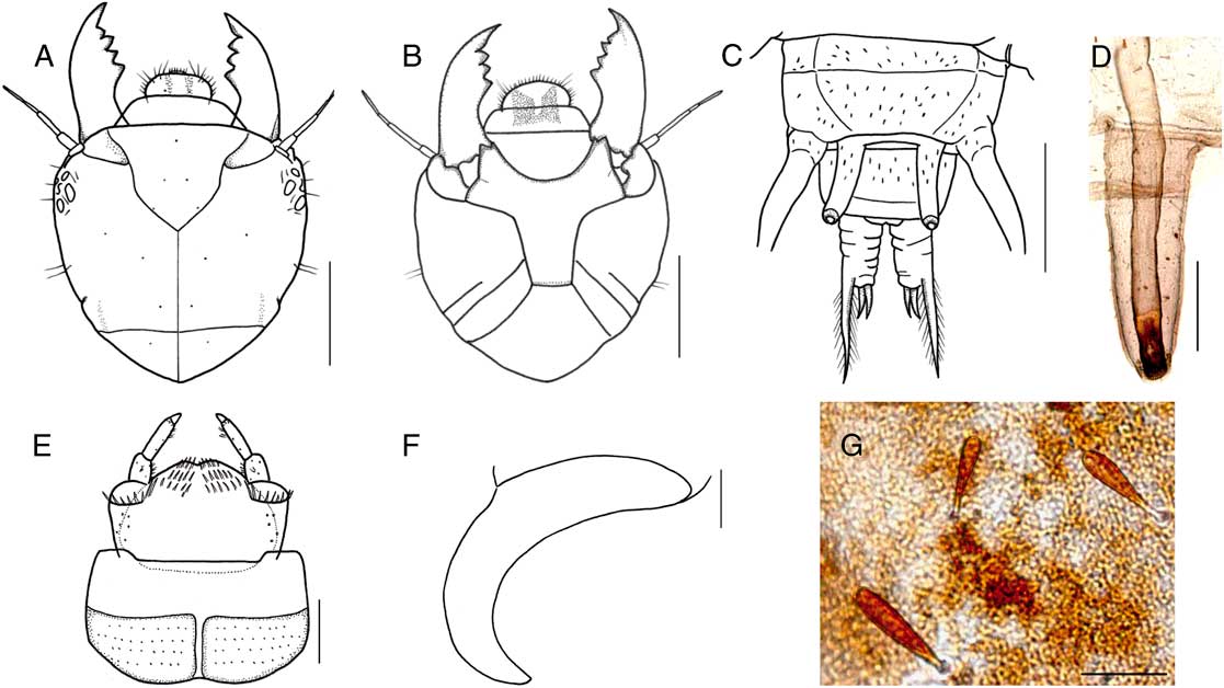
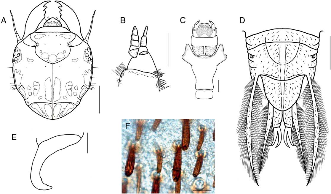
Larva of Neochauliodes formosanus. (A) Head, dorsal view; (B) head, ventral view; (C) abdominal segment VIII, dorsal view; (D) respiratory tube; (E) labium, ventral view; (F) anal claw; (G) macrosetae on abdominal segment VIII. Scale bars=2 mm for A–C; 0.5 mm for D, E; 0.2 mm for F; 0.05 mm for G.

Chauliodes formosanus Okamoto, Reference Okamoto1910: 263.
Chauliodes kawarayamanus Okamoto, Reference Okamoto1910: 262.
Neochauliodes formosanus (Okamoto): Liu et al. Reference Liu, Hayashi and Yang2007: 35; Jung and Bae Reference Jung and Bae2012: 4.
Parachauliodes continentalis Weele: Yoon Reference Yoon1988: 429 (Fig. 4) (misidentification).
Material examined. Korea: 1 larva, GG, Pocheon-si, Ildong-myeon, Suip-ri, Sampalgyo, 14.x.1997, Y.J. Bae (KU); 1 larva, GG, Yeoju-gun, Geumsa-myeon, Dogok-ri, 13.v.2010, S.W. Jung, DNA voucher: 017-PCL01 (# KJ155795) (KU); 2 larvae, GG, Suwon-si, Changan-gu, 5.vi.2012, S.W. Jung, (KU); 1 larva, JB, Jinan-gun, Bugwi-myeon, Suhang-ri, 1.x.1984, Y.J. Bae (KU); 1 larva, JN, Yeongam-gun, Yeongam-eup, Gaesin-ri, 6.iii.2010, S.W. Jung (KU); 1 female, GW, Samcheok-si, Miro-myeon, Hwalgi-ri, 30.viii.2011, S.W. Jung, DNA voucher: 016-PCa01 (# KJ127499) (KU); 1 larva, CB, Chungju-si, Eomjeong-myeon, Geodong-ri, 2.vi.2012, S.W. Jung (KU); 1 larva, CB, Jecheon-si, Geumseong-myeon, Seongnae-ri, 13.vi.2012, S.W. Jung (KU).
Description. Adult (male and female). See Liu et al. (Reference Liu, Hayashi and Yang2007) for full description.
Larva. Body length 42.4–47.6 mm (n=3), elongate and distinctly flattened. Head, prothorax reddish brown without pale markings. Mandibles, labium dark brown. Mesothorax and metathorax, legs brown. Abdomen grey with small yellow markings and lateral filaments.
Head (Fig. 4A) 5.0–5.7 mm in length, 5.0–5.5 mm in width (n=3), subquadrate but more or less round, narrowing posteriorly without markings; frontal suture lyriform, epicranial suture distinct, 3/5× length of head; genal carinae distinct and short. Antennae long and slender, about 2.3 mm in length, with five antennomeres; gradually slender apically; approximate ratio of antennomeres (n=1) as 1.0:3.2:7.5:2.2:5.0. Labrum round, slightly prominent medially with two thick darkish brown markings and several setae in anterior margin. Clypeus three times wider than long, brown without pale markings. Mandibles asymmetrical, dark or reddish brown, well developed, left mandible with four teeth; third tooth more or less long but blunt, right mandible with three teeth; second tooth wide and round. Maxillae reddish brown, cardo small; stipes elongate with several strong setae on inner margin; galea with three galeomeres; galeomere 1 wide; galeomere 2 stout, smaller (about 3/5) than galeomere 3; galeomere 3 long and slender; maxillary with five palpomeres; palpomere 3 shortest; palpomere 4 over 3×length of palpomere 3; approximate ratio of palpomeres (n=1) as 5.0:1.5:1.0:3.2:1.2. Labium (Fig. 4B) pale brown submentum brown, but anterolateral margin black, more or less narrow in posterior part; mentum entirely brown, but laterally blackish brown, and medially yellow; prementum blackish brown with several setae anteriorly; ligula concave at apex; labial palpomere (Fig. 4E), blackish brown; approximate ratio of palpomere (n=1) as 2.5:4.5:1.0.
Thorax. Prothorax slightly wider than long, 5.0–5.5 mm in length, 5.0–5.8 mm in width (n=3). Mesothorax and metathorax nearly equal to length and width, 2.2–2.5 mm in length, 5.0–6.0 mm in width (n=3), brown with several dark brown markings in dorsum. Legs brown, with six tarsomeres, with two claws and arolium; tarsi nearly equal to length of tibiae; prothoracic and mesothoracic claws with different in length, short outer and long inner side; metathoracic claws nearly equal to length.
Abdomen elongate and flattened, 10-segmented, with thin sparse microsetae (Fig. 4G) on dorsal part. Lateral filaments on abdominal segments I–VIII without setae; abdominal segments I–VII with round spiracles on lateral portion; abdominal segment VIII (Fig. 4C) with respiratory tubes on posterior margin; respiratory tubes (Fig. 4D) about 1.5 mm in length, slender and long with spiracle at apex; abdominal segment X with a pair of anal prolegs, each bearing two elongate claws (Fig. 4F) and anal proleg filaments with sparse setae.
Distribution. South Korea, China, Japan
Remarks. The larvae of N. formosanus were collected from the riffle areas from the midstream to downstream reaches (Fig. 2B) and they often occurred with P. asahinai. The larvae can be distinguished from P. asahinai by the combination of the following characters: generally grey body with reddish brown head and pronotum, narrow clypeus without pale marking, and presence of long respiratory tubes on the abdomen apex.
Based on the examination of preserved specimens at KU, the previous recording of larva of Parachauliodes continentalis Weele by Yoon (Reference Yoon1988) was a misidentification, in reality these larvae belong to N. formosanus. The long and slender respiratory tubes on the abdominal segment VII (Yoon Reference Yoon1988: 429, Fig. 4) are characteristic of the genus Neochauliodes.
Parachauliodes asahinai Liu et al., Reference Liu, Hayashi and Yang 2008
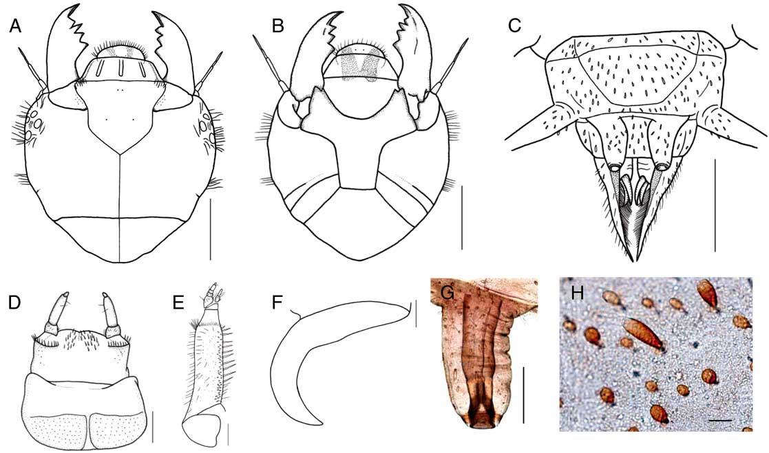
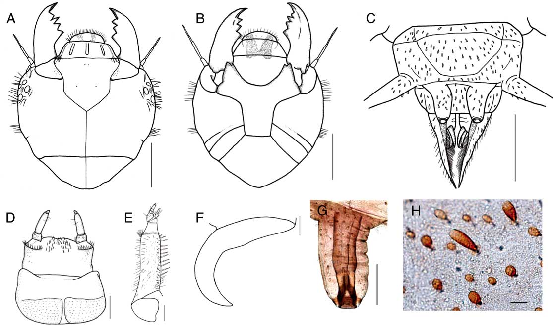
Larva of Parachauliodes asahinai. (A) Head, dorsal view; (B) head, ventral view; (C) abdominal segment VIII, dorsal view; (D) labium, ventral view; (E) left maxilla; (F) anal claw; (G) respiratory tube; (H) macrosetae on abdominal segment VIII. Scale bars=2 mm for A–C; 0.5 mm for D, E, G; 0.2 mm for F; 0.05 mm for H.
Parachauliodes asahinai Liu et al., Reference Liu, Hayashi and Yang2008: 563; Jung and Bae Reference Jung and Bae2012: 5.
Material examined. Korea: 3 larvae, JB, Jinan-gun, Bugwi-myeon, Suhang-ri, 1.x.1984, Y.J. Bae (KU); 1 larva, JN, Yeongam-gun, Yeongam-eup, Gaesin-ri, 6.iii.2010, S.W. Jung, DNA voucher: 018-PCL02 (# KJ155796) (KU); 1 larva, GN, Changwon-si, Dong-eup, Museong-ri, 22.vi.2011, S.W. Jung (KU); 1 male, GG, Gapyeong-gun, Buk-myeon, Jeokmok-ri, 19.v.2012, S.W. Jung and Y.J. Bae, DNA voucher: 001-MPA01 (# KJ127498) (CAU); 1 larva, JN, Gangjin-gun, Seongjeon-myeon, Wolpyeong-ri (34°42'16.0''N, 126°40'41.4''E), 18.vii.2013, M.C. Kim (KU).
Description. Adult (male and female). See Liu et al. (Reference Liu, Hayashi and Yang2008) for full description.
Larva. Body length 45.3–49.2 mm (n=3), elongate and distinctly flattened.
Head, mandibles, and prothorax dark brown without pale markings. Antennae, mouth parts and legs brown. Abdomen grey with green brindle markings and lateral filaments.
Head (Fig. 5A) 5.5–6.0 mm in length, 5.2–6.0 mm in width (n=3), subquadrate but more or less round and wide posteriorly without markings; frontal suture lyriform, epicranial suture distinct, 3/5×length of head; genal carinae distinct and short. Antennae long and slender, about 2.3 mm in length, with five antennomeres; antennomere 1 short and wide; antennomere 2 thicker than antennomere 3; antennomere 3 much thinner and longest, 2×length of antennomere 2; approximate ratio of antennomeres (n=1) as 1.0:4.0:8.0:2.5:5.0. Labrum round, reddish brown with several setae in anterior margin. Clypeus three times wider than long, brown with three longitudinal yellow markings, 2×length of labrum. Mandibles asymmetrical, dark or reddish brown, well developed, left mandible with four teeth; teeth 1, 2, and 4 wide; third tooth long and sharp, right mandible with three teeth; third tooth widest; second tooth smallest. Maxillae (Fig. 5E) blackish brown, cardo small; stipes elongate with several strong setae on inner margin; galea with three galeomeres; galeomere 1 wide and short; galeomere 2 stout, smaller than galeomere 3; galeomere 3 long and slender; maxillary with five palpomere; palpomere 4 much longer than palpomere 3; approximate ratio of palpomeres (n=1) as 5.0:1.0:1.0:2.5:1.0. Labium (Fig. 5B) pale brown submentum brown, but anterolateral margin black, more or less wide in posterior part; mentum entirely yellow, laterally blackish brown, but medially yellow; prementum blackish brown with several setae anteriorly; ligula broad margin, slightly concave at apex; labium with three palpomeres (Fig. 5D), blackish brown; approximate ratio of palpomeres (n=1) as 2.5:6.0:1.0.
Thorax. Prothorax slightly wider than long, 5.5 mm in length, 6.0–6.8 mm in width (n=3), blackish brown with brindle markings in posterior margin. Mesothorax and metathorax nearly equal to length and width, 2.8–3.0 mm in length, 6.0–6.5 mm in width (n=3), brown with several dark brown markings in dorsal part and brindle markings in anterior and posterior margin. Legs brown, with six tarsomeres, with two claws and arolium; tarsi nearly equal to length of tibiae; prothoracic and mesothoracic claws with different in length, short outer and long inner side; metathoracic claws nearly equal to length.
Abdomen elongate and flattened, 10-segmented, with short and long microsetae (Fig. 5H) densely scattered on dorsal part. Lateral filaments on abdominal segments I–VIII without setae; abdominal segments I–VII with round spiracles on lateral portion, abdominal segment VIII (Fig. 5C) with respiratory tubes on posterior margin; respiratory tubes (Fig. 5G) about 1.2 mm in length, more or less stout with spiracle at apex; abdominal segment X with a pair of anal prolegs, each bearing two elongate claws (Fig. 5F) and anal proleg filaments covered with dense setae.
Distribution. South Korea, Japan.
Remarks. The larval stage is described for the first time here. This species inhabits streams in rhithral zones preferring habitats with aquatic vegetation (Fig. 2C). The larvae of P. asahinai are morphologically similar to those of P. continentalis (Hayashi, Reference Hayashi2005: 381, figs. 2–4) in having clypeus with three longitudinal yellow markings, and short and stout respiratory tubes on abdominal segment VIII. However, P. asahinai differs from this species and all other described Korean Corydalidae by the following characters: wider head and pronotum, broad and round labrum, maxillary palpomere 4 much longer than palpomere 3, labial palpomere 2 about three times longer than first palpomere, abdomen with microsetae (elongate shape with smooth surface), stout and closely located respiratory tubes on the abdominal segment VIII, and proleg filaments covered with dense setae on the inner side.
Protohermes xanthodes Navás, Reference Navás 1913
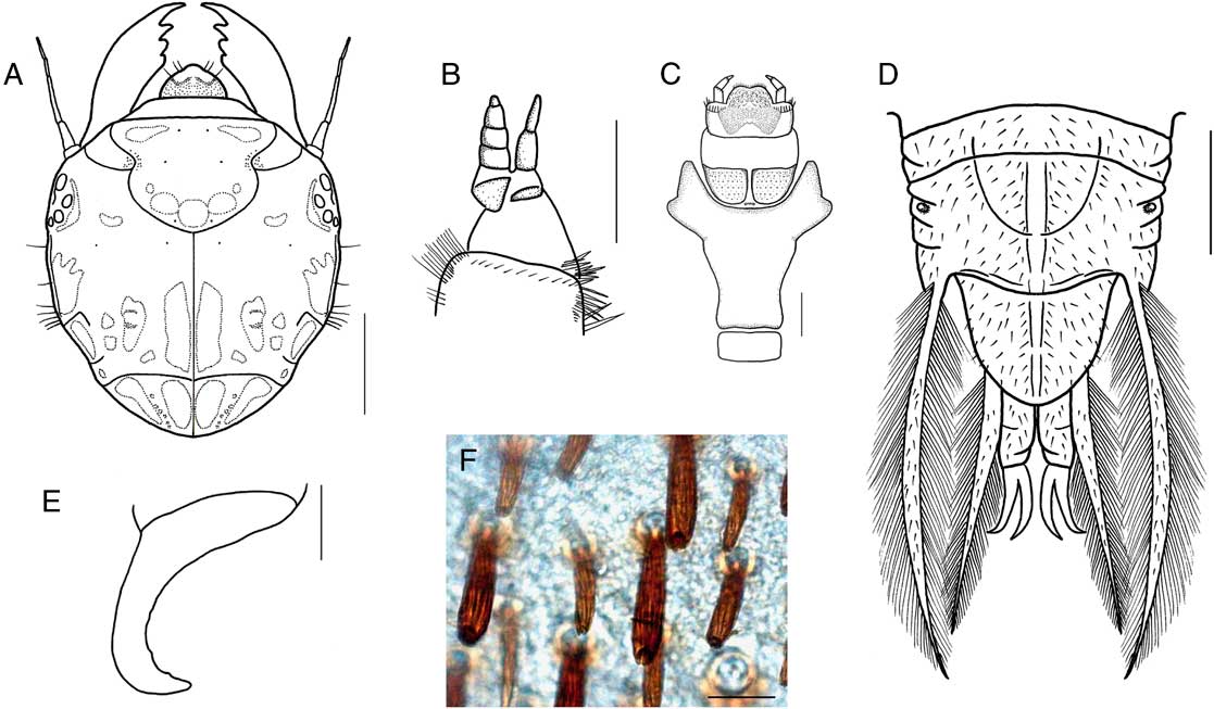
Larva of Protohermes xanthodes. (A) Head, dorsal view; (B) left maxilla; (C) labium, ventral view; (D) abdominal segment VIII, dorsal view; (E) anal claw; (F) macrosetae on abdominal segment VIII. Scale bars=2 mm for A, D; 1 mm for C; 0.5 mm for B, E; 0.05 mm for F.
Protohermes xanthodes Navás, Reference Navás1913: 427; Liu et al. Reference Liu, Hayashi and Yang2006: 405; Jung and Bae Reference Jung and Bae2012: 7.
Parachauliodes grandis (Thunberg): Yoon Reference Yoon1988: 428 (Fig. 3) (misidentification).
Material examined. Korea: 3 larvae, GG, Gapyeong-gun, Ha-myeon, Sinsang-ri, 11.vii.1993, Y.J. Bae (KU); 2 larvae, GG Gapyeong-gun, Buk-myeon, Dodae-ri, 10.vii.1995, Y.J. Bae (KU); 1 larva, GG, Yeoju-gun, Geumsa-myeon, Geumsa-ri, Geumsa 2-gyo (Br.), 13.v.2010, DNA voucher: 024-PGL01 (# KJ155799) (KU); 2 larvae, CB, Jechoen-si, Bongyang-eup, Jupo-ri, 6.x.2010, S.W. Jung and J.M. Hwang (KU); 3 females, 1 larva, GW, Hongcheon-gun, Seo-myeon, Dumi-ri, 20.vi.2011, S.W. Jung, DNA voucher: 020-PGa02 (# KJ155797), 021-PGa03 (# KJ155798), 023-PGa05 (# KJ127500) (KU); 4 larvae, GB, Gimcheon-si, Jirye-myeon, Dogok-ri, 18.iv.2012, S.W. Jung, DNA voucher: 006-MPL01 (# KJ155793), 007-MPL03 (# KJ155794) (KU); 2 larvae, GB, Uljin-gun, Seo-myeon, Wangpi-ri, 26.iv.2012, S.W. Jung (KU).
Description. Adult (male and female). See Liu et al. (Reference Liu, Hayashi and Yang2006) for full description.
Larva. Body length 52.0–55.8 mm (n=3), elongate and distinctly flattened. Head and thorax sclerotised, dark or reddish brown with several yellow markings, legs and abdomen brown, with lateral filaments.
Head (Fig. 6A) 6.2–7.0 mm in length, 5.5–6.5 mm in width (n=3), subquadrate, with several yellow markings and spots in dorsal and lateral view; frontal suture lyriform, more or less round laterally, epicranial suture distinct, 2/3 times as long as head; genal carinae distinct and long; frons with three large round yellow markings posteriorly, two oblong markings anteriorly; postoccipital suture slightly curved laterally. Antennae long and slender, 2.8 mm in length, with five antennomeres; antennomere 1 short and wide; antennomere 2 thicker than antennomere 3; antennomeres 4 and 5 much thinner and nearly equal in length; approximate ratio of antennomeres (n=1) as 1.0:3.5:5.5:2.0:2.0. Labrum protruding anteriorly, reddish brown, with paler anterior margin bearing two long setae laterally. Clypeus pale, 1/2×length of labrum. Mandibles dark or reddish brown, well developed, with three teeth; apical tooth blunt, twice length of third tooth; first and second teeth wide and round. Maxillae (Fig. 6B) blackish brown, cardo small; stipes elongate with several strong setae on inner margin; galea with three galeomeres; galeomere 1 wide and short; galeomere 2 stout; galeomere 3 slender and equal length of galeomere 2; maxillary with five palpomeres; palpomere 4 more or less longer than palpomere 3; approximate ratio of palpomeres (n=1) as 2.6:1.0:1.0:1.3:1.0. Labium (Fig. 6C) from pale brown to yellow, submentum brown, but with black margin, mentum entirely yellow, laterally blackish brown, prementum blackish brown; labium with three palpomeres, blackish brown; approximate ratio of palpomeres (n=1) as 2.0:1.6:1.0.
Thorax. Prothorax nearly as long as wide, 6.0–6.5 mm in length, 5.8–6.5 mm in width (n=3), blackish brown with yellow marking, laterally with dense short setae and long strong setae. Mesothorax and metathorax nearly equal to length and width, 2.8 mm in length, 5.5 mm in width (n=3), blackish brown with a few yellow markings. Legs yellow, with six tarsomeres, with two claws and triangular arolium; protarsi 1/2×length of tibiae with a row of protuberances on outer side, two curved claws with different length; mesotarsi and metatarsi as long as tibiae, two claws equal in length and strongly curved with one tooth located basally.
Abdomen elongate and flattened, 10-segmented, with dense microsetae (Fig. 6F) on dorsal part. Lateral filaments on abdominal segments I–VIII covered with setae; lateral filaments 7 and 8 longer than other and denser setae (Fig. 6D); abdominal segments I–VII ventrally bearing tufts of tracheal gills; abdominal segments I–VIII with round spiracle; abdominal segment X with a pair of anal prolegs, each bearing two elongate claws (Fig. 6E) and anal proleg filaments covered with long and dense setae.
Distribution. South Korea, China
Remarks. The larvae of P. xanthodes are common and widespread in the riffles of downstream reaches (Fig. 2A). The larvae resemble P. grandis Thunberg (Hayashi Reference Hayashi2005: 381, figs. 2–3) in having ventral gill tufts on abdominal segments I–VII and absence of respiratory tubes on abdominal segments VIII, but can be distinguished from the latter by their generally reddish brown head and pronotum with light markings, wider pronotum, short labial palpomere 2, thick and distinct gular suture, lower number of setae on the lateral filaments, and denser setation of the proleg filaments. Based on the examination of preserved specimens at KU, the previous recording of larva of P. grandis by Yoon (Reference Yoon1988) was a misidentification of P. xanthodes.
The close species Parachauliodes martynovae Vshivkova is recorded from the south of the Russian Far East: Primorye Territory (the last record is dated 1960) and recently from Khabarovsky District, Bolshekhekhtsyrskii Federal Nature Reserve (1 male and 1 female). Liu et al. (Reference Liu, Hayashi and Yang2006) considered this species as a junior synonym of P. xantodes. In order to solve the problem of P. martynovae taxonomic status it is necessary to carry out careful comparative study of species morphology (wing venation, non-genital, and genital structure) as well as the DNA data. (Vshivkova and Dubatolov Reference Vshivkova and Dubatolov2010). Also the geographical data should be revised along with reidentification of P. xantodes material from different areas.
Taxonomic accounts: Sialidae
Sialis annae Vshivkova, Reference Vshivkova 1979
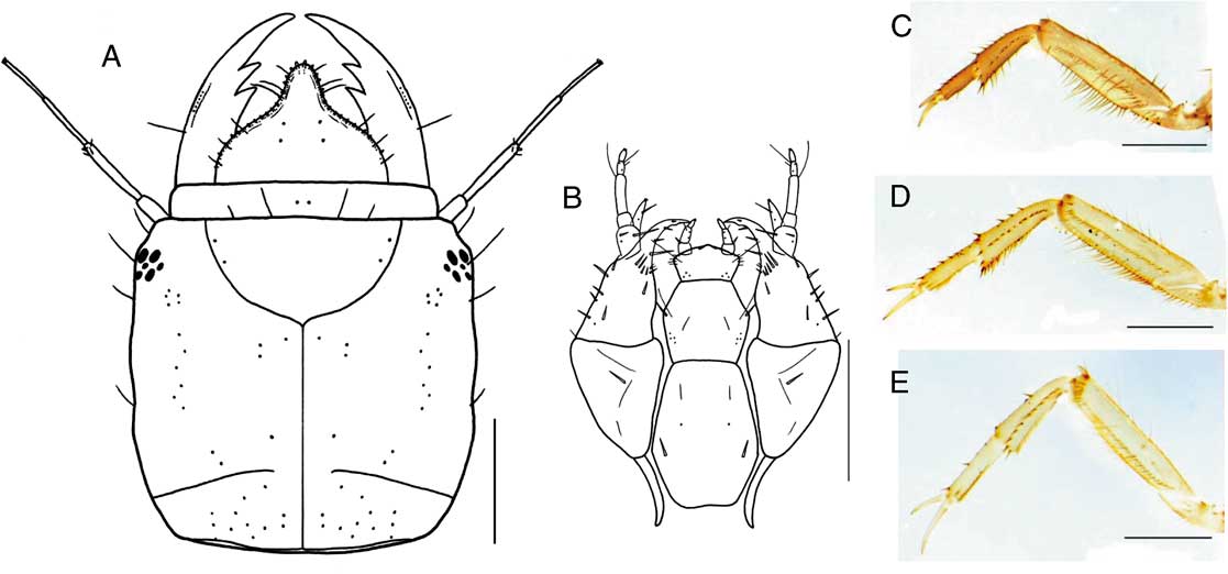
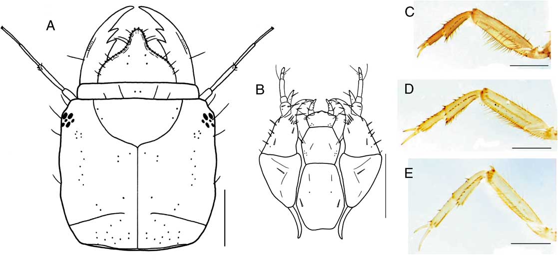
Larva of Sialis annae. (A) Head, dorsal view; (B) mouth parts, ventral view; (C) left proleg; (D) left mesoleg; (E) left metaleg. Scale bars=1 mm.
Sialis annae Vshivkova, Reference Vshivkova1979: 79; Vshivkova Reference Vshivkova1995: 33; Vshivkova Reference Vshivkova2001: 379, 779 (Fig. 8); Jung and Bae Reference Jung and Bae2012: 11.
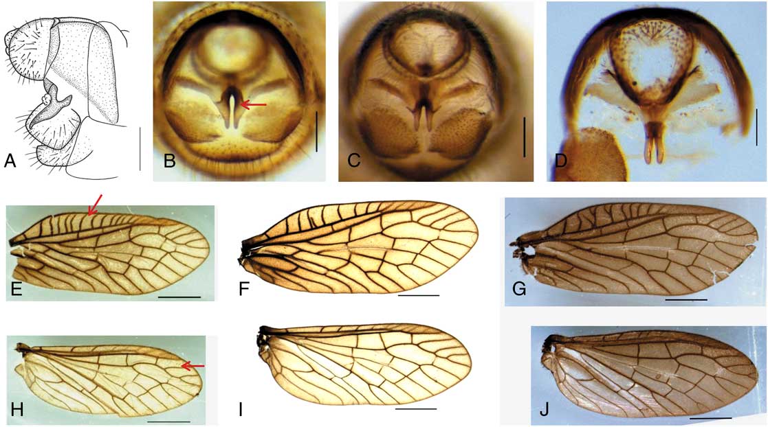
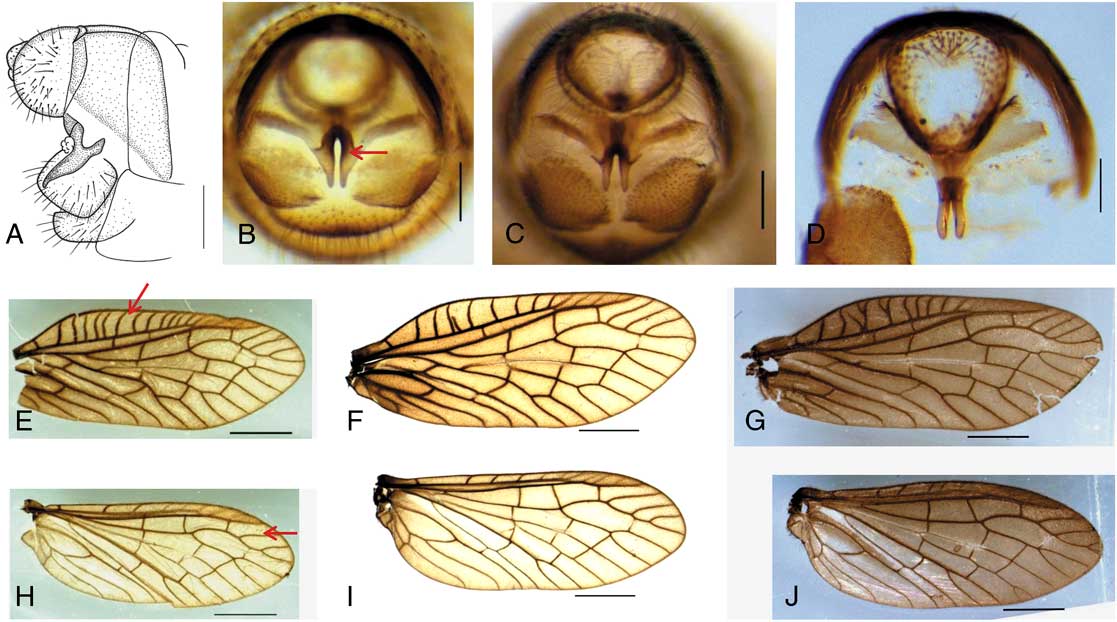
Male of Sialis longidens. (A) Genitalia, lateral view (South Korea, JN); (B) genitalia, caudal view (South Korea, JN); (C) genitalia, caudal view (South Korea, CB); (D) genitalia caudal view (Russia); (E) fore wing, right (South Korea, JN); (F) fore wing, right (South Korea, CB); (G) fore wing, right (Russia); (H) hind wing, right (South Korea, JN); (I) hind wing, right (South Korea, CB); (J) Hind wing, right (Russia). Scale bars=0.2 mm for A–D; 2 mm for E–J.
Material examined. Korea: 1 male, GG, Gapyeong-gun, Buk-myeon, Jeokmok-ri, Seungcheonsa (Temple), 20.v.2010, S.W. Jung, DNA voucher: 008-SSa04 (# KJ127487) (KU); 3 larvae, GG, Gapyeong-gun, Buk-myeon, Jeokmok-ri, 26.ii.2012, S.W. Jung, DNA voucher: 009-SSL04 (# KJ127488), 010-SSL05 (# KJ127489), 011-SSL06 (# KJ127490) (KU).
Description. Adult (male and female). See Vshivkova (Reference Vshivkova1979) for full description.
Larva. Body length 16.1–19.1 mm (n=3); caudal filaments length 5.1–5.2 mm (n=3). Body elongate, slightly flattened. Head and thorax sclerotised, brown with yellowish several markings, legs yellowish brown (without medial longitudinal light stripe), abdomen soft and pink brown, with pale lateral filaments.
Head (Fig. 7A) 2.1–2.8 mm in length, 2.5–3.1 mm in width (n=3), subquadrate, with several yellow markings and spots in dorsal and lateral view; Y-formed epicranial suture distinct; frons with four setae laterally; postoccipital suture slightly arched, interrupted at coronal suture. Antennae long and slender, 1.8 mm in length, with four antennomeres; antennomere 1 more or less stout; antennomere 2 thicker than antennomeres 3 and 4, with four short setae posteriorly, terminal antennomere with four sensorial appendages; approximate ratio of antennomeres (n=1) as 1.5:4.3:3.5:3.0. Labrum subtriangular and protruding anteriorly with anterior margin crenulate, abruptly narrow at 1/3 apical part. Mandibles well developed, with two teeth, long setae on outer margin. Maxillae (Fig. 7B) with large cardo; stipes slightly smaller than cardo; galea conical; lacinia falciform, with two strong long setae posteriorly and two strong small setae anteriorly; maxillary with five palpomeres; palpomere 1 stout, with two setae; palpomere 3 slender and longest, with long seta anteriorly; palpomere 4 short, with long seta; terminal palpomere with sensorial appendages; approximate ratio of palpomeres (n=1) as 1.6:1.1:1.9:0.7:0.6. Labium elongate; submentum large, with two strong setae posteriorly and two fine setae anteriorly, widest at posterior 2/5 part; mentum widest at middle part, as long as half submentum, with strong setae laterally; prementum short, subtriangular between palpi; labial palp short, with three palpomeres; terminal palpomere with sensorial appendages.
Thorax yellowish brown; prothorax large, 2.1–2.2 mm in length, about twice as long as mesothorax; mesothorax 1.2 mm in length, 2.5–3.1 mm in width; metathorax partially sclerotised, 1.1–1.3 mm in length, 2.4–2.8 mm in width (n=3). Legs (Figs. 7C–7E) long and slender, with six tarsomeres, with two claws in different length.
Abdomen elongate, 10-segemented, with 7 lateral filaments (from I to VII segment). Abdominal segment X small and emarginate at apex, with long caudal filament. Abdominal terga dorsally uniformly pink each with two distinct pale submedial bracket-like figures, which apically became smaller and stick-like; sublaterally at the base of lateral filament in each tergite there are slightly curved pale stripes. Sternites uniformly pink-brown, without any black pattern (Fig. 1D).
Distribution. South Korea, northeast China, Far East Russia (Primorye Territory)
Remarks. Adults of this species were recently recorded by Jung and Bae (Reference Jung and Bae2012) from South Korea. We collected immature larvae in a water-filled pool area with fallen leaves near a mountain stream (Fig. 2E). The substrate was composed mostly of fine sand or silt (<0.5 mm). Aquatic insects such as Chironomidae (Diptera) and the mayfly Siphlonurus chankae Tshernova (Ephemeroptera: Siphlonuridae) were dominant there; the caddisfly Semblis phalaenoides Linnaeus (Trichoptera: Phryganeidae) was also common. The larvae of S. annae are similar to the larvae of S. longidens and S. koreana in general, but can easily be distinguished from these species by slightly arched postoccipital suture, antennomere 3 as long as antennomere 2, protruding labrum, smaller sclerotisation of metathorax with yellow markings, more remarkably pink abdominal tergites and sternites without any obvious black longitudinal or transverse stripes or pattern.
Sialis koreana Jung and Bae, Reference Jung and Bae 2012
Sialis koreana Jung and Bae, Reference Jung and Bae2012: 8
Material examined. Korea: 3 larvae, GB, Gimcheon-si, Buchang-myeon, Picheon-ri, 22.ix.2011, S.W. Jung, DNA voucher: 028-SSL01 (# KJ127484), 029-SSL02 (# KJ127485), 030-SSL03 (# KJ127486) (KU); 1 male, GW, Inje-gun, Seohwa-myeon, Simjeok-ri, Suribong, 3.vi.2009, J.K. Choi and H.M. Lim, DNA voucher: 002-MSK01 (# KJ127477) (KU); 4 larvae, GW, Inje-gun, Seohwa-myeon, Seoheung-ri, Yongneup (swamp), 16.viii.2011, M.C. Kim and D.G. Kim, DNA voucher: 012-SSL07 (# KJ127480), 013-SSL08 (# KJ127481), 014-SSL09 (# KJ127482), 015-SSL10 (# KJ127483) (KU); 4 larvae, GW, Inje-gun, Seohwa-myeon, Seoheung-ri, Yongneup (swamp), 1.vi.2012, Y.J. Bae and S.W. Jung (KU); 1 male (paratype), 1 female (paratype), GW, Inje-gun, Seohwa-myeon, Seoheung-ri, Yongneup (swamp), 10.vi.2012, Y.J. Bae and S.W. Jung, DNA voucher: 003-MSK02 (# KJ127478), 004-MSK03 (# KJ127479) (KU).
Description. Adult (male and female). See Jung and Bae (Reference Jung and Bae2012) for full description.
Larva. See Jung and Bae (Reference Jung and Bae2012) for full description.
Diagnosis. The larva (Fig. 1A) of this species can be distinguished from congeners by the following characters: elongate body, almost square head with several yellow markings, distinct Y-shaped epicranial suture, slightly round postoccipital suture disconnected to the coronal suture, apically more or less emarginate abdominal segment X, and abdominal terga with light median longitudinal stripe and light paired small lateral spots; The important distinguish characters are also: the characteristic pattern of sternites, which on the periphery bordered with intense black stripe so that the centre of the sternum is presenting an oblong light field. This pattern would indicate that it is in the Sialis sibirica species group, however, S. koreana does not have two sublateral black round spots on sternum (Fig. 1B), which is different from the S. sibirica species group (Vshivkova Reference Vshivkova2001: 379, 779, plate 196, fig. 6).
Distribution. South Korea.
Remarks. This species is endemic to South Korea (Gangwon Province) and found only in high mountain swamps (above 1200 m altitude) (Fig. 2D) near the demilitarised zone (DMZ). The larval specimens from the southeastern area (Gyeongsangbuk Province) of the Korean Peninsula and the specimens collected together with the holotype adult of S. koreana (Jung and Bae Reference Jung and Bae2012) were confirmed to be conspecific based on the COI gene sequences (Table 1, Fig. 3). It is interesting to note that the separation the speciation process in the genus Sialis in some cases was caused by altitude factor: for example, in Caucasus two sister species S. abchasica Vshivkova and S. zhiltzovae Vshivkova in their distribution divided by altitude – one species is found in the highlands, the second inhabits foothills waters.
Sialis longidens Klingstedt, Reference Klingstedt 1932
Sialis longidens Klingstedt, Reference Klingstedt1932: 1; Hayashi and Suda Reference Hayashi and Suda1995: 6; Vshivkova and Ito Reference Vshivkova and Ito1993: 108; Vshivkova Reference Vshivkova1995: 33; Vshivkova Reference Vshivkova2001: 380, 769 (Figs. 1, 4, larva final stage), 773 (Figs. 1–4, larva first stage), 775 (Fig. 1, pupa); Liu and Yang Reference Liu and Yang2006: 396; Jung and Bae Reference Jung and Bae2012: 11.
Sialis KUa: Yoon Reference Yoon1988: 427 (Fig. 2).
Material examined. Korea: 3 males, JN, Naju-si, Noan-myeon, Gyelim-ri (35°03'52.77''N, 126°42'50.98''E), 16.iv.2009, J.K. Choi and D.H. Lee, DNA voucher: 005-MSA01 (# KJ127491), 031-MSA02 (# KJ127496), 033-MSA04 (# KJ127492) (KU); 3 males, 1 female, CB, Yeongdong-gun, Hwanggan-myeon, Nangok-ri (36°14'50.96"N, 127°57'19.92"E), 13.iv.2011, S.W. Jung, DNA voucher: 025-SSa01 (# KJ127493), 026-SSa02 (# KJ127494), 027-SSa03 (# KJ127495), 034-MSA05 (# KJ127497) (KU). Russia: 3 males, Primorye Territory, Lazovsky Nature Reserve, Kordon Sokolchi, 33.vi.1978, G. Zapolina and T.S.Vshivkova (KU).
Description. Adult (male and female). See Klingstedt (Reference Klingstedt1932) for full description.
Larva. See Vshivkova (Reference Vshivkova1995, Reference Vshivkova2001), Yoon (Reference Yoon1988), and Hayashi (Reference Hayashi2005) for diagnostic characters with habitus drawing and photograph.
Diagnosis. Adults of S. longidens can be distinguished from all the other Sialis species by the following combination of characters: black pronotum without markings, somewhat broad brown wings; round tergum X which is as long as 2/3 of the width of tergum IX and subquadrate from the lateral view, elongate sternum X, and subquadrate gonostylus IX. The larvae are similar to those of S. koreana, but can be distinguished from the latter by having abdominal terga with wider light median longitudinal stripe (Vshivkova Reference Vshivkova1995: 17, fig. 4–1; Vshivkova Reference Vshivkova2001: 769, figs. 1, 4) or without distinct median stripe; if medial stripe present, lateral margins of median stripe straight (Hayashi Reference Hayashi2005: 383, fig. 5).
Variations. The populations of S. longidens distributed in the Korean Peninsula and the Russian Far East show variations in male genitalia. The specimens from the southwestern part of the Korean Peninsula (Naju-si) have a basally broader and apically gradually tapering sternum X (Figs. 8A, 8B) compared with the specimens from the central part of the Korean Peninsula (Yeongdong-gun) (Fig. 8C) or those from the Russian Far East (Fig. 8D). Some other variations are also found in the pattern of several yellow markings on the head, antennomeres (35–39), and wing venation (e.g., number of costal crossveins in the forewings and R2 and R3 branches in the hindwings) (Figs. 8E–8J). The larvae also show geographical variations in abdominal terga with a wide light median longitudinal stripe (Russia) or without a median longitudinal stripe (Japan). The status of Japanese species “S. longidens having larvae without longitudinal black stripes” should be confirmed on DNA level, probably it may be another sister species of S. longidens species group.
Distribution. South Korea, northern China, Japan (Hokkaido), Mongolia, eastern Russia (southern Siberia and southern Russian Far East).
Remarks. This species is widely distributed in Japan (Hayashi and Suda Reference Hayashi and Suda1995), China (Liu and Yang Reference Liu and Yang2006), Russia, and Mongolia (Vshivkova Reference Vshivkova1980, Reference Vshivkova2001; Vshivkova and Ito Reference Vshivkova and Ito1993), and was first recorded in the middle and northern parts of South Korea by Jung and Bae (Reference Jung and Bae2012). Although the male adults collected from the southwestern part of the Korean Peninsula (Naju-si) showed variations in the male genitalia, their genetic distance based on COI gene sequences was relatively low (<2.0%).
Hayashi and Suda (Reference Hayashi and Suda1995) showed that two Sialis species (S. melania melania Nakahara and S. sinensis Banks) from Japan, China, and Taiwan showed geographic variations in male genitalia; the sternum X is pointed to a greater degree at the apex or slightly narrower among conspecific individuals. According to Bowles and Mathis (Reference Bowles and Mathis1992), the genital plate of the male of Neohermes concolor (Davis) differs in size and shape (rounded to pointed) among conspecific individuals. From this evidence, we concluded that the genital plate of the male is probably flexible and weakly sclerotised with a broad range of phenetic variation in this species.
Although we were unable to examine fresh larval specimens of this species in South Korea, we identified the undetermined larva of Sialis KUa in Yoon (Reference Yoon1988: 427, fig. 2) as S. longidens, as it has distinct smooth dark paired longitudinal stripes on the abdomen. This larva will be sequenced and compared to adult COI barcode references available in GenBank (Table 1) in order to confirm the conspecificity of the larva and adult. Future study should be focussed on the geographical variation in this species using extensive adult and larval materials and, if required, additional gene markers. Probably, some so-called “S. longidens” in reality may represent a complex of closely related sister species.
Key to the larvae of Megaloptera (Corydalidae and Sialidae) in South Korea
1. Body large (42–55 mm), 8 lateral filaments on abdomen with anal prolegs (Fig. 1E) (Corydalidae) 2
– Body small (12–19 mm), 7 lateral filaments on abdomen without anal prolegs (Fig. 1A) (Sialidae) 4
2. Abdominal segments VIII without respiratory tubes (Fig. 6D), ventral gill tufts present. Protohermes xanthodes Navás
– Abdominal segments VIII with respiratory tubes, ventral gill tufts absent 3
3. Respiratory tubes stout and short (Fig. 5G), clypeus with three longitudinal yellow markings Parachauliodes asahinai Liu, Hayashi, and Yang
– Respiratory tubes slender and long (Fig. 4D), clypeus without markings Neochauliodes formosanus (Okamoto)
4. Abdomen uniformly pink dorsally and ventrally (Fig. 1C) without dark stripes in ventral part (Fig. 1D) Sialis annae Vshivkova
– Abdomen black dorsally with medial longitudinal light stripe 5
5. Abdominal terga with narrow light median longitudinal stripe (Fig. 1A); light stripe typically rhomboid-shaped on each tergum (Jung and Bae Reference Jung and Bae2012: 9, fig. 7D); with sternites bordered on the periphery with intense black stripe and light oblong field in the centre of sternum (Fig. 1B) Sialis koreana Jung and Bae
– Abdominal terga with wide light median longitudinal stripe (Vshivkova Reference Vshivkova1995: 17, fig. 4–1); variation (or sibling species) without distinct median stripe (Hayashi Reference Hayashi2005: 383, fig. 5) Sialis longidens Klingstedt
Acknowledgements
The authors thank Dr. Xingyue Liu (China Agricultural University) for valuable comments on the variation in Megaloptera; Dr. Fumio Hayashi (Tokyo Metropolitan University) for the information on the larvae of Sialis longidens; Dr. Ahn Soo-Jeong (Erang Bio-Environment Research System) for providing an excellent photograph of Parachauliodes asahinai. This research was supported by a grant from the National Institute of Biological Resources (NIBR), funded by the Ministry of Environment (MOE) of the Republic of Korea (NIBR 201401203).