Introduction
Congenital hydrocephalus is a common central nervous system disorder in humans. In the US, congenital hydrocephalus occurs in one to two out of 1000 births and is one of the most common birth defects (Vintzileos et al., Reference Vintzileos, Ingardia and Nochimson1983; Retrieved February 14, 2008, from http://www.hydrocephalus.org/facts.htm).
During the development of the brain, a multitude of genetically controlled processes can be responsible for malformations (Forestier, Reference Forestier2003). Not surprisingly therefore, it has been difficult to find single genes responsible for hydrocephalus in humans despite the evidence for genetic causes. The exception is one gene on the X-chromosome, L1-CAM, of which the associated protein and its function are known (Dahme et al., Reference Dahme, Bartsch, Martini, Anliker, Schachner and Mantei1997; Forestier, Reference Forestier2003).
Further research on the genetic and epigenetic causes of hydrocephalus is needed since this malformation causes serious discomfort and permanent disabilities in humans (Jones et al., Reference Jones, Yehia, Chen and Carter2004). About 75% of all children with hydrocephalus have certain motor disabilities (http://www.hydrocephalus.org/facts/htm). Animal models of this disease are necessary to make further progress in the detection of causes and therapies of hydrocephalus in humans.
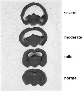
Hydrocephalus internus was diagnosed in dead golden hamsters in our laboratory during routine dissections (Figure 1). The incidence of hydrocephalus in our breeding stock was high. In one batch of nine litters only one litter was free of affected animals (Edwards et al., Reference Edwards, Gebhardt-Henrich, Fischer, Hauzenberger and Steiger2006). Two hamsters died before they reached 10 weeks of age. Another animal with hydrocephalus had to be euthanized because of severe neurological disease. Most hamsters with mild to severe hydrocephalus showed no apparent symptoms. No viral antibodies were detected in our animals and no toxins were found in the livers of affected hamsters. Test matings provided evidence for a genetic cause but the lack of sufficient data did not permit conclusions regarding the mode of inheritance or whether a single gene was likely to cause the condition (Edwards et al., Reference Edwards, Gebhardt-Henrich, Fischer, Hauzenberger and Steiger2006).
Brain sections of golden hamsters without hydrocephalus (normal) and with mild, moderate, and severe hydrocephalus.

Hydrocephalus also occurs in pet hamsters. A pet golden hamster (mottled color pattern, ours are wild-type) was brought to the Klinik für kleine Heimtiere at the Vetsuisse Faculty Bern recently and had to be euthanized due to loss of balance. The post mortem exam revealed hydrocephalus internus (C. Geigy, personal communication). Presumably most cases remain undetected, because the brain is usually not examined.
In this study, we investigated the mode of inheritance of the hydrocephalus without obvious symptoms and if a single locus of large effect played a role in our model of inheritance. Since images of hydrocephalic brains indicated that the hippocampus was affected, Timm staining was used to investigate the hippocampus.
Material and methods
Animals and housing
Group 1 consisted of golden hamsters which were progeny of breeders of the outbred strain ‘Laklbm:FUME’ obtained from RCC Ltd, (Füllinsdorf, BL, Switzerland). Group 2 was progeny of the outbred strain Crl:LVG(SYR) (until 31 December 2004: Lak:LVG(SYR)) obtained from Charles River (Sulzfeld, Germany), as well as adult Crl:LVG(SYR) directly obtained from Charles River (Sulzfeld, Germany). Group 3 was golden hamsters of the wild strain (were obtained from Prof. Gattermann (Gattermann et al., Reference Gattermann, Fritzsche, Neumann, Al-Hussein, Kayser, Abiad and Yakti2001 and Reference Gattermann, Fritzsche, Weinandy and Neumann2002)). Group 4 consisted of golden hamsters of the aura strain (RjHan:AURA), which were bought from Janvier (Le GenestSt-Isle, France). All animals were kept singly in cages (97 × 57 × 10 cm plastic bottom and a 45-cm-high wire top) and bedding consisted of wood shavings (Allspan®, D-76151 Karlsruhe, Germany). Food (Witte Molen™ (No. 3005301)) and water were offered ad libitum. Cat food as a protein source and mineral supplementation (Marienfelde Vitakalk®, D-22761 Hamburg, Germany) were offered once a week. Lighting was artificial (12 h light/12 h dark) with the light period between 0130 and 1330 h CEST (Central European Summer Time). Temperatures were between 21°C and 23°C, relative humidity varied from 30% to 55%. Our animal facility was approved by the Cantonal Veterinary Office.
Pathological examinations
The brain was immersion-fixed in buffered formalin and processed for routine, light-microscopic examination. One section of each visceral organ and four coronal sections of each brain were examined (at approximately levels 15 to 20, 26 to 30, 35 to 40, and 45 to 50 (Morin and Wood, Reference Morin and Wood2001)). Brains were graded as normal, mildly hydrocephalic (if lesions were barely visibly macroscopically or only noted histologically), moderately or severely hydrocephalic (if lesions were macroscopically obvious). Examinations were performed by several pathologists at the Vetsuisse Faculty Bern, as well as at the Institut für Labortierkunde, Vetsuisse Faculty, Zurich. The examined hamsters were between 18 days and 2 years old. All degrees of hydrocephalus were found at all ages and we could not detect a systematic influence of age on the degree of hydrocephalus (unpublished results).
Breeding
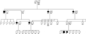
Inbreeding was generally avoided; however, the relatedness of the purchased animals was unknown. Breeding was done within lines except that two males of the wild derived stock were mated with two affected Crl:LVG(SYR) sisters. One of their female offspring was mated to an affected Crl:LVG(SYR) male, one was mated to an unaffected Crl:LVG(SYR) male. Two of their male offspring were mated to two affected Crl:LVG(SYR) females (Figure 2).
Pedigree of the test matings. Hamsters with hydrocephalus are filled circles (females) or squares (males) and hamsters without hydrocephalus are empty symbols. REP: These animals occur at more than one place in the pedigree. All animals were from Group 2 except the two males with the letter ‘w’ which were from Group 3 (derived from wild animals caught in 2001 by Gattermann et al. (Reference Gattermann, Fritzsche, Neumann, Al-Hussein, Kayser, Abiad and Yakti2001)).

Statistic analyses
For body masses the value closest to an age of 160 days (but at least that age) was taken since after that age no systematic changes in body mass occurred. Body masses were log-transformed. For litter sizes only first litters were considered. Analyses on body masses and litter sizes were performed with PROC GLM SAS 9.1.2 (SAS Institute, Cary, NC, USA).
Genetic analysis
Two hundred and ninety nine hamsters from one family (i.e. connected by pedigree) were used for the genetic analysis. One hundred and eighty seven of them had been examined for hydrocephalus. They belonged to Group 2.
Estimation of heritability
Continuous approach
Evaluation of the condition of hydrocephalus followed an ordered categorical score (e.g. normal, mild, moderate), which can be approached statistically as a continuously distributed trait. Though not ideal, scoring each category as 0, 1, 2, 2.5, 3 and 4, and treating this trait as a continuously distributed character, does permit a simple approach to the estimation of heritability. Specifically, we used this ordered score for the estimation of heritability making use of all known relationships in a linear model that included a fixed effect for sex and a random effect for the additive genetic contribution of each animal to its own hydrocephalus score. The null hypothesis of no genetic contribution (i.e. h 2 = 0) was tested with a likelihood ratio test, comparing the full model likelihood with that of the restricted model where h 2 = 0. Heritability is expressed as the mean ± s.e. of the mean. In addition, a test of the effect of sex on the hydrocephalus score was also tested through the likelihood ratio test. Estimation of the unknown genetic and residual variances was based on maximizing the multivariate normal likelihood and was implemented with the public domain computer program SOLAR (Almasy and Blangero, Reference Almasy and Blangero1998; Blangero et al., Reference Blangero, Almasy, Goring, Williams, Dyer and Peterson2005).
Binary
A second approach for estimating the heritability of hydrocephalus in the golden hamster is to consider the disease as a strict binary trait, where all animals with any sign of disease are grouped as affected. Hence, a threshold model for the liability to disease was used. This method assumes that an animal can be assigned to a specific disease class (unaffected/affected) when an underlying, unobservable risk (or liability) for disease exceeds a threshold of τ = 0. The distribution of the unobservable liability was assumed to be multivariate normal. The correlation in liability of two animals i and j was modelled to be  where ρij is the correlation in liability to disease between animals i and j; aij is the additive relationship between animals i and j; h 2 is the narrow sense heritability of liability to disease; δij is the coefficient for the random environmental component for animals i and j such that δij equals 1 if i equals j and zero otherwise; and
 , with no loss of generality. The null hypothesis of no genetic contribution (i.e. h 2 = 0) was tested with a likelihood ratio test, comparing the full model likelihood with that of the restricted model where h 2 = 0. Heritability is expressed as the mean ± s.e. of the mean. In addition, a test of the effect of sex on the liability of hydrocephalus was also tested through the likelihood ratio test. Calculations were implemented through the computer program SOLAR (Almasy and Blangero, Reference Almasy and Blangero1998; Blangero et al., Reference Blangero, Almasy, Goring, Williams, Dyer and Peterson2005), making use of the binary trait analysis first described in Duggirala et al. (Reference Duggirala, Williams, Williams-Blangero and Blangero1997).
Complex segregation analysis
The possibility that hydrocephalus in golden hamsters is influenced by the action of a single segregating locus of large effect can also be examined. Complex segregation analysis, developed by Bonney (Reference Bonney1986), is intended to integrate Mendelian transmission genetics at a single locus with the patterns of covariance expected in polygenic inheritance. Lynch and Walsh (Reference Lynch and Walsh1998) provide a more complete description of complex segregation analysis. Elston et al. (Reference Elston, Namboodiri, Glueck, Fallat, Tsang and Leuba1975) outlined the criteria that must be satisfied before acceptance of the single major locus model so as to reduce the risk of false-positive declarations of a major locus model. Evaluation of the models necessary for complex segregation analysis was conducted with the Bayesian software package iBay (2006, v. 1.0; Janss Biostatistics, Leiden, Netherlands). The iBay software is an extension of MaGGic (Janss, 1998) rewritten to accommodate complex segregation analysis in binary traits for pedigrees that include inbreeding. Accordingly, we can evaluate the actions of a major locus in hydrocephalus as a binary trait or with the ordered categorical score.
The software selected to conduct the complex segregation analysis is built upon a Bayesian foundation, making use of Monte Carlo Markov chains (MCMC) through a Gibbs sampler. Accordingly, point estimates of unknown parameters are not derived, but rather estimates of the posterior density for unknown parameters. The iBay (2006) package was recently used to evaluate the contribution of a major locus to osteochondral diseases in pigs (Kadarmideen and Janss, Reference Kadarmideen and Janss2005), where a more complete outline of the MCMC approach is detailed. The goal of this strategy was to simultaneously estimate the posterior density for a polygenic contribution to hydrocephalus along with the contributions of a putative Mendelian locus. Specifically, for this mixed-inheritance model, the strategy allowed the evaluation of a polygenic variance component, the additive and dominance contributions of a single locus (the parameters −a, d and a for the putative major locus genotypes AA, AB and BB, respectively) and the frequency of allele A of the putative major locus (defined as ‘q’). Given our scoring of phenotypes, where affected is 1 and unaffected scored as zero, the ‘B’ allele represents the putative disease-enhancing allele. Note also that the iBay software models the unobservable scale of this threshold trait such that the residual variance is fixed at 1.0 (i.e. σ e2 = 1).
Creation of the Gibbs sample requires several key assumptions about the behavior of these unknown parameters. Although a variety of models can be considered, all are some variant of the following: sex as a fixed effect with a flat (i.e. uniform) prior density, the polygenic variance component with a flat prior density, as well as flat prior densities for the additive, dominance and allele frequency parameters. A Gibbs sample of 5000 was generated, beginning with the creation of 300 000 total samples, a ‘burn-in’ of 50 000 and a sampling rate of every 100th Gibbs value. This process was repeated a second time, to create two replicate chains. From the 5000 Gibbs samples, the mean, standard deviation, mode and the upper and lower limits of a 95% highest density region (HDR) were computed for each of the unknown parameters.
Timm staining
Timm’s stain is visualizing the distribution of heavy metals in the brain (Timm, Reference Timm1958 and Reference Timm1962). Perfusion with a sulfide solution results in the formation of insoluble metallosulfide complexes. These complexes trigger a catalytic deposition of silver analogous to physical development as used in photography. Large metallosulfide complexes result in massive silver deposits appearing as black stain, while finely distributed small complexes yield yellowish or brown staining. In the hippocampus, the synaptic boutons of the mossy fibers (the axons of the dentate granule cells) stain deeply black with this method, because of their high content of presynaptic zinc. Timm’s stain is traditionally used to visualize the cytoatchitectonic boundaries of the hippocampal subregion CA3, and provides a rapid judgement about anomalous layering of the synaptic inputs onto apical and basal dendrites of pyramidal neurons.
Timm staining was done on six hamsters from Group 2 that had a hydrocephalus and on two hamsters from a cross between Group 2 and Group 3, which were free of hydrocephalus.
Results
Of 225 macroscopically and histologically examined animals of our breeding stock (Groups 1 and 2, i.e. hamsters derived from stock Lak:LVG(SYR)), only about half of the hamsters (108) were normal. Mild to severe forms of hydrocephalus were found in 117 hamsters. This high incidence occurred despite selection against hydrocephalus during part of the breeding period. As a control, 18 golden hamsters derived from a recently captured ‘wild stock’ from Syria (Group 3) (see Gattermann et al., Reference Gattermann, Fritzsche, Neumann, Al-Hussein, Kayser, Abiad and Yakti2001) as well as six heads of the strain Zoh:GOHA (Gattermann, Reference Gattermann1986) were diagnosed to be free of hydrocephalus. All hamsters of the AURA strain (Group 4, 45 animals) were unaffected.
Test matings with the wild derived males
All nine hybrids were unaffected. This is consistent with an autosomal recessive inheritance. When hybrids were mated to affected hamsters, eight offspring resulted and none had a hydrocephalus. This is not consistent with the presumed inheritance (χ 21 = 8, P < 0.005). When a female hybrid was mated to an unaffected male, five of the offspring were unaffected and two were hydrocephalic. Under the hypothesis of an autosomal recessive inheritance none of the offspring could have been hydrocephalic.
Matings of one unaffected male with affected and unaffected females
When the unaffected male was mated to unaffected females seven offspring were unaffected and nine were affected. When the same unaffected male was mated to affected females 10 offspring were unaffected and 28 were affected. Assuming an autosomal recessive inheritance, both the unaffected male and the unaffected females had to be heterozygous. Otherwise, no affected offspring could have arisen in the mating with the unaffected female. The hypothesis that hydrocephalus was autosomal recessively inherited can be rejected again (with the unaffected female: χ 21 = 8.33, P < 0.005; with the affected female: χ 21 = 8.52, P < 0.005).
Genetical analysis
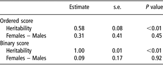
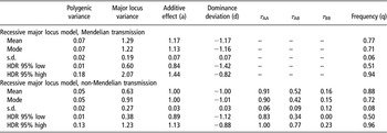
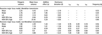
Table 1 presents the heritability of hydrocephalus under the two possible scoring systems for the disease. As both heritability values from this Table indicate, this character is clearly inherited, although the value of 1.0 reported for the binary analysis is obviously excessive. Such inflated values for heritability often result from the action of a single locus with a large effect (Lynch and Walsh, Reference Lynch and Walsh1998). In this light, we see that Tables 2 and 3 offer important evidence that a single recessive allele is acting on the expression of hydrocephalus. Specifically, the fact that the highest density regions for the major locus parameters (i.e. a, d and frequency) do not overlap 0.0 is a first step in providing statistical evidence that a locus with a major impact on hydrocephalus is segregating in this population.
Estimate of heritability and gender contrast in a linear model for the evaluation of hydrocephalus on ordered scores or as a binary evaluation of disease

Marginal posterior means, modes, standard deviations and limits to the 95% highest density regions (HDRs) of model parameters for hydrocephalus as an ordered score in golden hamsters in a Bayesian mixed-inheritance model with a completely recessive major locus, with and without Mendelian transmission

Marginal posterior means, modes, standard deviations and limits to the 95% highest density regions (HDRs) of model parameters for binary hydrocephalus in golden hamsters in a Bayesian mixed-inheritance model with a completely recessive major locus, with and without Mendelian transmission

In addition, it is interesting to compare the heritability estimate of the ordered score of hydrocephalus (the value of 0.58) with the estimate of the polygenic variance of the ordered score in Table 2. Specifically, we see a dramatic decline in the genetic variance attributable to the unexplained polygenic variance. Concomitantly, as the results of Table 2 show, this drop in polygenic variance is accompanied by a significant amount of genetic variance accounted for by the putative hydrocephalus major locus. Such behavior in the estimate of heritability is exactly that anticipated in the presence of a putative major locus before accommodation (result of Table 1) and after the inclusion of this Mendelian term (results of Table 2).
Also presented in Table 2, however, are tests of the significance of the Mendelian transmission patterns of this putative major allele. Elston et al. (Reference Elston, Namboodiri, Glueck, Fallat, Tsang and Leuba1975) outlined the criteria that must be met for the declaration of a significant major locus in complex segregation analysis. One of these criteria was a test for Mendelian transmission at the putative major locus. Under Mendelian transmission, the probability that a putative AA parent passes on an ‘A’ allele is 1.0, with values of 0.5 and 0 for genotypes AB and BB, respectively. Not shown are the results for the estimation of the transmission probabilities, which, for the ordered score measure of hydrocephalus, were not significantly different from the values expected under Mendelian transmission (the highest density regions overlap with 1, 0.5 and 0, respectively). Accordingly, we can consider the hydrocephalus score to be under the influence of a single recessive locus. Not so for the binary measure of hydrocephalus. In that case, the highest density regions for the transmission probabilities presented in Table 3 do not include the expected Mendelian values. As a final comment, we did not consider the possibility that a putative major locus was anything other than an autosomal locus with two alleles. The software employed for the complex segregation analysis permitted only such a single locus model.
Hippocampus in hamsters with hydrocephalus
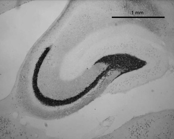
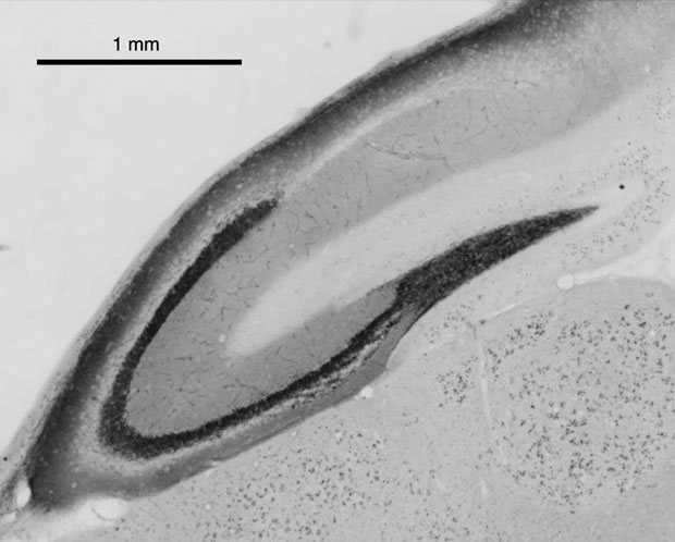
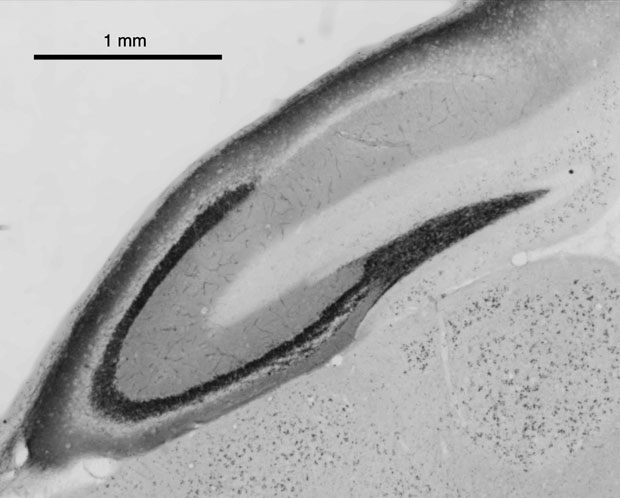
The Timm staining revealed a dorsally compressed but a ventrally intact hippocampus. Often, the frontal areas of the cortex were less compressed. In hamsters with a mild hydrocephalus, the hippocampus appeared normal (Figure 3). In hamsters with a severe hydrocephalus the hippocampus was dorsally compressed, but ventrally normal (Figure 4). Likewise, in Timm staining of hydrocephalic hamsters, mossy fibers were visible in the compressed part, implying at least partial functionality. The dentate gyrus and its hilus (CA4) were most compressed. However, the overall area covered by hippocampal tissue appeared about equal in hydrocephalic and control hamsters (see Figures 3 and 4).
Timm staining of the hippocampus of a mildly hydrocephalic hamster. The horizontal section from the mid-septotemporal level is shown.

Timm staining of the hippocampus of a severely hydrocephalic hamster. The horizontal section from the mid-septotemporal level is shown.
Reproduction and body mass
Dams with hydrocephalus had significantly smaller litters than dams without hydrocephalus (with h.: 7.4 ± 2.6 n = 18, without h.: 8.5 ± 2.3 n = 32). The presence of hydrocephalus in the sire did not influence the size of the litter (dam: F 1,26 = 4.5, P = 0.04, sire: F 1,26 = 0.79, ns, interaction: F 1,26 = 1.59, ns). Since more data on females were available, the body masses of fully grown (older than 160 days) female hamsters with (n = 74) and without (n = 56) hydrocephalus were compared. The presence of a hydrocephalus had no effect on body mass (F 1,128 = 0.26, ns, n = 130).
Discussion
After Yoon and Slaney (Reference Yoon and Slaney1972), Wahnschaffe et al. (Reference Wahnschaffe, Fredow, Heintz and Löscher1990) and Edwards et al. (Reference Edwards, Gebhardt-Henrich, Fischer, Hauzenberger and Steiger2006), we report a hereditary hydrocephalus in golden hamsters.
Several mutations in mice are associated with the occurrence of hydrocephalus (reviewed by Jones et al., Reference Jones, Lopman, Jones, Carter, Depelteau and Morel2000). These autosomal recessive mutations lead to different pathologies (Lyon and Searle, Reference Lyon and Searle1990). A gene of the mutation ch was localized, which induces several skeletal developmental defects in addition to a lethal hydrocephalus (Hong et al., Reference Hong, Lass and Chakravarti1999). Mutant mice were generated by gene targeting to obtain an animal model of the human CRASH phenotype, which includes a hydrocephalus (Dahme et al. Reference Dahme, Bartsch, Martini, Anliker, Schachner and Mantei1997).
There are two known mutations in rats: one line (H-Tx) with a high rate of inherited hydrocephalus was derived from one mating (Cai et al., Reference Cai, McGraw, Pattisapu, von Kalm, Willingham, Socci and Gibson2000; Jones et al., Reference Jones, Lopman, Jones, Carter, Depelteau and Morel2000, Reference Jones, Delpeteau, Carter, Lopman and Morel2001 and Reference Jones, Delpeteau, Carter and Somera2002) and the other strain (LEW/Jms) was derived from an inbred strain of Wistar-Lewis rats (Jones et al., Reference Jones, Carter and Morel2003). Both mutations seem to have a complex mode of inheritance probably involving more than one gene (Jones et al., Reference Jones, Carter and Morel2003). It was suspected that the inheritance mode of LEW/Jms changed over time (Jones et al., Reference Jones, Carter and Morel2003). Both mutations were normally lethal at an early age, but a mild form of H-Tx rats was described (Kiefer et al., Reference Kiefer, Eymann, von Tiling, Müller, Steudel and Booz1998).
Yoon and Slaney (Reference Yoon and Slaney1972) described an autosomal recessive mutation in golden hamsters with a dome-shaped head. Most did not live past 3 weeks of age and the few that survived long enough did not reproduce. Therefore this mutation must be different from our cases. It is not clear from which strain their hamsters were derived. This mutation was linked with cream coat color (Yoon and Peterson, Reference Yoon and Peterson1977). Wahnschaffe et al. (Reference Wahnschaffe, Fredow, Heintz and Löscher1990) claimed that hydrocephalus in golden hamsters was inherited like an autosomal recessive trait without presenting data on it. This mutation was detected by coronal brain sections and did not induce apparent symptoms (Wahnschaffe et al., Reference Wahnschaffe, Fredow, Heintz and Löscher1990). Therefore, it is likely a different mutation from the one Yoon and Slaney (Reference Yoon and Slaney1972) described and resembled our cases. It involved the same strain. The strain Laklbm:FUME was derived from Lak:LVG(SYR) and may therefore share the same mutation.
Much work has been devoted to the experimental induction of hydrocephalus in golden hamsters. Hydrocephalus could be induced by in utero exposure to a virus (Takano et al., Reference Takano, Takikita and Shimada1999), toxins (Hood et al., Reference Hood, Naughton and Hayes1976), chromium trioxide (Gale, Reference Gale1978a) and lead (Gale, Reference Gale1978b). It could also be induced by intracerebral inoculation with various viruses (Johnson and Johnson, Reference Johnson and Johnson1968; Kilham and Margolis, Reference Kilham and Margolis1969; Breschkin et al., Reference Breschkin, Haspel and Rapp1976; Davis, Reference Davis1981; Lagace-Simard et al., Reference Lagace-Simard, Descoteaux and Lussier1982), bacteria (Kohn et al., Reference Kohn, Kirk and Chou1977 and Reference Kohn, Chinookoswong and Wang1984) and chemicals (Mantovani et al., Reference Mantovani, Maranghi, Ricciardi, Macri, Stazi, Attias and Zapponi1998; Azzi et al., Reference Azzi, Canady, Ham and Mitchell1999).
We found evidence of an autosomal recessive mutation for the gradual hydrocephalus score but not for the binary score. Thus, the disease seems to be acting like a quantitative trait and on this scale the influence of a major locus could be demonstrated. Using the yes/no score, the major locus was masked and the test matings did not yield results consistent with Mendelian inheritance. The reason could be the complex development of the brain, which has made the identification of major loci difficult in humans, as well (Forestier, Reference Forestier2003). It is likely that the effect of the mutation was dependent on the genetic background. Alternatively, the distinction between no and mild hydrocephalus by the pathologists might not have been accurate enough. Since the study of Wahnschaffe et al. (Reference Wahnschaffe, Fredow, Heintz and Löscher1990) was done in the US in the end of the 1980s, this relatively benign mutation could be deeply embedded in this strain worldwide. The absence of an easy diagnosis in live animals will prevent the elimination of the incidence of hydrocephalus in this strain. Therefore, researchers using this strain should be aware of the fact that their hamsters likely have a hydrocephalus with a compressed hippocampus. Much of the research on environmentally induced hydrocephalus in golden hamsters of this strain will have to be critically reexamined and reevaluated. Since litter sizes of hydrocephalic females were reduced compared with litters from unaffected females their fertility was suppressed. However, since the status of the sire did not influence fertility, the genotype of the fetus did not likely cause mortality in utero. Results of the Timm staining revealed that the hippocampus was compressed in hydrocephalic brains. The potential influences of hydrocephalus on learning and memory, as well as implications for the well-being of severely hydrocephalic hamsters are unknown. Since well-being is reduced in humans suffering from hydrocephalus, this should be investigated further. In mice, the dorsal hippocampus is important for spatial learning (Zhu et al., Reference Zhu, Yee, Nyffeler, Winblad, Feldon and Mohammed2006), which may be impaired in golden hamsters with severe hydrocephalus. High-incidence genetic animal models are needed. We believe that these hamsters offer a great potential for such research.
Acknowledgements
We appreciate the expert help of Rosmarie Lang for neurohistology. H.-P.L. was supported by the Swiss National Science Foundation and the NCCR ‘Neural Plasticity and Repair’.