Introduction
An ecological niche is the sum of all the interactions of a species with the biotic and abiotic factors of its environment (Polechová and Storch, Reference Polechová, Storch and Lek2008). Niche theory predicts that two species with major niche overlap cannot coexist in the same environment, or in the case of plant pathogens, on the same host, indefinitely. Eventually one will be eliminated through competitive exclusion. In order to persist together, two species must exhibit some degree of niche specialization relative to each other. Many studies of niche specialization in fungi focus on closely related species, with the emerging generalization that related species often occupy divergent niches (Taylor et al., Reference Taylor2014). In some cases differences in niche dimensions can be difficult to ascertain, especially for sibling species on the same host, but close examination has revealed niche differentiation based on separation in time, in space, or in resource use even in these cases (Fitt et al., Reference Fitt2006). In other cases, where two plant pathogens are not closely related and have markedly different temporal or spatial patterns of resource use, the unique dimensions of species niche space are more easily determined.
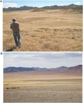
In this paper we use the framework of niche theory to explore how two of the most important fungal pathogens in seed banks of the invasive annual grass Bromus tectorum can coexist at high abundance while apparently exploiting a common resource. Bromus tectorum is an invasive annual grass that forms monocultures over very large areas in the semi-arid western United States (Fig. 1A; Chambers et al., Reference Chambers2014). It provides an abundant resource for a diverse community of fungal pathogens, including the two pathogens discussed here (Meyer et al., Reference Meyer, Beckstead, Pearce, Chambers, Germino and Brown2016). Pyrenophora semeniperda (the ‘black fingers of death’ pathogen) is a well-understood seed pathogen that is abundant in B. tectorum seed banks but rarely causes direct population-level effects (Meyer et al., Reference Meyer2007, Reference Meyer, Beckstead, Pearce, Chambers, Germino and Brown2016). The Fusarium seed rot pathogen (Fusarium sp.) also occurs regularly in B. tectorum seed banks and is the likely causal agent of B. tectorum stand failure or ‘die-off’ (Fig. 1B; Meyer et al. Reference Meyer2014a). We first briefly describe B. tectorum seed ecology and seed bank dynamics as a framework for the discussion of these two seed pathogens. We then examine pathogen life history and infection modality, focusing on contrasts that could indicate niche differentiation. Lastly we consider the interacting effects of these two pathogens on host population ecology in order to begin to elucidate the phenomenon of B. tectorum stand failure and recovery.
(A) Bromus tectorum monoculture near Dugway in Skull Valley, western Utah, USA. (B) Bromus tectorum die-off area (foreground) in Buena Vista Valley, central Nevada, USA.

Host seed ecology
Bromus tectorum monocultures present a relatively simple ecological setting for seed bank studies. The species has large seeds (mean floret mass ca 3 mg; Meyer, Reference Meyer, Pendleton, Meyer and Schultz2010) that are easily retrieved from the seed bank by screening and hand selection, and there are few, if any, other grass seeds in the samples that could confound identification. There is no regular mechanism for deep burial in wildland settings, and most viable seeds are found in the litter layer or in the top 2 cm of mineral soil (J. Beckstead, unpublished data). The seeds form a short-persistent seed bank, with an exponential decline in numbers of viable seeds in a seed cohort through time (Meyer et al., Reference Meyer2007). Very few seeds survive in the soil seed bank for more than two years (Smith et al., Reference Smith, Meyer and Anderson2008).
The seed ecology of B. tectorum has been well studied by our group, including ecophysiological modelling of seed dormancy loss, germination, and secondary dormancy induction (Christensen et al., Reference Christensen, Meyer and Allen1996; Bauer et al., Reference Bauer, Meyer and Allen1998; Bair et al., Reference Bair, Meyer and Allen2006; Meyer and Allen, Reference Meyer and Allen2009; Hawkins et al., Reference Hawkins, Allen and Meyer2017) and examination of patterns of seed bank persistence in the field (Beckstead et al., Reference Beckstead2007; Meyer et al., Reference Meyer2007; Smith et al., Reference Smith, Meyer and Anderson2008). These investigations have shown that seeds of this species are at least conditionally dormant at dispersal in early summer, lose dormancy through dry after-ripening under summer conditions, and are then poised to germinate rapidly in response to autumn rains. If autumn precipitation is insufficient to trigger full germination, the remaining viable seeds can enter secondary dormancy under partially hydrated conditions in late autumn or early winter. These seeds generally remain dormant through spring, lose dormancy over the following summer, and become germinable in time for autumn rains the following year. In exceptionally dry years, seeds may remain ungerminated but non-dormant through the winter, then germinate in response to spring precipitation. This phenological cycling of seeds between dormant and non-dormant states has important implications for seed pathogen attack.
Pyrenophora semeniperda
The Ascomycete genus Pyrenophora belongs to the family Pleosporaceae (order Pleosporales, class Dothidiomycetes). Pyrenophora semeniperda is the only known seed pathogen in the genus, and is only distantly related to species that are important foliar pathogens on cereal crops (Zhang and Berbee, Reference Zhang and Berbee2001). Pyrenophora semeniperda is a common pathogen of cosmopolitan distribution (Yonow et al., Reference Yonow, Kriticos and Medd2004; Stewart et al., Reference Stewart, Allen and Meyer2009) and has a very wide host range, particularly among cool-season grasses (Beckstead et al., Reference Beckstead and Meyer2014, Reference Beckstead2016). This fungus is an obligate pathogen known to reproduce only on seeds.
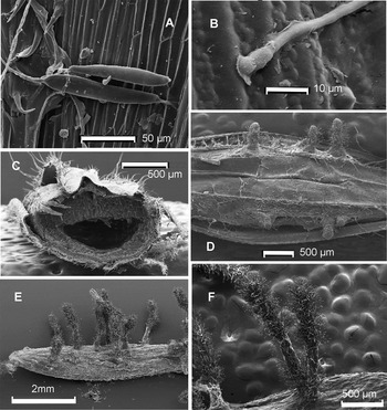
Pyrenophora semeniperda is an excellent candidate for study as a model system for understanding pathogenesis in soil seed banks. It forms macroscopic fruiting structures on seeds that make quantification in field seed bank samples a straightforward process (Fig. 2). It is most effective at attacking dormant B. tectorum seeds, either in summer before primary dormancy is lost (Hawkins, Reference Hawkins2013) or after secondary dormancy is induced in late autumn in the carry-over seed bank (Meyer et al., Reference Meyer2007; Hawkins et al., Reference Hawkins, Allen and Meyer2017). Pyrenophora semeniperda reproduces asexually through the production of septate conidial spores that are produced on the tips of branched conidiophores formed along the margins of elongate, black stromatal structures (Fig. 2 F). These conidial spores are produced under dry conditions in spring or early summer, and remain in the seed bank until rains stimulate their germination. The spores can germinate anywhere on the surface of the floret coverings or the naked caryopsis wall (Fig. 2A; Finch-Boekweg et al., Reference Finch-Boekweg2016). They apparently do not require an immediate stimulus from the host seed in order to initiate growth. The mycelial network ramifies on the surface, forming club-shaped appressorial structures at each point of penetration into the seed (Fig. 2B). These structures enable the hyphae to penetrate directly through the caryopsis wall and proliferate in the testa layer. The fungus then sends hyphae directly into the heart of the endosperm, which is digested first, resulting in a hollowing out of the interior of the seed and dense ramification of melanized hyphae throughout the testa, making killed seeds rigid (Fig. 2C). The fungus then produces the characteristic ‘black fingers’ or stromata that protrude through the coverings at any location on the surface (Fig. 2D–F). The pathogen does not target the embryo directly, and embryo dissolution is slower than the digestion of the endosperm, possibly explaining how a non-dormant seed that is infected and shows disease signs can often produce a healthy seedling (Beckstead et al., Reference Beckstead2007). Eventually, however, the embryo of a dormant seed is consumed, resulting in seed death and abundant stromatal production, which typically occurs ca 14 days following inoculation.
Stages in the life cycle of Pyrenophora semeniperda. (A) Germination of club-shaped conidia on the surface of a B. tectorum floret. (B) Formation of an appressorium for penetration into the floret tissue. (C) Floret cross-section at 10 days post-inoculation showing the hollowing out of the endosperm by the pathogen. (D) Pathogen stromata beginning to protrude from the killed seed. (E) Mature stromata protruding from the surface of a killed host seed. (F) Close-up of stromata with bristle-like conidiophores that bear the conidial spores. Photographs by Heather Finch-Boekweg; see Finch-Boekweg et al. (Reference Finch-Boekweg2016) for details.

The propensity for P. semeniperda to attack mostly dormant seeds in the seed bank can be explained by two features of this life cycle. First, the pathogen is able to effectively penetrate the seed coverings and does not require any signal from the germinating seed to initiate growth. This means that it can attack and kill dormant seeds. On the other hand, the fungus is relatively slow to develop once inside the seed, and primarily targets the endosperm rather than the embryo. This makes it possible for most non-dormant seeds under optimum germination conditions to escape mortality and develop into seedlings, which deprives the pathogen of resources that it can easily obtain from a dormant seed. Even dormant seeds can apparently defend themselves from attack, however, as disease development time is much reduced when killed seeds rather than living dormant seeds are inoculated, with stromata appearing within 5–6 days post-inoculation (Finch-Boekweg et al., Reference Finch-Boekweg2016).
Beckstead et al. (Reference Beckstead2007) coined the phrase ‘race for survival’ to describe the importance of dormancy status and thus seed germination rate in determining outcomes of seed infection by P. semeniperda. Fully after-ripened seeds germinated rapidly (<2 days) and escaped mortality, even though they could be infected and show disease signs on seeds that produced healthy seedlings. In contrast, dormant or slow-germinating seeds were almost always killed.
In spite of its adaptations for attacking dormant seeds, there is evidence that P. semeniperda can cause mortality of non-dormant seeds under some scenarios. One possible explanation is that there is genetic variation among strains in the ability to kill non-dormant seeds at high inoculum loads (Meyer et al., Reference Meyer, Stewart and Clement2010). Interestingly, the slowest-growing strains were better able to kill non-dormant seeds than faster-growing strains, in seeming contradiction to the ‘race for survival’ hypothesis. It was already known that this pathogen can produce large quantities of the cytotoxin cytochalasin B (Evidente et al., Reference Evidente2002). This fungal secondary metabolite is well known for its ability to disrupt mitosis through its effect on actin spindle fibre formation (MacClean-Fletcher and Pollard, Reference MacLean-Fletcher and Pollard1980). We determined that slow-growing strains of P. semeniperda make significantly more cytochalasin B than fast-growing strains, probably because the metabolic cost of producing this complex molecule limits resources for growth (Masi et al., Reference Masi and Evidente2014; Meyer et al., Reference Meyer2015). Increased cytochalasin B production might then be associated with increased ability to disable and kill germinating seeds, explaining why slow-growing strains caused higher mortality.
We also determined that cytochalasin B production by the fungus was induced only in solid culture on seeds or in liquid culture that contained seed constituents, further evidence of its role in pathogenesis (Meyer et al., Reference Meyer2015). In addition, inoculation into living dormant host seeds reduced cytochalasin B production by a factor of three relative to inoculation into killed host seeds, showing that living host seeds were able to suppress the production of this powerful toxin (Meyer et al., Reference Meyer2015).
In contrast to the results for non-dormant seeds, we showed that fast-growing strains of P. semeniperda were more likely than slow-growing strains to cause mortality on dormant B. tectorum seeds at low inoculum loads (Meyer et al., Reference Meyer2015), whereas at moderate to high inoculum loads mortality is very high regardless of strain (e.g. Beckstead et al., Reference Beckstead2007, Reference Beckstead2016; Finch et al., Reference Finch, Allen and Meyer2013). We reasoned that faster mycelial proliferation might be similar to increased inoculum load in its effect on seed mortality. We also used molecular-genetic techniques to show that a faster-growing strain had a competitive advantage over a slow-growing strain in terms of number of stromata produced in co-inoculations on dormant seeds, whereas the slow-growing strain produced more stromata than the fast-growing strain in competition on non-dormant seeds (Meyer et al., Reference Meyer2015). Based on these results we proposed that temporally varying selection on different seed dormancy phenotypes could maintain the growth rate polymorphisms observed in natural populations of the fungus by favouring strains with slow growth rates in the transient autumn seed bank but strains with fast growth rates in the dormant seed bank.
Another mechanism that could explain how P. semeniperda causes non-dormant seed mortality in spite of the ‘race for survival’ is related to the intermittent autumn precipitation regime and resulting seed bed water stress characteristic of semi-arid habitats where this pathogen is most successful. Hydrothermal time models developed for B. tectorum seed germination indicate that few non-dormant seeds are germinable at water potentials <2 MPa (Christensen et al., Reference Christensen, Meyer and Allen1996). This fungus, on the other hand, can germinate and grow at water potentials as low as –10 MPa (Barth et al., Reference Barth2015). This suggests that the pathogen may have an advantage under conditions of intermittent water stress. Experiments with pre-incubation for 7 days at germination-suppressive water potentials resulted in seed mortality at optimum temperature (20°C) of 22% in water after the –2 MPa treatment and 47% after the –1.5 MPa treatment (Finch et al., Reference Finch, Allen and Meyer2013). Seeds incubated in free water without a water stress pre-incubation treatment suffered no mortality. In further experiments, we showed that pathogen inoculum within seeds incubated in water for 24 h post-inoculation prior to transfer to much lower water potentials could complete most steps of pathogenesis over a 3-week period and then produce stromata almost immediately upon transfer to water, causing seed mortality (Allen et al., in press). This process could take place at water potentials as low as –40 MPa.
Fusarium seed rot pathogen
The Ascomycete genus Fusarium belongs to the family Nectriaceae (order Hypocreales, class Sordariomycetes). The genus is extremely diverse, including plant pathogens that produce diseases with widely varying signs and symptoms on a broad range of hosts, including many important crop diseases. Based on molecular genetic analysis, the Fusarium seed rot pathogen found in B. tectorum seed banks belongs to one or more undescribed species within the Fusarium tricinctum species group (O'Donnell et al., Reference O'Donnell2013; Franke et al., Reference Franke, Geary and Meyer2014; S.E. Meyer, unpublished data). This group consists mainly of grass pathogens such as F. tricinctum, F. avenaceum and F. acuminatum that cause various head blight, seed rot and pre- and post-emergence seedling diseases. The process of naming one or more new Fusarium species from B. tectorum seed banks is underway; they will subsequently be referenced in this review as the Fusarium seed rot pathogen or simply as Fusarium.
The Fusarium seed rot pathogen was first identified in 2009 from greenhouse studies aimed at determining the causal organisms involved in B. tectorum stand failure or ‘die-off’ at two field sites (Meyer et al., Reference Meyer2014a). This phenomenon manifests itself as total emergence failure in an area that supported a full stand of B. tectorum in the previous year. We hypothesized that this emergence failure might be caused by a fungal pathogen that could kill germinating seeds or germinants that had not yet emerged from the soil. In isolations from killed seeds in these experiments, 80% of the isolates obtained from the unsterilized soil treatment belonged to the genus Fusarium and were later shown to be members of the F. tricinctum species group. This strongly suggested that Fusarium might be implicated as a key die-off organism.
In pathogenicity trials with 16 of the isolated Fusarium strains, we found wide variation in the ability to kill non-dormant B. tectorum seeds, while there was low mortality on dormant seeds regardless of strain (Meyer et al., Reference Meyer2014a; S.E. Meyer, unpublished data). Mortality on non-dormant seeds inoculated and incubated in free water averaged 24%, whereas on dormant seeds mortality averaged less than 5%. As with P. semeniperda, adding a 7-day water stress pretreatment that imitated intermittent water availability in the field greatly increased mortality, especially of non-dormant seeds. Mean mortality was increased to 55%, and the most aggressive strains caused mortality as high as 83%. Dormant seed mortality was also slightly increased with water stress (mean 12% mortality). These results showed that some of the Fusarium isolates were strongly pathogenic on B. tectorum seeds and could potentially play a role in stand failure in the field. The results also demonstrated that this pathogen targets non-dormant B. tectorum seeds, in direct contrast to P. semeniperda.
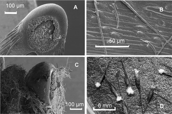
We used scanning electron microscopy to examine in detail the process of seed infection by the Fusarium seed rot pathogen (Franke et al., Reference Franke, Geary and Meyer2014). As with P. semeniperda, fungal spores could germinate at any location on the surface of the floret or naked caryopsis (Fig. 3B), but instead of penetrating the seed coverings directly, the hyphae exhibited clear directional surface growth towards the floret attachment scar that would later be the point of radicle emergence (Fig. 3A). The mechanism driving this directional growth has not been determined with certainty, but the most likely explanation is that the germinating seeds produce a chemical cue that emanates from the tip of the embryo through the porous tissue of the attachment scar (Fig. 3A; Nelson, Reference Nelson, Keister and Cregan1991). The fungus could detect this cue and follow the gradient to the point where the radicle will emerge. It then formed a conspicuous infection cushion over the attachment scar, visible macroscopically in inoculation trials as a ‘white tuft’ of mycelium (Fig. 3C,D; Franke et al., Reference Franke, Geary and Meyer2014). With incubation under water stress, this infection cushion remained at a relatively constant size indefinitely, apparently unable to penetrate the embryo under these conditions. However, once the seeds were transferred to free water, the fungus could rapidly invade the embryo through the weakened endosperm cap, killing the seed while undergoing massive mycelial proliferation. In later experiments, we determined that a water stress treatment as short as 24 h was sufficient to give Fusarium the head start it needs to cause high mortality of germinating seeds (J. Beckstead, unpublished data).
Stages in the life cycle of the Fusarium seed rot pathogen. (A) The floret attachment scar where pathogen attack will occur. (B) Adhesion and germination of sickle-shaped macroconidia on the surface of a B. tectorum floret. (C) Formation of the pathogen infection cushion over the floret attachment scar. (D) Infected florets in a Petri dish, showing the macroscopic infection cushions visible as ‘white tufts’. Photographs by JanaLynn Franke Pearce; see Franke et al. (Reference Franke, Geary and Meyer2014) for details.

The Fusarium seed rot pathogen does not develop a specialized appressorial structure for penetrating host tissue, relying instead on an infection cushion (Franke et al., Reference Franke, Geary and Meyer2014). This limits its ability to infect directly through more resistant host tissues, including seed coverings. Adaptations for locating zones of weakness where this penetration method can be effective are thus under strong selection, including a sensing mechanism for assuring that the infection cushion forms over the most vulnerable tissue. Because this sensing mechanism apparently depends on exudates from the radicle end of a seed that has initiated germination, it largely restricts attack by this pathogen to germinable, i.e. non-dormant, seeds. Occasionally a ‘white tuft’ can form at a point on the surface where a seed has suffered mechanical damage; this may be one way that dormant seeds can be attacked.
The Fusarium seed rot pathogen also appears to be a generalist on seeds of many grasses Host range testing has not been systematically performed for these strains, but some strains are known to attack seeds of at least two native cool-season grass species (Meyer et al., Reference Meyer2014b; J. Beckstead, unpublished data).
Pathogen niche specialization
When the adaptations exhibited by the two major pathogens on B. tectorum seeds in wildland seed banks are examined together, it is apparent that they occupy distinct niches in the B. tectorum seed bank pathogen community and probably experience limited competitive interaction, in spite of exploiting the same seed resource. The Fusarium seed rot pathogen targets B. tectorum seeds in the autumn when they are non-dormant and ready to germinate. It is a relatively fast-growing fungus that can achieve success even in environments with reliable autumn rains that elicit fast seed germination. The pathogen is likely to experience even greater success in drier areas with less reliable precipitation, given the advantage it enjoys under intermittent precipitation scenarios. It is clearly specialized to detect and respond to germinating seeds by targeting its attack to the point of radicle emergence where it can quickly destroy the embryo.
In contrast, the slower-growing pathogen P. semeniperda targets dormant seeds in the seed bank through its ability to form powerful penetration structures that can puncture seed coverings and reach endosperm reserves directly from any position on the exterior of the floret. By attacking dormant seeds, it ensures that all seed resources will be available for its use in spite of its relatively slow growth rate and the fact that it does not directly target the embryo. Intermittent precipitation scenarios in autumn also potentially increase P. semeniperda success, but because of its slow development, it requires much longer periods of water stress to achieve high seed mortality, whereas Fusarium can cause high mortality after a single relatively short water stress period. This would appear to give Fusarium a competitive advantage under intermittent autumn precipitation scenarios, which would in turn tend to select for even more specialization on dormant seeds in P. semeniperda. The exact environmental conditions for successful exploitation of water stress to increase the infection window may be sufficiently different for the two pathogens that interspecific competition even in this narrowly defined part of the niche space is minimized.
Die-off causes and consequences
Our current hypothesis for how these two pathogens interact to influence B. tectorum stand failure and recovery is that the Fusarium seed rot pathogen is the primary organism responsible for the large-scale seed death that results in stand failure, while P. semeniperda plays a role in the rate of post-stand failure recovery through its impacts on the persistent seed bank.
We first tested the hypothesis that P. semeniperda was directly implicated in stand failure by examining pathogen loads in both die-off areas and adjacent areas with current-year stands (Baughman and Meyer, Reference Baughman and Meyer2013). We found no evidence that this pathogen could cause stand failure, even though it can potentially reduce stand density through its impact on non-dormant seeds. Seed densities in the B. tectorum autumn seed bank can number in the tens of thousands per square metre (Meyer et al., Reference Meyer2007; Smith et al., Reference Smith, Meyer and Anderson2008), so that even significant mortality can leave ample densities of germinating seeds for stand establishment. This makes it even more remarkable that any pathogen can cause complete stand failure.
The case for Fusarium as the primary die-off pathogen is also still not conclusive (Meyer et al., Reference Meyer2014a), but its proven ability to kill germinating B. tectorum seeds in pathogenicity trials coupled with its ability to reduce stand density in field inoculum addition experiments (S.E. Meyer, unpublished data) is strong evidence that it is one of the primary players. Another line of evidence that Fusarium is implicated in die-offs comes from Baughman and Meyer (Reference Baughman and Meyer2013). Post-die-off viable seed densities in carry-over seed banks were not statistically different in paired die-off and control sites, indicating that the pathogen killed germinating seeds but did not attack dormant seeds.
Stand recovery the year following B. tectorum stand failure is the most common pattern observed in the field (Weisberg et al., Reference Weisberg2017). The presence of a persistent seed bank not affected by the die-off causal agent, as mentioned above, is the most likely explanation for this stand recovery. Most die-offs are therefore transient phenomena. However, sometimes stand recovery does not occur so quickly. Two alternative hypotheses have been proposed to explain this. First, the organism that caused the die-off may still be active the following year. Precision seeding experiments (using toothpick-mounted seeds that could be followed individually) in die-off areas and adjacent areas with intact stands at two sites showed no significant difference in B. tectorum emergence the year following a die-off (Nicholson, Reference Nicholson2014). This makes residual effects of the pathogen an unlikely explanation for a persistent die-off. A second hypothesis is that there was insufficient carry-over seed to establish a post-die-off stand. We examined this possibility with a precision seeding experiment into an unrecovered die-off, and demonstrated that seed addition was the only requirement for stand establishment (Nicholson, Reference Nicholson2014). This supported the idea that lack of a persistent seed bank following the die-off was the cause of slow stand recovery. While we have no direct evidence that the carry-over seed bank was eliminated due to P. semeniperda attack, this remains the most likely explanation.
Conclusions
Mechanistic or even descriptive studies of natural pathosystems involving seeds as hosts are uncommon, in spite of their potential importance as mediators of host population dynamics (Chambers and MacMahon, Reference Chambers and MacMahon1994; Gilbert, Reference Gilbert2002). The seed bank pathogen community is more often treated as a ‘black box’, that is, an assemblage of multiple pathogens that act in concert to affect seeds. These are manipulated as a group, for example, through fungicide applications (Blaney and Kotanen, Reference Blaney and Kotanen2001; Orrock and Damschen, Reference Orrock and Damschen2005) or through the measurement of soil legacy effects (Bever, Reference Bever1994; Klironomos, Reference Klironomos2002). Our investigations have made a unique contribution to the study of seed bank pathosystems in natural settings by focusing on the biology and ecology of specific pathogens, enabling us to more fully understand their effects on host seed ecology.
Acknowledgments
We thank the many research collaborators and students who contributed to this effort over the years. We also thank the organizers of the ISSS meeting in Monterey, California, September 2017 for the opportunity to include this synthesis as an invited presentation and for providing funding for meeting participation. Thanks to Heather Finch-Boekweg and JanaLynn F. Pearce for the use of unpublished photographs, and to Bettina Schultz for formatting the graphics.
Financial support
The preparation of this review received no specific grant from any funding agency, or commercial, or not-for-profit sector. Funding sources for the primary research upon which this review is largely based are included in the original publications.