Introduction
Single-sided deafness is characterised by unilateral sensorineural hearing loss (SNHL) in the presence of normal hearing in the contralateral ear. Single-sided deafness was undervalued for a long time, as it was presumed that one intact ear is adequate for normal language development.Reference Ead, Hale, DeAlwis and Lieu1 With increasing awareness of the functional impact of single-sided deafness in developing children, recent studies have focused on the aetiology of unilateral SNHL and relevant management strategies. Although there are well-established protocols for bilateral SNHL in the current literature, there is no consensus regarding the management of single-sided deafness in paediatric cases.Reference Arndt, Prosse, Laszig, Wesarg, Aschendorff and Hassepass2–Reference Mertens, De Bodt and Van de Heyning4 The issue of which radiological imaging modality is the preferred method is still under debate, and radiological imaging in single-sided deafness cases is a novel subject in the literature.
Inner-ear malformations are common in children with unilateral or bilateral hearing loss, and their identification directly affects the management of hearing loss.Reference Sennaroğlu and Bajin5 Cochleovestibular malformations and cochlear nerve deficiency have been described in paediatric unilateral SNHL cases.Reference Friedmann, Ahmed, McMenomey, Shapiro, Waltzman and Roland6 It is highly recommended that all children with single-sided deafness undergo radiological imaging, in addition to audiological evaluation, in order to shed light on the aetiology of the condition. For instance, in certain inner-ear malformations, such as cochlear nerve aplasia, cochlear aplasia, internal auditory canal atresia or cochlear aperture atresia, cochlear implantation is not beneficial. In order to make a decision on the direction of the treatment for single-sided deafness patients, establishing a final radiological diagnosis is a must.
This study aimed to elucidate the prevalence and distribution of inner-ear malformations in congenital single-sided deafness. Details of malformation type are crucial for the treatment and management of the disease, and to increase awareness of the functional impact of single-sided deafness and its radiological findings, which may influence the treatment of this condition.
Materials and methods
A retrospective review was conducted of the medical charts and radiological findings of patients with congenital single-sided deafness, who were younger than 18 years, and diagnosed between 2011 and 2017 at a tertiary referral centre. A total of 90 patients were included in this study. Cases with acquired aetiology such as trauma or chronic otitis media, and those who displayed conductive components of hearing loss, were excluded. Institutional ethical committee approval for the study was obtained (number: GO1052).
Audiological evaluation
Patients’ files were reviewed to evaluate hearing status, utilising otoacoustic emissions (OAE), auditory brainstem response (ABR) and audiogram data if available. The diagnosis of single-sided deafness was initially established based on the findings of pure tone audiometry, or OAE and ABR. Hearing loss was graded as follows: 71–95 dB represents severe hearing loss, and greater than 95 dB HL reflects profound hearing loss. Pure tone average was the average of hearing thresholds at 0.5, 1 and 2 kHz. If the pure tone average was equal to or less than 71 dB in one ear, the patient was defined as having single-sided deafness and included in the study.
When pure tone audiometry could not be performed because of the patients’ age, the diagnosis of hearing loss was made by OAE and ABR. The ABR recordings were obtained while the children were sleeping or under sedation. If no V waves were elicited by a click stimulus at 105 dB HL, the response was considered negative and the patient was classified as having single-sided deafness. The OAE tests were performed using the GSI-60 system (Grason-Stadler, Eden Prairie, Minnesota, USA). Patients with insufficient audiological documentation were not included in the study.
Radiological evaluation
The study included patients who had been diagnosed in our clinic, and whose temporal bone computed tomography (CT) and magnetic resonance imaging (MRI) scans were available in our picture archiving system. Normally, when a patient is diagnosed with single-sided deafness, we routinely perform both temporal bone CT and MRI in our clinic. Patients for whom there were not scans for both CT and MRI scans were excluded from this study. In recent years, because of radiation concerns, only MRI has been requested for diagnosis; CT scans are obtained if a surgical intervention is planned.
The CT and MRI images of all patients were investigated by two otolaryngologists. The CT imaging was performed with 0.5 mm slice thickness (using a Somatom Plus 4 Volume Zoom; Siemens, Erlangen, Germany). The MRI was conducted using 3 Tesla scanner (a Magnetom Allegra, Siemens, or an Ingenia, Royal Philips, Amsterdam, Netherlands).
The measurements were conducted manually, using calipers, on the picture archiving system screen (GE Healthcare, Chicago, Illinois, USA), and measurements were rounded up to the nearest 0.1 mm. The focus of interest was the existence of any inner-ear malformations that might be the cause of single-sided deafness.
Inner-ear malformations were grouped according to the classification system suggested by Sennaroğlu and Bajin.Reference Sennaroğlu and Bajin5 Inner-ear malformations were classified as: Michel deformity, rudimentary otocyst, common cavity, cochlear aplasia, incomplete partition, cochlear hypoplasia, cochlear aperture abnormality or enlarged vestibular aqueduct.
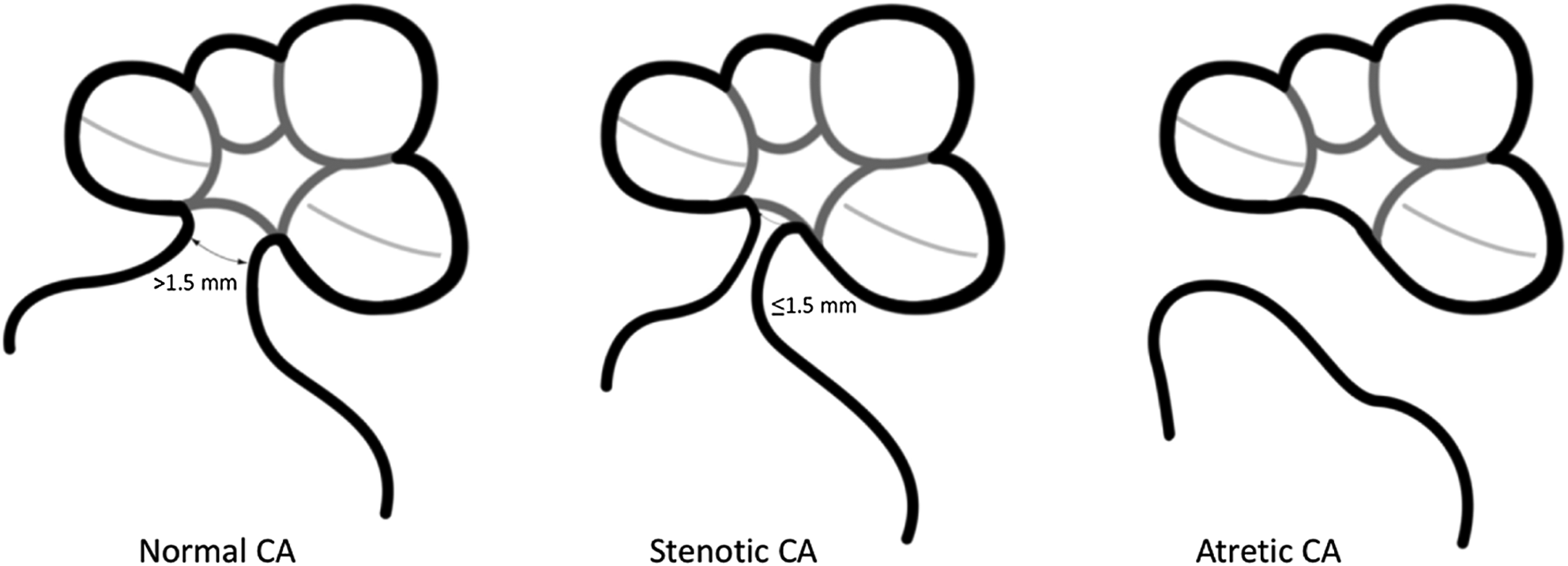
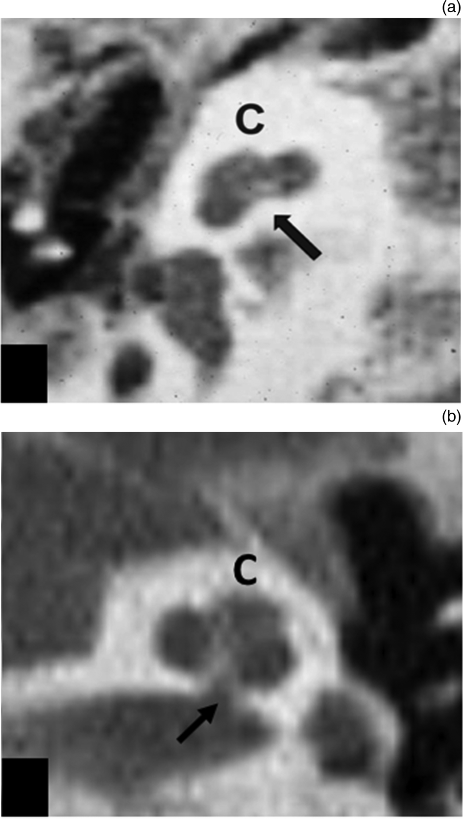
The width of the cochlear aperture was measured at its midportion at the mid-modiolar level on the axial images. Based on the previous literature, the cochlear aperture was considered stenotic when its mid-modiolar diameter was less than 1.5 mm. The cochlear aperture structures were grouped as normal (over 1.5 mm), stenotic (1.5 mm or less) or atretic (0 mm) (Figure 1). When the cochlear aperture was atretic with an otherwise normal cochlear structure and dimensions, it was described as ‘isolated aperture atresia’ (Figure 2a). If a stenotic cochlear aperture accompanied a normal cochlear structure and dimensions, the condition was described as ‘isolated aperture stenosis’ (Figure 2b). The internal auditory canal was also measured, and was defined as ‘narrow’ when its midpoint diameter was less than 2 mm on CT axial images.
Illustrations of normal, stenotic and atretic cochlear apertures (CA).

Axial, high-resolution computed tomography images showing (a) isolated cochlear aperture atresia (arrow) and (b) isolated cochlear aperture stenosis (arrow), with otherwise normal cochlear structure and dimensions. C = cochlea

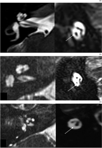
For the MRI evaluation, the authors consulted a neuroradiologist on the definitive status of the cochlear nerve. In the MRI studies, the cochlear nerve was examined on axial and especially on oblique sagittal planes. The cochlear nerve width was compared with the ipsilateral facial nerve, and classified as normal, hypoplastic or aplastic according to its size compared to ipsilateral facial nerve (Figure 3). When a cochlear nerve was hypoplastic or aplastic, this situation was described as ‘cochlear nerve deficiency’.
Axial (left) and oblique sagittal (right) magnetic resonance imaging sections showing: (a) a normal cochlear nerve, (b) a hypoplastic cochlear nerve and (c) an aplastic cochlear nerve. Dotted white arrows indicate the cochlear nerve. C = cochlea

Contralateral ears were also examined radiologically and the presence of an anomaly was determined.
Statistical analyses
Descriptive statistics were presented as frequency, percentage, mean or median, standard deviation, and minimum and maximum values. The distribution of the data was determined by Kolmogorov–Smirnoff and Shapiro–Wilk tests, and the continuous variables that were not normally distributed were analysed using the Mann–Whitney U test. Categorical data were analysed by Fisher's exact test. The digital data of the three groups were compared using a Kruskal–Wallis test; the Bonferroni–Dunn procedure was followed to perform paired comparisons among statistically significant results. SPSS Statistics for Windows, version 21.0 software (IBM, Armonk, New York, USA) was used for the statistical analyses. The results were considered statistically significant when p < 0.05.
Results
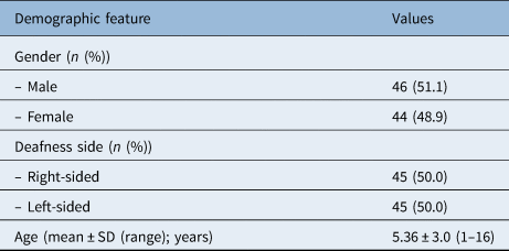
This study comprised 90 cases of congenital single-sided severe or profound SNHL. Patients’ age ranged from 1 to 16 years, with a mean age of 5.36 ± 3.0 years. There were 46 males (51.1 per cent) and 44 females (48.9 per cent). The right and left ear distribution was even: 45 patients (50 per cent) had right-sided deafness and the remaining 45 (50 per cent) had left-sided deafness (Table 1). Among the 90 children in this study, 42 (46.7 per cent) had abnormal CT and/or MRI findings in the affected ear.
Demographic features of study patients

SD = standard deviation
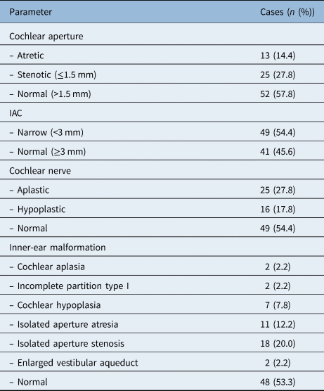
Examination of the distribution of inner-ear malformations in the affected ear revealed that: 2 cases (2.2 per cent) had cochlear aplasia, 2 cases (2.2 per cent) had incomplete partition type I, 2 cases (2.2 per cent) had an isolated enlarged vestibular aqueduct, 7 cases (7.8 per cent) had cochlear hypoplasia, 11 cases (12.2 per cent) had isolated aperture atresia and 18 cases (20 per cent) had isolated aperture stenosis. The remaining 48 cases (53.3 per cent) had a normal cochlear structure. The distributions of inner-ear malformations, status of the cochlear aperture, width of the internal auditory canal and anomalies of the cochlear nerve can be seen in Table 2.
Distributions of inner-ear malformations, cochlear nerve status, IAC width and cochlear aperture anomalies in study group

IAC = internal auditory canal
There were no significant differences in demographic features between patients with normal cochlear anatomy (26 males and 22 females, mean age of 5.42 ± 2.53 years) and patients with inner-ear malformations (20 males and 22 females, mean age of 5.29 ± 3.49 years). In addition, the right to left ear ratio was similar in the normal cochlear group (24 right ears and 22 left ears) and inner-ear malformation group (23 right ears and 19 left ears).
The cochlear nerve was evaluated in all cases and was found to be aplastic in 25 ears (27.8 per cent), hypoplastic in 16 ears (17.8 per cent) and normal in 49 ears (54.4 per cent); hence, 41 ears (45.6 per cent) had cochlear nerve deficiency. Cochlear nerve deficiency was the most common anomaly in the affected ears, with a frequency of 45.6 per cent. The distributions of cases with cochlear nerve deficiency (18 male and 23 females, mean age of 5.27 ± 3.51 years) and those with a normal cochlear nerve (28 males and 21 females, mean age of 5.43 + 2.52 years) were similar in terms of demographic features and right to left ear ratio (cochlear nerve deficiency = 28 right ears and 21 left ears; normal cochlear nerve = 23 right ears and 26 left ears) (p > 0.05).
The mean internal auditory canal width in the study group as a whole was 2.78 ± 0.85 mm (minimum = 1.33 mm, maximum = 4.35 mm). Forty-nine ears (54.4 per cent) had a narrow internal auditory canal (less than 2 mm) and the remaining 41 cases (46.9 per cent) had a normal internal auditory canal (2 mm or over). Cases with a narrow internal auditory canal (23 males and 26 females, mean age of 5.22 ± 3.32 years) and a normal internal auditory canal (23 males and 18 females, mean age of 5.46 ± 2.59 years) did not display differences in terms of demographic features and right to left ear distribution (narrow internal auditory canal = 27 right ears and 22 left ears; normal internal auditory canal = 18 right ears and 23 left ears) (p > 0.05).
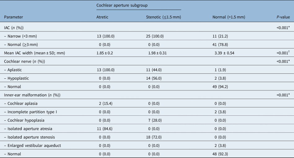
Another important parameter was the width of the cochlear aperture. Among the study group as a whole, 13 cases had an atretic cochlear aperture and 25 cases had a stenotic cochlear aperture (1.5 mm or less), while the remaining ears had a normal cochlear aperture width (over 1.5 mm). Out of 13 cases with an atretic cochlear aperture, 2 had cochlear aplasia. The remaining 11 cases had normal cochlear anatomy with an atretic cochlear aperture, which can be termed as isolated aperture atresia. In those with a stenotic cochlear aperture, seven cases had cochlear hypoplasia in addition to cochlear aperture stenosis. The remaining 18 cases had otherwise normal cochlear anatomy with a stenotic cochlear aperture, which can be termed as isolated aperture stenosis. The distributions of inner-ear malformations, status of the cochlear nerve, and width of the internal auditory canal in the cochlear aperture atresia or stenosis and normal cochlear aperture subgroups can be examined in Table 3.
Distributions of inner-ear malformations, cochlear nerve status and IAC width in cochlear aperture subgroups

*Fisher's exact test; †Kruskal–Wallis analysis. IAC = internal auditory canal; SD = standard deviation
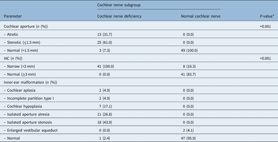
The relationship between cochlear aperture and cochlear nerve was another parameter investigated. In the normal aperture group (over 1.5 mm), there were only 3 cases of cochlear nerve deficiency (1 aplastic, 2 hypoplastic); the remaining 49 cases (94.2 per cent) had a normal cochlear nerve. Cochlear nerve deficiency was strongly correlated with aperture width (p < 0.001, Fisher's exact test). In the stenotic aperture group (1.5 mm or less), 11 cases (44 per cent) had an aplastic cochlear nerve and 14 cases (56 per cent) had a hypoplastic cochlear nerve. In other words, cochlear nerve deficiency was always accompanied by a stenotic aperture. Eleven cases with isolated aperture atresia were examined; all cases were found to have cochlear nerve aplasia, as well as a stenotic or atretic internal auditory canal (p < 0.001, Fisher's exact test). Cochlear nerve deficiency and its associations with cochlear aperture anomalies, and internal auditory canal width in subgroups with a cochlear nerve deficiency or normal cochlear nerve, are demonstrated in Table 4.
Distributions of inner-ear malformations, cochlear aperture anomalies and IAC width in cochlear nerve subgroups

*Fisher's exact test. IAC = internal auditory canal
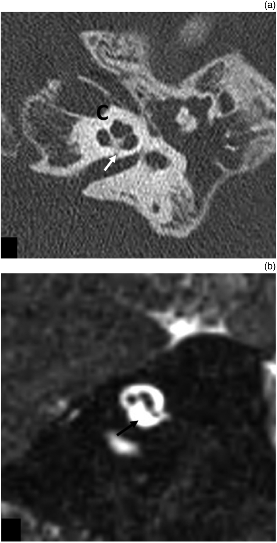
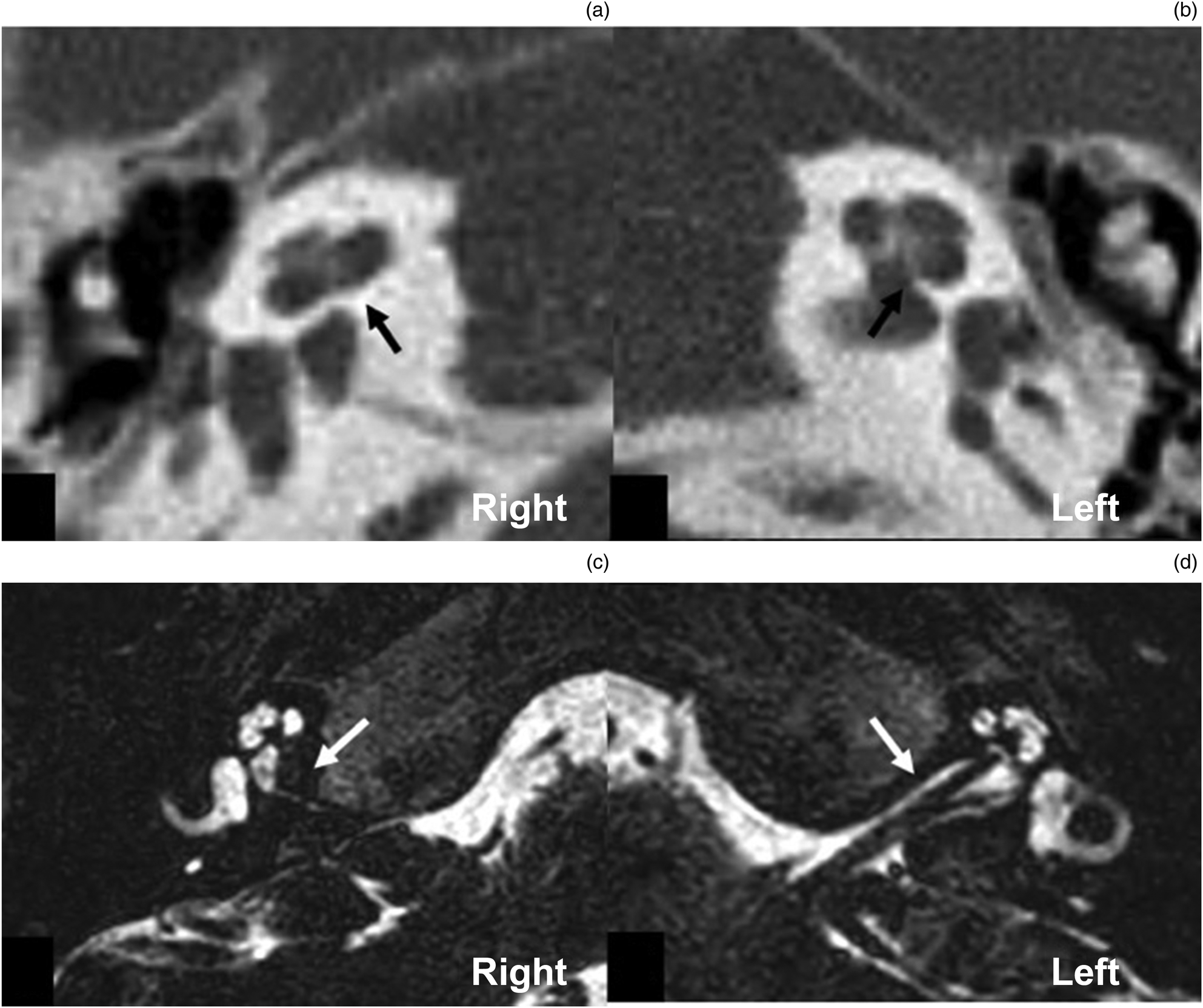
Figure 4 shows the CT and MRI scans of a patient with isolated aperture atresia and an aplastic cochlear nerve in the left ear. Figure 5 demonstrates the CT and MRI scans of a patient with isolated aperture stenosis and a hypoplastic cochlear nerve.
(a) Axial, high-resolution computed tomography image showing isolated cochlear aperture atresia (white arrow indicates cochlear aperture), and (b) oblique sagittal magnetic resonance imaging section showing an aplastic cochlear nerve (black arrow), in the left ear of the same patient. C = cochlea

Axial, high-resolution computed tomography images showing (a) isolated cochlear aperture stenosis in the right ear and (b) a normal cochlear aperture in the left ear of the same patient (black arrows indicate cochlear aperture). Axial magnetic resonance imaging sections showing (c) a hypoplastic cochlear nerve in the right ear and (d) a normal cochlear nerve in the left ear of the same patient (as in parts a & b) (white arrows indicate the cochlear nerve).

The relationship between internal auditory canal width and the cochlear nerve was also analysed. All ears with a normal internal auditory canal had a normal cochlear nerve. In ears with a narrow internal auditory canal, 25 ears (51 per cent) had an aplastic cochlear nerve, 16 ears (32.7 per cent) had a hypoplastic cochlear nerve and 8 ears (16.3 per cent) had a normal cochlear nerve. The internal auditory canal width was found to be a strong predictive parameter in estimating the status of the cochlear nerve.
Cochlear aperture width and internal auditory canal width were found to be strongly correlated to each other, and this relationship was bidirectional. The mean cochlear aperture width was 0.97 ± 0.65 mm in the narrow internal auditory canal group and 1.74 ± 0.15 mm in the normal internal auditory canal group (p < 0.001, Mann–Whitney U test). When the cochlear aperture was atretic or stenotic, the mean internal auditory canal width was 1.85 ± 0.20 mm and 1.98 ± 0.31 mm, respectively. In cases with a normal cochlear aperture width, the mean internal auditory canal width was 3.39 ± 0.54 mm (p < 0.001, Mann–Whitney U test). In other words, when the cochlear aperture is atretic or stenotic, the internal auditory canal is stenotic, and vice versa.
The likelihood of a deficient cochlear nerve was reduced in cases where the cochlear anatomy, aperture width and internal auditory canal width were normal. Forty-eight cases had a normal cochlear anatomy and cochlear aperture width, and only one case (2.1 per cent) demonstrated cochlear nerve hypoplasia (p < 0.001).
Discussion
Single-sided deafness has been an underestimated entity because, given normal contralateral hearing, it was not considered to affect normal language development. However, since the 1980s, several studies have shown that single-sided deafness may be associated with educational and behavioural handicaps in children.Reference Bess, Klee and Culbertson7 Audiological deficits in children with single-sided deafness negatively influence brain development, language performance and socio-emotional development. The duration of deafness is crucial for audiological development and cortical plasticity, especially in children. Therefore, prompt and detailed evaluation of paediatric single-sided deafness patients is mandatory to provide the best modality of auditory rehabilitation to these children.Reference Welsh, Welsh, Rosen and Dragonette8
Previously, single-sided deafness was attributed to viral infections, especially mumps. However, the accumulation of knowledge on this subject led researchers to investigate other possible causes, and it has since been demonstrated that inner-ear malformations may be the primary cause of single-sided deafness.Reference Song, Choi, Oh, Chang, Kim and Lee9–Reference Simons, Mandell and Arjmand11
In this study, we evaluated the CT and MRI scans of 90 cases with single-sided deafness. The overall prevalence of inner-ear malformations was 46.7 per cent, which is consistent with previous reports. The most frequent inner-ear malformation was isolated aperture stenosis (20 per cent), followed by isolated aperture atresia (12.2 per cent) and cochlear hypoplasia (7.8 per cent), and the prevalence of cochlear nerve deficiency was 45.6 per cent.
In 1995, Bamiou et al. emphasised the importance of CT scanning in the aetiological diagnosis of unilateral SNHL.Reference Bamiou, Savy, O'Mahoney, Phelps and Sirimanna12 In their study, 11 out of 35 CT scans were abnormal, and labyrinthine ossificans and an enlarged vestibular aqueduct were the most common findings. That study can be regarded as a pioneer in highlighting the importance of radiological investigation in single-sided deafness to explore inner-ear malformations.
Song et al. investigated 322 single-sided deafness cases.Reference Song, Choi, Oh, Chang, Kim and Lee9 Ninety-three of the cases (28.9 per cent) had radiologically detectable inner-ear malformations, predominantly incomplete partition type I and internal auditory canal stenosis. However, in that study, MRI images of the cases were not evaluated. Thus, possible cochlear nerve deficiency cases may have been missed. In addition, the cochlear aperture was not measured in that study, further contributing to the possibility of cochlear nerve deficiency being overlooked. Our radiological assessment can be deemed more accurate than that conducted by Song et al., as we recorded cochlear nerve, internal auditory canal and cochlear aperture measurements, and utilised both CT and MRI scans.
Cochlear nerve deficiency is a recently recognised cause of SNHL. As a result of improvements in MRI technology, cochlear nerve deficiency is now detected and diagnosed more frequently, and is a much more common condition than once thought.Reference McClay, Booth, Parry, Johnson and Roland13
In their investigation of the aetiological factors causing single-sided deafness, Usami et al. analysed the radiological examinations of 199 patients with unilateral SNHL.Reference Usami, Kitoh, Moteki, Nishio, Kitano and Kobayashi14 They too classified inner-ear malformations according to Sennaroğlu and Bajin's classification system. These authors found that cochlear nerve deficiency was the most common anomaly in patients with unilateral SNHL (n = 87, 43.7 per cent), as in our study (n = 41, 45.6 per cent). The limitation of their study was the availability of only one imaging modality, rather than complete investigation, using both CT and MRI, of the entire cochlear nerve, internal auditory canal and bony labyrinth. They found only eight cases (3.8 per cent) with inner-ear malformations (five cases with incomplete partition type I and three cases with a common cavity). However, they studied viral serology (such data were not available in our study), and they detected mumps (6.2 per cent) and cytomegalovirus (6.2 per cent) as causative agents in their case series. We can assert that cochlear nerve deficiency and inner-ear malformations were underestimated as aetiologies of single-sided deafness, but they have recently been diagnosed more frequently with the help of advanced radiological devices.
The bony cochlear nerve canal, the cochlear aperture in other words, is a bony canal that carries the cochlear nerve fibres from the spiral ganglion to the internal auditory canal.Reference Fatterpekar, Mukherji, Alley, Lin and Castillo15 A narrow cochlear aperture is a newly defined anomaly causing SNHL, with or without accompanying inner-ear malformation.Reference Masuda, Usui and Matsunaga16 It is usually seen together with a narrow internal auditory canal and an aplastic or hypoplastic cochlear nerve. The authors of the current study would like to emphasise the term ‘isolated’, which means that the cochlear aperture can be abnormal in a normal cochlear morphology. Careful assessment of this type of anomaly is crucial as it is frequently associated with cochlear nerve deficiency, which constitutes a contraindication for cochlear implantation. This is the most important reason for including the cochlear aperture abnormalities as a separate group of inner-ear malformations, indicating that they can be the only abnormality when all other inner-ear structures are normal.Reference Sennaroğlu and Bajin5
Cochlear aperture abnormalities are recognised as being among the more common findings in congenital single-sided deafness, but the underlying cause is unknown.Reference Yi, Lim, Kang, Park, Park and Lee17 Cochlear aperture stenosis is known to be strongly correlated with SNHL.Reference Yi, Lim, Kang, Park, Park and Lee17 There are now a few published studies focusing on the causal relationship between aperture stenosis and cochlear nerve status. In our study, when the cochlear aperture was atretic, the cochlear nerve was always aplastic; furthermore, a stenotic cochlear aperture was associated with an aplastic cochlear nerve in 44 per cent of cases. In addition, the cochlear aperture width and internal auditory canal width were strongly correlated, and a narrow internal auditory canal resulted in a deficient cochlear nerve.
Nakano et al. showed the differences in the aetiology and mechanisms of cochlear nerve deficiency in children with bilateral versus unilateral SNHL.Reference Nakano, Arimoto and Matsunaga18 The prevalence of cochlear nerve deficiency was much higher in unilateral SNHL cases (50 per cent) than in bilateral ones, as the results of our study also indicate. A small or absent cochlear aperture was a major cause of unilateral SNHL in their cohort. However, they used only CT scans to estimate cochlear nerve deficiency. Although cochlear aperture stenosis is a strong indicator of cochlear nerve deficiency, MRI should also be performed for a more accurate diagnosis.
Our study is superior in this respect, as we included only those cases with both CT and MRI scans. Imaging is of particular diagnostic importance in children compared to adults, as various inner-ear malformations can be detected in paediatric cases, and imaging is crucial for a definitive diagnosis. The most ideal algorithm for the use of both CT and MRI in children is, however, controversial. In our clinic, both CT and MRI were initially employed in all paediatric cases of unilateral or bilateral SNHL. However, because of radiation concerns, we subsequently started to investigate paediatric single-sided deafness cases with MRI only and to reserve CT for cases requiring surgery.
Purcell et al. focused on the progression of unilateral SNHL in children with cochlear aperture stenosis.Reference Purcell, Shinn, Coggeshall, Phillips, Paladin and Sie19 In their series, 17 per cent of the patients had aperture stenosis and the overall prevalence of inner-ear malformation was 42 per cent, which is similar to our findings. Their investigation was one of the leading studies on the cochlear aperture and unilateral SNHL. They identified an enlarged vestibular aqueduct in 33 per cent of their cases;Reference Purcell, Shinn, Coggeshall, Phillips, Paladin and Sie19 in contrast, just 2.2 per cent of our cohort (n = 2) had an enlarged vestibular aqueduct as the cause of single-sided deafness.
• The cause of single-sided deafness differs between congenital and adult cases
• Inner-ear malformations, especially cochlear aperture anomalies, are involved in the aetiology of single-sided deafness
• Cochlear nerve deficiency has a high prevalence in single-sided deafness cases, compared with other inner-ear malformations
• All children with single-sided deafness should undergo radiological evaluation
• Prognosis and management of single-sided deafness depends on the type of inner-ear malformation
The pathophysiology of cochlear nerve deficiency is not yet known; it is unclear whether the cochlear nerve is formed first and then degenerates because of cochlear abnormalities, or whether the cochlear nerve cannot be formed in the first place because of such abnormalities. There is an ongoing debate over whether the loss of growth factors because of the maldeveloped cochlea causes cochlear nerve deficiency, or whether a deficient nerve and diminished neural signalling interrupts the development of the cochlea; cochlear aperture stenosis or atresia may be a manifestation of the developmental insult of the otic capsule.Reference van Beeck Calkoen, Sanchez Aliaga, Merkus, Smit, van de Kamp and Mulder20–Reference Clemmens, Guidi, Caroff, Cohn, Brant and Laury22 It has been speculated that bilateral cochlear nerve deficiency is a result of an early developmental error that also affects brain and other neural structures, whereas unilateral cochlear nerve deficiency is attributed to a later developmental error which is localised in the cochlea and cochlear nerve.Reference Huang, Roche, Buchman and Castillo23
Additional research is required to elucidate this complex relationship between the cochlear aperture, cochlear nerve and cochlea. In our study, when cochlear anatomy, aperture width and internal auditory canal width were normal, the presence of a deficient cochlear nerve was unlikely. Forty-eight cases had a normal cochlear anatomy and cochlear aperture width, and only one of these cases (2.1 per cent) was found to have cochlear nerve hypoplasia. A narrow internal auditory canal (less than 3 mm) was also a common finding (54.4 per cent) in our study, which was correlated with cochlear nerve deficiency and cochlear aperture atresia or stenosis.
Birdane et al. investigated the aetiological factors of unilateral SNHL, together with vestibular system evaluation findings, in 33 patients.Reference Birdane, İncesulu, Özüdoğru, Cingi, Caklı and Gürbüz24 They found that 51.5 per cent of their cases had inner-ear malformations, predominantly lateral semicircular canal aplasia (n = 11, 33.3 per cent). They focused on vestibular structures, and did not investigate the status of the cochlear nerve and other inner-ear malformations. As lateral semicircular canal aplasia may be a part of other inner-ear malformations such as cochlear hypoplasia, this should have been elaborated on further in that paper. The authors conducted a detailed vestibular examination, and documented canal paresis and absent vestibular-evoked myogenic potential responses in cases with abnormal radiological findings. The use of vestibular tests in inner-ear malformation cases is novel and deserves attention. Detailed identification of the vestibular structures in single-sided deafness cases might be included in the next part of our research.
Our results suggest differences in the aetiology and mechanisms of unilateral and bilateral hearing loss in children compared with adults. In our study, in accordance with previous investigations,Reference Song, Choi, Oh, Chang, Kim and Lee9,Reference Nakano, Arimoto and Matsunaga18 anomalies detected by CT were significantly more prevalent among children with single-sided deafness compared to those with bilateral hearing loss.
We did not include mild-to-moderate SNHL cases; all children in this study had severe or profound SNHL, which is different from previously published studies.Reference Song, Choi, Oh, Chang, Kim and Lee9,Reference Purcell, Shinn, Coggeshall, Phillips, Paladin and Sie19,Reference van Beeck Calkoen, Sanchez Aliaga, Merkus, Smit, van de Kamp and Mulder20 Milder anomalies may also be associated with milder hearing loss; thus, the high prevalence of inner-ear malformations and cochlear nerve deficiency in our cases may be because of the inclusion of patients with severe or profound hearing loss only.
Rehabilitative options for single-sided deafness involve either stimulation via contralateral routing of signal (‘CROS’) devices, or osseointegrated implant systems such as a bone-anchored hearing aid (BAHA), but binaural hearing is not maximally achieved because the brain receives input only from one side.Reference Dumper, Hodgetts, Liu and Brandner25 Cochlear implants have been suggested as an alternative treatment option in cases with single-sided deafness. Cochlear implantation for single-sided deafness was first introduced for the treatment of intractable tinnitus, but recently it has also become popular in the auditory rehabilitation of single-sided deafness cases to achieve binaural hearing.Reference Mertens, De Bodt and Van de Heyning4 Cochlear implants are superior to BAHAs and contralateral routing of signal devices in terms of the localisation of sound and hearing in noise.Reference Vermeire and Van de Heyning26 However, if there is a non-functional deficient cochlear nerve, cochlear implantation is apparently contraindicated.
The most complicated aspect of single-sided deafness is determining if and when treatment is indicated. Another complex issue is how to manage single-sided deafness. The nature, prognosis and management of inner-ear malformations are greatly dependent on the type of the malformation. For instance, an enlarged vestibular aqueduct is a well-known anomaly associated with progressive hearing loss, so that family counselling and strict follow up of the patients is crucial.Reference Alemi and Chan27 In isolated aperture stenosis cases, we might expect a stable hearing loss as the auditory fibres are deficient. However, in some cases, aperture stenosis accompanies other malformations such as cochlear hypoplasia, which may worsen the hearing loss. In our study, we detected five ears with cochlear nerve deficiency and three ears with isolated aperture stenosis in the contralateral ear, but these did not cause a profound SNHL. This finding might suggest that a deficient cochlear nerve may not be the only cause of profound SNHL, and the auditory deficit may be related to the number of dysfunctional auditory fibres.
The appropriate definition of the malformation is of critical importance when counselling families regarding the risk of progressive impairment. Moreover, isolated aperture atresia and internal auditory canal stenosis do not always constitute a contraindication to cochlear implantation, as they may be related to a hypoplastic but functional cochlear nerve. Given that the non-functioning hypoplastic or aplastic cochlear nerve, together with cochlear aperture atresia, is a contraindication for cochlear implantation, a detailed evaluation of the cochlear aperture and cochlear nerve should be performed in patients with single-sided deafness prior to implantation. In cases of cochlear nerve deficiency, the functional status of the cochlear nerve should be assessed with the help of detailed audiological evaluation before proceeding to any implantation surgery. The use of contralateral routing of signal devices and BAHAs should be considered in cases where stimulation of the brainstem is impossible on the deaf side because of pathologies such as cochlear nerve aplasia or cochlear aplasia.
As some inner-ear malformations are associated with gene mutations, abnormal inner-ear findings on CT may require genetic counselling. The unilateral predominance of cochlear nerve deficiency and isolated aperture atresia or stenosis is an unexpected finding of this study, and this condition should be a subject of future genetic studies. The high rate of cochlear aperture anomalies and cochlear nerve deficiency in single-sided deafness cases indicates that detailed genetic mapping is required, which may lead to the discovery of new genes responsible for hearing loss. Understanding the nature of this anomaly can be the base of gene therapy in the future.
The most unexpected finding of this study was the exceptionally high prevalence of cochlear nerve deficiency accompanied by isolated cochlear aperture stenosis or atresia as a cause of single-sided deafness, compared with other inner-ear malformations. The high rate of inner-ear malformations and its statistical significance in single-sided deafness patients highlights the necessity of meticulous radiological evaluation.
The main limitations of our study include the retrospective nature of the study, which limits the number of cases, as those patients who did not attend audiological or radiological follow-up appointments were excluded. Another limitation is the inclusion of only the unilateral hearing loss cases. A comparison of unilateral versus bilateral cases would yield more conclusive results. In addition, prolonged audiological follow-up data would provide information about the progression of any hearing loss in the contralateral ear, which can be a topic for a future study.
In conclusion, inner-ear malformations are involved in single-sided deafness more than expected, and the aetiology of single-sided deafness differs greatly between congenital and adult cases. Adults with single-sided deafness often undergo MRI for evaluation of retrocochlear abnormalities such as acoustic neuroma, but it is unusual to encounter a tumour in children with single-sided deafness. In congenital single-sided deafness cases, the causative malformation can usually be identified if an appropriate set of radiological investigations is carried out. All children with single-sided deafness should undergo radiological evaluation, as not only the aetiology but also the prognosis and management of these cases may be significantly influenced by the type of inner-ear malformations.
Competing interests
None declared