Introduction
The culture of preantral follicles has been developed over nearly two decades, with the aim of improving the reproductive potential of genetically superior female animals. However, only in mice has the culture of primordial follicles resulted in the birth of healthy offspring (O'Brien et al., Reference O'Brien, Pendola and Eppig2003). In farm animals, such as ruminants, results have thus far been limited to the production of a few embryos in bubaline (Gupta et al., Reference Gupta, Ramesh, Manjunatha, Nandi and Ravindra2008), ovine (Arunakumari et al., Reference Arunakumari, Shanmugasundaram and Rao2010) and caprine (Saraiva et al., Reference Saraiva, Rossetto, Brito, Celestino, Silva, Faustino, Almeida, Bruno, Magalhães, Matos, Campello and Figueiredo2010b) species. The small number of embryos obtained in these species is mainly due to the general lack of understanding of the regulatory mechanisms of follicle growth in vitro, which involves the action of a multitude of substances added to the culture media. As a result, currently available media only partially supply the follicles’ biological needs in vitro, thus restricting their growth as well as the subsequent development of potentially fertilizable oocytes.
Accordingly, in addition to studying the effect, it is important to understand how several growth factors and hormones influence follicular development in vitro. Among the important growth factors that regulate folliculogenesis, activin-A has been identified as a substance that is capable of inducing preantral follicle development in different species in vitro (caprine: Silva et al., Reference Silva, van den Hurk, van Tol, Roelen and Figueiredo2004; bovine: McLaughlin et al., Reference McLaughlin, Bromfield, Albertini and Telfer2010; human: Telfer et al., Reference Telfer, McLaughlin, Ding and Thong2008). The growth factor activin-A is a member of the TGF-β superfamily and consists of two β subunits (A or B). It is present as a homo- or heterodimer in mammalian ovaries, the homodimer being the predominant form (Telfer et al., Reference Telfer, McLaughlin, Ding and Thong2008). Activin-A interacts with two closely related types of receptors, type I and type II. Each receptor is expressed as two different isoforms. These constitute the activin-A receptor type IA (ActR-IA), IB (ActR-IB), IIA (ActR-IIA) and IIB (ActR-IIB) (Silva et al., Reference Silva, van den Hurk, van Tol, Roelen and Figueiredo2004).
Increasing evidence suggests that activin-A has important functions during follicular development in mammals (Ethier & Findlay, Reference Ethier and Findlay2001). The presence of activin-A receptors in different species’ ovarian follicles supports this hypothesis. In goats, the mRNAs that encode activin-A (A subunit), ActR-IA, ActR-IIA, ActR-IB and ActR-IIB, have been detected at all stages in the follicular compartment. The exception is ActR-IIB, which was not found in follicles that had not developed an antrum (Silva et al., Reference Silva, van den Hurk, van Tol, Roelen and Figueiredo2004). In addition, the receptors for activin-A have been detected in the cumulus cells of tertiary ovarian follicles in women (Rabinovici et al., Reference Rabinovici, Spencer, Doldi, Goldsmith, Schwall and Jaffe1992), rhesus monkeys (Rabinovici et al., Reference Rabinovici, Goldsmith, Roberts, Vaughan, Vale and Jaffe1991), sheep (McNatty et al., Reference McNatty, Fidler, Juengel, Quirke, Smith, Heath, Lundy, O'Connell and Tisdall2000), cows (Hulshof et al., Reference Hulshof, Figueiredo, Beckers, Bevers, Vanderstichele and van den Hurk1997) and rats (Andreone et al., Reference Andreone, Velásquez, Abramovich, Ambao, Loreti, Croxatto, Parborell, Tesone and Campo2009).
In the ovary, activin-A acts on the regulation and differentiation of granulosa cells (Pangas et al., Reference Pangas, Jorgez, Tran, Agno, Li, Brown, Kumar and Matzuk2007), increases follicular growth (Zhao et al., Reference Zhao, Taverne, van der Weijden, Bevers and van den Hurk2001), promotes oocyte maturation and stimulates steroidogenesis (Knight & Glister, Reference Knight and Glister2001). Recently, in cattle, the addition of activin-A to culture medium maintained the morphology of oocytes recovered from secondary follicles after isolation and a short culture period (8 days) (McLaughlin et al., Reference McLaughlin, Bromfield, Albertini and Telfer2010).
Although these studies are related to the role of activin-A in follicular development, it is important to emphasize that no previous study has assessed the effect of activin-A on the in vitro development of isolated goat secondary follicles cultured long term (18 days). In addition, there are no studies that have evaluated the effect of activin-A on the expression of mRNA for P450 aromatase, follicle stimulating hormone (FSH) receptors and activin-A's own receptors. Thus, the objectives of this study were to evaluate the effect of activin-A on goat secondary follicle survival and growth in vitro, estradiol production and mRNA levels for activin-A, ActR-IA, ActR-IB, ActR-IIA, ActR-IIB, FSH-R and P450 aromatase after 18 days of culture.
Materials and methods
All chemicals utilized were purchased from Sigma (Sigma-Aldrich Corp., St. Louis, Missouri, USA), except when specified.
Ovary collection and isolation, selection and culture of secondary follicles
Ovaries were collected at a local abattoir from 28 adult crossbred goats aged 1–3 years. Sixteen goats (four replicates) were used for Experiment 1, and 12 goats (three replicates) were used for Experiment 2. Four animals were used for each replicate. Immediately post mortem, the ovaries were washed in 70% ethanol and then rinsed twice in Minimum Essential Medium (MEM) supplemented with 100 μg/ml penicillin, 100 μg/ml streptomycin and 25 mM HEPES. The ovaries were transported within 1 h to the laboratory at 4°C in MEM. At the laboratory, fat and connective tissue surrounding the ovaries were removed. Goat ovarian cortical slices of 1–2 mm in thickness were cut from the ovarian surface using a surgical blade under sterile conditions. Then, the ovarian cortex slices were placed in medium consisting of HEPES-buffered MEM. Secondary follicles of ≥150 μm in diameter were visualized under a stereomicroscope (SMZ 645 Nikon, Tokyo, Japan) and manually dissected from strips of the ovarian cortex using 26-gauge needles. After isolation, the follicles were transferred to 100 μl drops of fresh medium under mineral oil for further evaluation of the follicular quality. Follicles with a visible oocyte surrounded internally by granulosa cells and an intact basement membrane, but without an antral cavity, were selected for polymerase chain reaction (PCR) analysis and culture. Follicles were cultured individually in 100 μl droplets of medium in Petri dishes (60 × 15 mm, Corning, NY, USA), and incubated at 39°C and in an atmosphere of 5% CO2 in air for 18 days. The basic culture medium, α-MEM+, consisted of α-MEM (pH 7.2–7.4) supplemented with 3 mg/ml bovine serum albumin (BSA), ITS (10 μg/ml insulin, 5.5 μg/ml transferrin, 5 ng/ml selenium), 2 mM glutamine, 2 mM hypoxanthine and 50 μg/ml ascorbic acid under mineral oil. Additionally, increasing concentrations of rbFSH were applied throughout the culture period (100 ng/ml until day 6; 500 ng/ml until day 12 and 1000 ng/ml until day 18), as described previously by Saraiva et al. (Reference Saraiva, Celestino, Araújo, Chaves, Almeida, Lima-Verde, Duarte, Silva, Martins, Bruno, Matos, Campello, Silva and Figueiredo2010a). Fresh medium was prepared and incubated for 2 h prior to use. Every other day, 60 μl of the culture medium was replaced with fresh medium. On days 6 and 12, the total volume of the culture medium was replaced to ensure the appropriate concentration of FSH.
Experiment 1: The effects of activin-A on goat secondary follicles in vitro
Experimental design
After isolation, secondary follicles were cultured from day 0 to day 18 under one of the following three conditions: (1) α-MEM+ (cultured control); (2) α-MEM+ plus 50 ng/ml activin-A (activin-A 50); or (3) α-MEM+ plus 100 ng/ml activin-A (activin-A 100). The culture was replicated four times and at least 44 follicles were used for each treatment condition. The concentrations of activin-A were selected based on previous studies that have obtained satisfactory in vitro developmental results with secondary follicles in rat (Zhao et al., Reference Zhao, Taverne, van der Weijden, Bevers and van den Hurk2001), human (Telfer et al., Reference Telfer, McLaughlin, Ding and Thong2008) and bovine (McLaughlin et al., Reference McLaughlin, Bromfield, Albertini and Telfer2010).
Morphological evaluation of follicle development
Follicles were classified according to their morphological features. Those exhibiting morphological signs of degeneration, such as darkening of the oocytes and surrounding cumulus cells or misshapen oocytes, were classified as degenerated. In healthy follicles, the diameter was measured only in the x and y dimensions (90o) using an ocular micrometer (×100 magnification) inserted into a stereomicroscope. Measurements were taken after every 6 days of culture (at days 0, 6, 12 and 18). The daily follicular growth rate was calculated on every sixth day of culture as the final follicular diameter minus the initial follicular diameter divided by the culture period (6 days). Follicles that exhibited daily growth rates of ≤10 μm/day and >10 μm/day were classified as slow- and fast-growing follicles, respectively. We classified antral cavity formation as the presence of a visible translucent cavity within the granulosa cell layers.
Maturation of goat oocytes from cultured follicles
At the end of the 18-day culture period, all of the healthy follicles were carefully and mechanically opened with 26-gauge needles under a stereomicroscope to recover oocytes. Only oocytes with a diameter of ≥110 μm, homogeneous cytoplasm, and surrounded by at least one compact layer of cumulus cells were selected for in vitro maturation (IVM). The recovery rate was calculated by dividing the number of oocytes ≥110 μm in diameter by the number of total viable follicles at day 18 of culture and subsequently multiplying this value by 100. The selected cumulus–oocyte complexes (COCs) were washed three times in maturation medium. This medium was composed of TCM199 supplemented with 1 mg/ml BSA, 5 μg/ml luteinizing hormone (LH), 0.5 μg/ml rFSH, 1 μg/ml 17β-estradiol, 10 ng/ml epidermal growth factor (EGF), 50 ng/ml insulin-like growth factor-1 (IGF-1), 100 μM cysteamine and 1 mM pyruvate. After washing, the oocytes were transferred to 50 μl droplets of maturation medium under mineral oil and incubated for 40 h at 39°C in an atmosphere of 5% CO2 in air.
Assessment of oocyte viability and chromatin configuration by fluorescence
At the end of the maturation period, all oocytes were incubated with 4 μM calcein-AM and 2 μM ethidium homodimer-1. Oocyte viability was then assessed using a fluorescence microscope (Nikon, Eclipse 80i, Tokyo, Japan). The oocytes were considered viable if their cytoplasm was marked positively with calcein-AM (green) and if their chromatin was not labelled with ethidium homodimer-1 (red) (Molecular Probes, Invitrogen, Karlsruhe, Germany). The emitted fluorescent signals of calcein-AM and ethidium homodimer-1 were collected at a wavelength of 488 nm. In addition, oocytes were stained with 10 μM Hoechst 33342 (which emitted fluorescent signals at 568 nm). This dye was employed to analyse the oocytes’ chromatin configuration through observation of the intact germinal vesicle (GV), meiotic resumption, germinal vesicle breakdown (GVBD) and metaphase I (MI).
Experiment 2: mRNA expression of activin-A, ActR-IA, ActR-IB, ActR-IIA, ActR-IIB, FSH-R and P450 aromatase, ultrastructural analysis and estradiol measurements in goat secondary follicles
mRNA expression levels of activin-A, ActR-IA, ActR-IB, ActR-IIA, ActR-IIB, FSH-R and P450 aromatase in non-cultured and cultured follicles
For this procedure, 90 isolated secondary follicles were distributed randomly into the following three groups: (1) non-cultured control (day 0); (2) α-MEM+ (cultured control); or (3) α-MEM+ plus 50 ng/ml activin-A (activin-A 50), as determined in Experiment 1. The follicles were cultured as described above. To isolate RNA, three pools of 10 viable follicles were collected from each of the three experimental groups at culture day 0, 6, and 18. The samples were stored in 1.5 ml microcentrifuge tubes, frozen in liquid nitrogen and stored at –80°C until RNA extraction. Total RNA was isolated with a Trizol Plus Purification kit (Invitrogen, São Paulo, Brazil). The isolated RNA preparations were treated with DNase I and the RNeasy Micro kit (Invitrogen, São Paulo, Brazil). Complementary DNA (cDNA) was synthesized from the isolated RNA (0.15 μg from each sample) using Superscript™ II RNase H-Reverse Transcriptase (Invitrogen, São Paulo, Brazil).
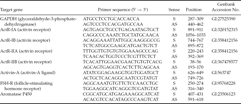
The quantitative PCR (qPCR) reactions had a final volume of 20 μl and contained the following components: 1 μl of each samples’ cDNA, 10 μl of 1× Power SYBR® Green PCR Master Mix, 7.4 μl of ultra-pure water and 0.4 μM (final concentration) of both the sense and antisense primers. The gene-specific primers used for the amplification of the different transcripts are shown in Table 1. The reference gene glyceraldehyde-3-phosphate-dehydrogenase (GAPDH) was selected as an endogenous control for normalisation and to investigate the expression stability of the samples. Primer specificity and amplification efficiency were verified for each gene. The cycle profile for the first PCR step consisted of an initial denaturation and polymerase activation step for 15 min at 94°C. This step was followed by 40 cycles of 15 s at 94°C, 30 s at 60°C and 45 s at 72°C. A final extension cycle was performed for 10 min at 72°C. The specificity for each primer set was tested using a melting curve performed between 60 and 95°C for all genes. All amplifications were performed using an i-cycler IQ5 system (Bio-Rad, Hercules, CA, USA). The ΔΔCT method was used to transform threshold cycle values into normalized relative expression levels (Livak & Schmittgen, Reference Livak and Schmittgen2001).
Oligonucleotide primers used for the analysis of goat secondary follicles by polymerase chain reaction

AS, antisense; S, sense.
Ultrastructural analysis of cultured goat secondary follicles
To examine follicular morphology, transmission electron microscopy (TEM) was used to analyse the ultrastructure of secondary follicles cultured in the cultured control or in 50 ng/ml activin-treated medium (this concentration of activin had previously demonstrated the best result in Experiment 1; activin-A 50). Isolated follicles were fixed in Karnovsky solution (4% paraformaldehyde and 2.5% glutaraldehyde in 0.1 M sodium cacodylate buffer, pH 7.2) for at least 3 h at room temperature (approximately 25°C). After fixation, cultured follicles were embedded in drops of 4% low-melting agarose and maintained in a sodium cacodylate buffer. Specimens were post-fixed in 1% osmium tetroxide, 0.8% potassium ferricyanide and 5 mM calcium chloride in a 0.1 M sodium cacodylate buffer for 1 h at room temperature. Specimens were then washed in sodium cacodylate buffer and counterstained with 5% uranyl acetate. The samples were then dehydrated in a graded series of acetone washes and embedded in epoxy resin (Epoxy-Embedding Kit, Fluka Chemika-BioChemika). Subsequently, semi-thin (1 μm) sections were cut, stained with toluidine blue and analyzed by light microscopy at ×400 magnification. Ultra-thin (60–70 nm) sections were obtained from secondary follicles classified as morphologically normal according to their corresponding semi-thin sections. Finally, the ultra-thin sections were contrasted with uranyl acetate and lead citrate and examined with a Morgani-FEI transmission electron microscope operating at 80 kV.
Estradiol secretion
To evaluate follicular steroidogenesis in vitro, estradiol levels were measured by a microparticle enzyme immunoassay (MEIA; Abbott Diagnostics AxSYM® SYSTEM) using a commercial kit (Axsym Estradiol, Abbott Japan Co., Ltd Tokyo, Japan). The sensitivity of the assay permitted the detection of molecules at 20 pg/ml concentrations. Samples of medium removed from follicles cultured in both treatment conditions (cultured control and activin-A 50) on days 6, 12 and 18 of the culture period were analyzed.
Statistical analysis
Data regarding follicular survival, antrum formation, follicular growth rate, number of fully grown oocytes, and meiotic resumption following culture were collected. These values were compared using the chi-squared test and the results were expressed as percentages. Due to the heterogeneity of the variance, days of culture and estradiol levels were compared using the non-parametric Kruskal–Wallis test. The results are expressed as the mean ± standard error of the mean (SEM), and differences were considered to be significant when P-values were <0.05. For qPCR, the treatment and control samples were randomly assigned to blocks, and the relative expression values (2−ΔΔCt) were subjected to the Shapiro–Wilk normality test using the UNIVARIATE procedure. The relative expression data were logarithmically transformed (log10 (X + 1)) for normal distribution adjustment. The log-transformed relative expression levels were evaluated using an analysis of variance (ANOVA), and the differences between the control and treatments were assessed with a T-test (P < 0.05). In all tests the software package SAS 9.0 was used.
Results
Experiment 1: The effects of activin-A on goat secondary follicles in vitro
Follicle survival, antrum formation and oocyte growth following culture with activin-A
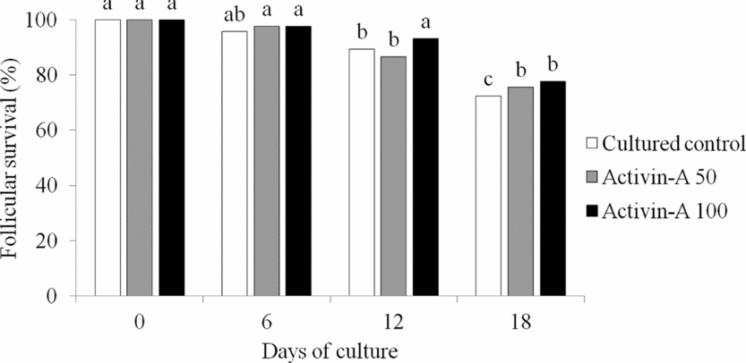
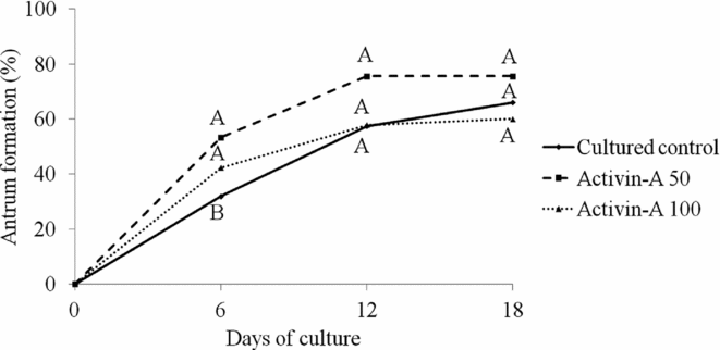
The percentage of surviving secondary follicles after 18 days of culture is shown in Fig. 1. After 18 days of culture, a high percentage of follicular survival was observed in all treatment conditions. However, a significant decrease in the percentage of surviving follicles was observed from day 0 to day 18 of culture. After 6 days of culture, both activin-A treatment conditions showed a higher percentage of antrum formation compared with the cultured control (P < 0.05). Nevertheless, from day 12 onward, all treatments were similar (Fig. 2).
Survival assessment of goat secondary follicles cultured for 18 days in α-MEM+ (cultured control), α-MEM+ plus 50 ng/ml activin-A (activin-A 50) or α-MEM+ plus 100 ng/ml activin-A (activin-A 100). a,bIndicates significant differences between culture periods treated with the same culture medium (P < 0.05).
Antrum formation in goat secondary follicles cultured for 18 days in α-MEM+ (cultured control), α-MEM+ plus 50 ng/ml activin-A (activin-A 50) or α-MEM+ plus 100 ng/ml activin-A (activin-A 100). a,bIndicates significant differences between culture periods treated with the same culture medium (P < 0.05). A,BIndicates significant differences between treatments in the same culture period (P < 0.05).
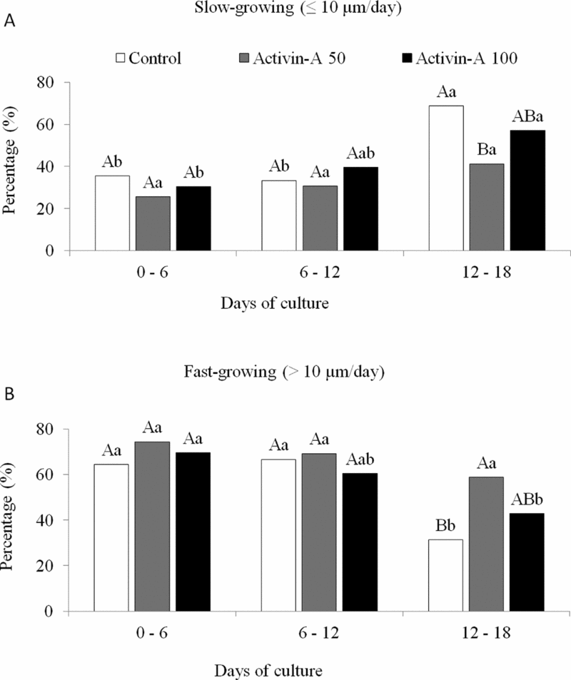
The proportion of fast- and slow-growing follicles during the 18 days of culture is represented in Fig. 3. After evaluating the goat secondary follicles growth rate in the cultured control and 100 ng/ml activin-A, a significant decrease in the proportion of fast-growing follicles between the first (0–6 days) and the final (12–18 days) third of the culture was observed (P < 0.05). However, the addition of 50 ng/ml activin-A to the culture medium promoted the maintenance of the proportion of fast-growing follicles during all time periods tested. The percentages of fast-growing follicles in the tested treatments over the same culture periods were also compared and it was observed that the addition of 50 ng/ml activin-A promoted a significantly greater proportion of fast-growing follicles in the final third of the culture than that observed in the cultured control group (P < 0.05).
Development of goat secondary follicles cultured for 18 days in α-MEM+ (cultured control), α-MEM+ plus 50 ng/ml activin-A (activin-A 50) or α-MEM+ plus 100 ng/ml activin-A (activin-A 100). (A) Proportion of slow-growing follicles (≤10 μm/day). (B) Proportion of fast-growing follicles (>10 μm/day). a,bIndicates significant differences between culture periods treated with the same culture media (P < 0.05). A,BIndicates significant differences between treatments in the same culture period (P < 0.05).
After measuring the diameters of the oocytes, no significant differences were observed among the tested treatments (cultured control, 127.11 ± 9.9 μm; 50 ng/ml activin-A, 131.76 ± 15.59 μm; and 100 ng/ml activin-A, 132.26 ± 12.80 μm).
Morphological evaluation and maturational competence of oocytes in vitro
Only oocytes with a diameter ≥110 μm were selected for IVM. After maturation, all oocytes were incubated with calcein-AM and ethidium homodimer-1 (to assess viability) and Hoechst 33342 (to evaluate chromatin configuration) (Fig. 4). Changes in the chromatin configuration of oocytes in the cultured control or activin-A (50 or 100 ng/ml) are shown in Table 2. The recovery rates of viable oocytes ≥ 110 μm in diameter did not differ among the treatments. However, the percentage of oocytes that resumed meiosis after the addition of 50 ng/ml activin-A was significantly higher than that in the other treatments tested (P < 0.05).
Goat oocytes after growth and maturation of secondary follicles in vitro. (A) Non-stained oocyte (diameter ≥ 110μm). (B) Viable oocyte labelled with calcein-AM (green). (C) Non-viable oocyte labelled with ethidium homodimer-1 (red). (D) Oocyte exhibiting a germinal vesicle (GV). (E) Oocyte exhibiting germinal vesicle breakdown (GVBD). (F) Oocyte in metaphase I (MI).
Meiosis resumption of oocytes from goat secondary follicles cultured for 18 days in α-MEM+ (cultured control), α-MEM+ plus 50 ng/ml activin-A (activin-A 50) or α-MEM+ plus 100 ng/ml activin-A (activin-A 100)
*Only oocytes (≥100 μm) were selected for the in vitro maturation procedure.
a ,b Values with different letters in the same column differ signficantly (P < 0.05).
Experiment 2: mRNA expression of activin-A, ActR-IA, ActR-IB, ActR-IIA, ActR-IIB, FSH-R and P450 aromatase, ultrastructural analysis and estradiol measurements in goat secondary follicles
mRNA expression levels of activin-A, ActR-IA, ActR-IB, ActR-IIA, ActR-IIB, FSH-R and P450 aromatase in non-cultured and cultured follicles
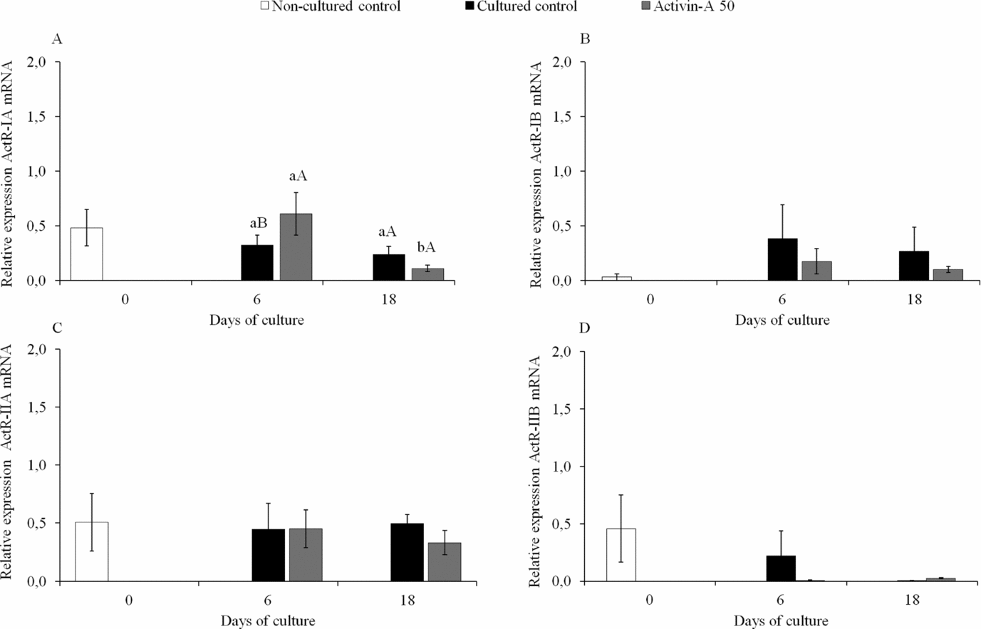
The mRNA levels of different activin-A receptors in fresh or cultured secondary goat follicles (after 6 or 18 days of culture) are represented in Fig. 5. After 6 days of culture, the addition of 50 ng/ml activin-A stimulated a significant increase in ActR-IA mRNA levels in comparison with the cultured control (P < 0.05). However, after 18 days of culture, this same treatment condition exhibited a significant decrease in ActR-IA mRNA levels when compared with levels at day 6 of culture (Fig. 5A). The mRNA expression levels for ActR-IB (Fig. 5B), ActR-IIA (Fig. 5C) and ActR-IIB (Fig. 5D) were not affected by medium supplementation with activin-A at any time during the culture period.
Relative mRNA expression of activin-A receptors ActR-IA (A), ActR-IB (B), ActR-IIA (C), ActR-IIB (D) in fresh (non-cultured control) or cultured goat secondary follicles. The cultured follicles were maintained for 6 and 18 days in α-MEM+ (cultured control) or α-MEM+ plus 50 ng/ml activin-A (activin-A 50). *Indicates significant differences from the non-cultured control (P < 0.05). a,bIndicates significant differences during culture day 6 and day 18 under the same treatment conditions (P < 0.05). A,BIndicates significant differences between treatments on each day of the culture (P < 0.05).
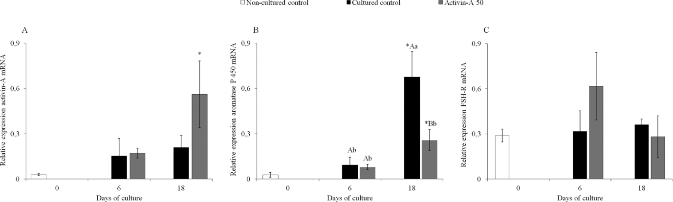
The relative mRNA expression level of activin-A was increased significantly in follicles cultured for 18 days in the presence of activin-A in comparison with the non-cultured control (Fig. 6A; P < 0.05). From day 6 to day 18, an increase in the levels of P450 aromatase mRNA was observed only in cultured control (Fig. 6B). After 18 days of culture, the addition of activin-A was observed to decrease significantly the levels of P450 aromatase mRNA in comparison with the cultured control (P < 0.05). However, in both cultured treatment conditions, the mRNA levels of P450 aromatase were significantly higher than those of the non-cultured control (P < 0.05). In contrast, FSH-R mRNA levels were not affected by activin-A supplementation during culture (P > 0.05; Fig. 6C).
Relative mRNA expression of activin-A (A), P450 aromatase (B), and follicle stimulating hormone receptor (FSH-R) (C), in fresh (non-cultured control) or cultured goat secondary follicles. The cultured follicles were maintained for 6 and 18 days in α-MEM+ (cultured control) or α-MEM+ plus 50 ng/ml activin-A (activin-A 50). *Indicates significant differences between the non-cultured control and other conditions (P < 0.05). a,b Indicates significant differences between culture day 6 and day 18 with the same treatment conditions (P < 0.05). A,BIndicates significant differences between treatments on each day of the culture (P < 0.05).
Ultrastructural analysis of goat secondary follicles after 18 days of culture
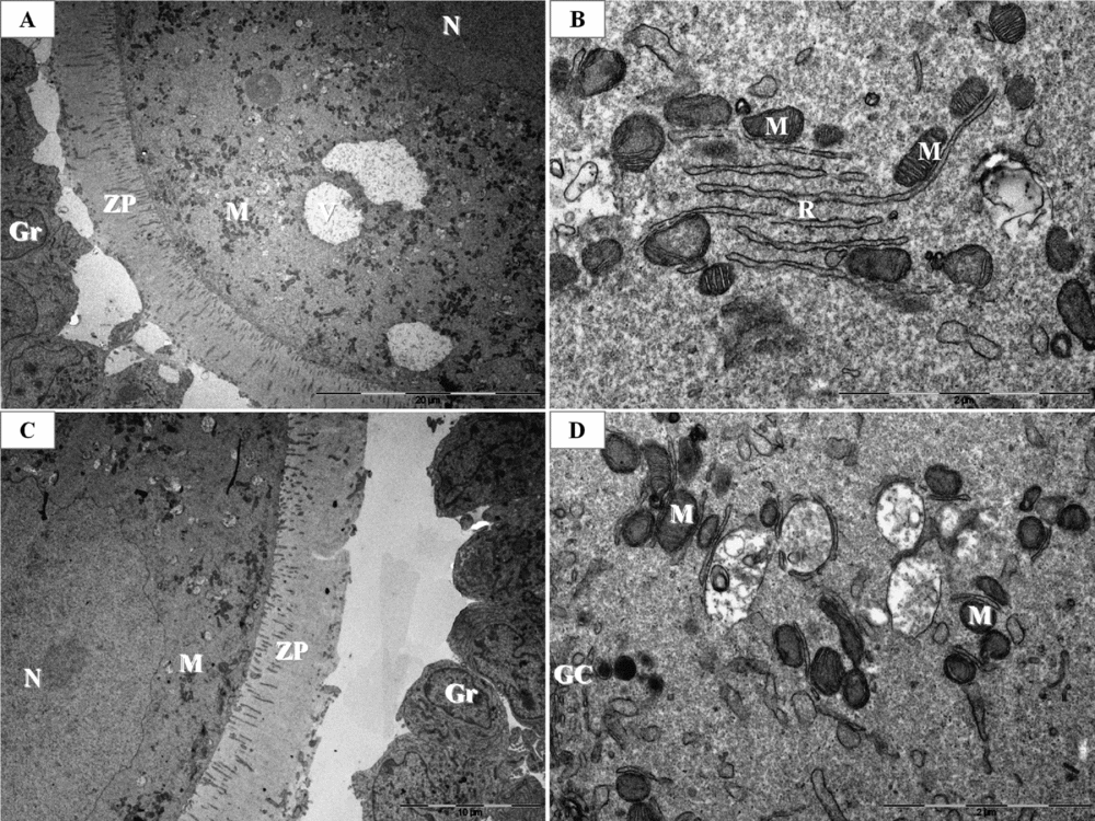
After 18 days of culture, the follicular ultrastructure was well preserved in both treatment conditions tested (cultured control and activin-A 50), (Fig. 7). This finding confirms the results on follicular survival in Experiment 1. Follicles from both treatment conditions exhibited intact oocytes with a typical size, nuclei with no abnormalities and zona pellucida microvilli essential for oocyte–granulosa cell interactions (Fig. 7A,C). Moreover, important organelles (e.g. mitochondria, endoplasmic reticulum and Golgi complex) were observed to be well preserved (Fig. 7B,D).
Ultrastructural analysis of goat secondary follicles after 18 days of culture. (A, B) α-MEM+ (cultured control). (C, D) α-MEM+ plus 50 ng/ml activin-A (activin-A 50). GC, Golgi complex; Gr, granulosa cell; M, mitochondria; Nu, nucleolus; R, endoplasmic reticulum; ZP, zona pellucida.
Estradiol secretion
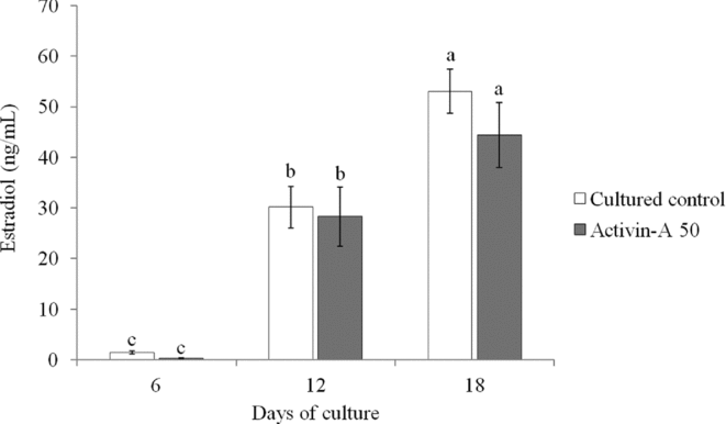
Culture medium from individual follicles in both treatments (cultured control and activin-A 50) were analyzed for estradiol content by MEIA every 6 days. Fifteen samples per treatment condition were analyzed on each selected day (days 6 (n = 15), 12 (n = 15) and 18 (n = 15) of culture). During the culture period, estradiol levels significantly increased (P < 0.05), but no significant difference was observed between the tested treatments (P > 0.05; Fig. 8).
Estradiol secretion at 6, 12 and 18 days in culture by goat follicles in either α-MEM+ (cultured control) or α-MEM+ plus 50 ng/ml activin-A (activin-A 50). a,b,cIndicates significant differences between treatment conditions during the progression of the culture (P < 0.05).
Discussion
The present study is the first to demonstrate that the addition of activin-A to culture medium stimulates antrum formation and early follicle development as well as improves meiosis resumption rates in isolated goat secondary follicles cultured over a long period of time.
Previous studies have demonstrated that activin-A stimulates preantral follicle survival in bovines (Hulshof et al., Reference Hulshof, Figueiredo, Beckers, Bevers, Vanderstichele and van den Hurk1997), ovines (Thomas et al., Reference Thomas, Armstrong and Telfer2003) and humans (Telfer et al., Reference Telfer, McLaughlin, Ding and Thong2008). However, in this study, no significant difference was observed in follicular survival between tested treatments. Such a result may be expected due to the medium used in the current study, which was chosen as the best medium based on the results from a previous study by our group in which the addition of increasing concentrations of FSH was reported to significantly improve follicular survival (Saraiva et al., Reference Saraiva, Celestino, Araújo, Chaves, Almeida, Lima-Verde, Duarte, Silva, Martins, Bruno, Matos, Campello, Silva and Figueiredo2010a). In addition, other substances such as ascorbic acid (Rossetto et al., Reference Rossetto, Lima-Verde, Matos, Saraiva, Martins, Faustino, Araújo, Silva, Name, Báo, Campello, Figueiredo and Blume2009; Silva et al., Reference Silva, Araújo, Duarte, Chaves, Silva, Lobo, Almeida, Matos, Tavares, Campelo and Figueiredo2011) and insulin (Chaves et al., Reference Chaves, Alves, Faustino, Oliveira, Campello, Lopes, Báo and Figueiredo2011) have been added to the base medium to successfully maintain the survival of goat preantral follicles in vitro. The efficiency of our medium in maintaining follicle survival was confirmed by ultrastructural analysis after 18 days of culture. It is well established that TEM analysis is an efficient method for the detailed study of ovarian follicle morphodynamics and this method can be considered to be an essential tool in the evaluation of follicular quality after culture (Nottola et al., Reference Nottola, Cecconi, Bianchi, Motta, Rossi, Continenza and Macchiarelli2011).
After 6 days of culture, compared with the cultured control, the addition of activin-A to the culture medium promoted earlier antrum formation followed by an increase in the mRNA levels of the ActR-IA receptor. These results indicate that antrum formation induced by the addition of activin-A may be associated with an early increase in the mRNA levels of the ActR-IA receptor in goat secondary follicles in vitro. Previous studies have shown a positive effect of the addition of activin-A on secondary follicles’ antrum formation in other species in vitro (rat: Zhao et al., Reference Zhao, Taverne, van der Weijden, Bevers and van den Hurk2001; cattle: McLaughlin et al., Reference McLaughlin, Bromfield, Albertini and Telfer2010; human: Telfer et al., Reference Telfer, McLaughlin, Ding and Thong2008).
The levels of mRNAs encoding the receptors ActR-IB ActR-IIA, ActR-IIB, and FSH-R were not found to be affected by supplementation with activin-A at any of the culture time points tested. Several studies have suggested that FSH is essential for the proper expression of numerous growth factors that regulate ovarian folliculogenesis (Ethier & Findlay, Reference Ethier and Findlay2001). Considering this function of FSH, it is possible that its initial presence in the culture medium was able to stimulate the expression of FSH-R and activin-A in follicular cells (granulosa/theca); therefore, any additional effects of activin-A on the expression of its own receptors or on FSH-R would not be observed during the culture. Similar results were recently noted by Cossigny et al. (Reference Cossigny, Findlay and Drummond2012), who found that the interaction between FSH and activin-A in rat preantral follicles did not affect the mRNA levels of follistatin, activin-A or FSH-R in comparison with the control cultured with only FSH.
According to Xu et al. (Reference Xu, Bernuci, Lawson, Yeoman, Fisher, Zelinski and Stouffer2010), the in vitro follicular growth rate in monkeys varies depending on the ability of the follicle to recognise and respond to exogenous stimuli. It is believed that some follicles are highly responsive to the addition of growth factors or hormones to the culture medium, whereas other follicles have little or no response. In the current study, estradiol levels were not related to the follicular growth rate. This finding indicates that estradiol synthesis and follicular growth pattern are not strictly dependent on each other. Our finding confirms previous results obtained by Cecconi et al. (Reference Cecconi, Barboni, Coccia and Mattioli1999).
Although the mRNA levels for the enzyme P450 aromatase were lower in the cultures containing 50 ng/ml activin-A than in the cultured control, estradiol production was not affected (after 18 days of culture). These results indicate that in the cultured control group, a fraction of the mRNA transcripts encoding P450 aromatase may not have been translated into protein. Thus, the activity of this enzyme remained similar in all cultured treatments and differences in the estradiol synthesis could not be observed.
After 18 days of culture, compared with the other treatments, there was a significant increase in meiosis resumption rates when the medium was supplemented with 50 ng/ml activin-A. This result is in agreement with prior studies that reported the involvement of activin-A in bovine (Silva & Knight, Reference Silva and Knight1998) and human (Alak et al., Reference Alak, Coskun, Friedman, Kennard, Kim and Seifer1998) oocyte maturation. We hypothesize that the positive effect of 50 ng/ml activin-A on the meiosis resumption rate is associated with the higher follicular growth rate (>10 mm/day) in the final third of the culture growth period (12–18 days). In this study, no oocyte reached metaphase II in any of the tested treatments. We hypothesize that this inability to complete nuclear maturation may be associated with the limited oocyte growth during culture. According to Crozet et al. (Reference Crozet, Dahirel and Gall2000), the successful growth of oocytes in vitro is directly related to the acquisition of meiotic competence and, potentially, due to the ability of oocytes to activate cyclin-dependent protein kinase (CDC2) and mitogen activated protein (MAP) kinases (Miyano & Manabe, Reference Miyano and Manabe2007). These kinases are essential for complete nuclear maturation in metaphase II. However, successful maturation of goat oocytes from secondary follicles grown in vitro remains a rare event (Magalhães et al., Reference Magalhães, Duarte, Araújo, Brito, Soares, Lima, Lopes, Campello, Rodrigues and Figueiredo2011; Saraiva et al., Reference Saraiva, Rossetto, Brito, Celestino, Silva, Faustino, Almeida, Bruno, Magalhães, Matos, Campello and Figueiredo2010b).
In conclusion, the addition of activin-A to our culture medium stimulated early antrum formation in goat follicles and increased the proportion of fast-growing follicles in the final third of the culture growth period, in turn resulting in a higher percentage of meiosis resumption. The results of this study may contribute knowledge to future research seeking to investigate the mechanisms involved in the developmental regulation of goat secondary follicles in vitro.
Conflict of interest
None of the authors has any conflicts of interest to declare.
Acknowledgements
This research was supported by grants from the National Council for Scientific and Technological Development (CNPq), Brazil (RENORBIO: grant number 554812 ⁄ 2006–1), Financiadora de Estudos e Projectos (FINEP) and Pronex–Brazil. Cleidson Manoel Gomes da Silva is a recipient of a grant from Coordenação de Aperfeiçoamento de Pessoal de Nível Superior (CAPES), Brazil.