Coagulase-negative staphylococci (CNS) have long been considered as minor mastitis pathogens. Even though their role in intramammary infections has not been fully clarified, they are reported to have become the most common bovine mastitis isolates in many countries and are regarded as emerging mastitis pathogens (Pyörälä & Taponen, Reference Pyörälä and Taponen2009). CNS mastitis is especially common in first-lactation cows (Myllys, Reference Myllys1995) and the course is usually subclinical or mildly clinical but affects milk quality by increasing somatic cell count (SCC) (Timms & Schultz, Reference Timms and Schultz1987; Sampimon et al. Reference Sampimon, Barkema, Berends, Sol and Lam2009; Taponen & Pyörälä, Reference Pyörälä and Taponen2009). In addition, CNS infection can damage udder tissue and lead to decreased milk production (Pyörälä & Taponen, Reference Pyörälä and Taponen2009). CNS have been isolated from different body parts of cows of all ages, from udder secretions and the cow environment (Devriese & De Keyser, Reference Devriese and De Keyser1980; Boddie et al. Reference Boddie, Nickerson, Owens and Watts1987; Mathews et al. Reference Matthews, Harmon and Langlois1992; Taponen et al. Reference Taponen, Björkroth and Pyörälä2008). The organisms are opportunistic and the source of mammary gland infection appears to be the environment (Oliver et al. Reference Oliver, Gillespie, Headrick, Lewis and Dowlen2005). More than ten different CNS species have been isolated from bovine milk. The most commonly reported species are a part of the normal skin flora and include Staphylococcus (Staph.) chromogenes, Staph. simulans, Staph. hyicus and Staph. epidermidis (Trinidad et al. Reference Trinidad, Nickerson and Alley1990; Matthews et al. Reference Matthews, Harmon and Langlois1992; Myllys, Reference Myllys1995; Thorberg et al. Reference Thorberg, Kuhn, Aarestrup, Brandstrom, Jonsson and Nielsson-Tham2006; Taponen et al. Reference Taponen, Björkroth and Pyörälä2008; Gillespie et al. Reference Gillespie, Headrick, Boonyayatra and Oliver2009; Fessler et al. Reference Fessler, Billerbeck, Kadlec and Schwarz2010).
Even though cases of subclinical and mild clinical mastitis due to CNS may resolve themselves spontaneously, national policies and strategies on the use of antimicrobial agents in the treatment of udder infections vary (Pyörälä & Taponen, Reference Pyörälä and Taponen2009). Despite the favourable situation regarding β-lactamase-positive isolates (Devriese et al. Reference Devriese, Baele, Vaneechoutte, Martel and Haesebrouck2002; Taponen et al. Reference Taponen, Simojoki, Harveri, Larsen and Pyörälä2006; Rajala-Schultz et al. Reference Rajala-Schultz, Torres, DeGraves, Gebreyes and Patchanee2009), increasing resistance of different CNS species to antimicrobial agents has been reported (Sawant et al. Reference Sawant, Gillespie and Oliver2009). In addition, CNS easily develop multiresistance (Taponen & Pyörälä, Reference Pyörälä and Taponen2009).
New identification methods based on molecular biology enable investigations at a DNA level and comparison of the strains from different sources. Pulsed-field gel electrophoresis (PFGE) has been frequently used for CNS strain differentiation in human medicine (Krause et al. Reference Krause, Haberl, Wölfler, Daxböck, Auner, Krejs, Wenisch and Reisinger2003; Garcia et al. Reference Garcia, Benitez, Lam, Salinas, Wirth, Espinoza, Garay, Depix, Labarca and Guzmán2004; Persson et al. Reference Persson, Strid, Tidefeld and Söderquist2006); in recent years, the method was applied also to CNS isolates from veterinary specimens (Taponen et al. Reference Taponen, Björkroth and Pyörälä2008; Gillespie et al. Reference Gillespie, Headrick, Boonyayatra and Oliver2009; Fessler et al. Reference Fessler, Billerbeck, Kadlec and Schwarz2010).
The main objective of this study was to characterize the genetic diversity of Staph. chromogenes and Staph. haemolyticus isolated from bovine milk samples.
Materials and Methods
Milk samples
Milk samples were collected during regular milk quality control in three geographical regions from herds with elevated bulk milk SCC (>400 000 cells/ml) and with a high prevalence of subclinical mastitis. A total of 231 dairy cattle from 12 herds were included in the study. All lactating cows from individual herd were sampled, regardless of SCC level. Milk samples from 912 udder quarters were investigated for the presence of CNS. After teat disinfection and discarding the first milk streams, samples from individual udder quarters were collected into sterile tubes and transported to the laboratory for bacteriological examination.
Somatic cell counting
Somatic cells in milk samples were counted by Fossomatic (Foss, Hillerød, Denmark).
Bacteriological examinations
Milk samples (a loop containing 10 μl per each investigated quarter) were inoculated onto blood agar plates (Columbia agar supplemented with 5% of ovine blood) and incubated for 48 h at 37°C. Culture result was regarded as positive if at least five colony-forming units (cfu) were grown from 10 μl of milk sample. The isolates were identified on the basis of Gram staining, morphological characteristics (colony shape, size and colour), haemolysis (presence and type), catalase test and coagulase test. Definitive identification was performed by commercial kit API Staph (bioMerieux, Marcy-l'Etoile, France), taking into account the probability limit of >90%. In case of ambiguous results, the isolates were subjected to additional biochemical tests as suggested by the manufacturer of API Staph. As a result of these criteria, 17 CNS isolates that could not be identified to the species level were declared as Staphylococcus spp. and excluded from further investigation.
Antibiotic susceptibility testing
Antibiotic susceptibility testing was performed using the agar disc diffusion method (Kirby-Bauer) following the recommendations of the National Committee for Clinical Laboratory Standards Institute (NCCLS) and Clinical Laboratory Standards Institute (CLSI), respectively. Colonies were suspended in 5 ml of sterile saline solution to obtain the turbidity of a 0·5 McFarland standard (bioMerieux, Marcy-l'Etoile, France). The suspension was streaked onto Mueller-Hinton agar plates (Merck, Darmstadt, Germany) using a sterile swab. Eleven antibiotics most commonly used in mastitis treatment were used. Antibiotic discs (Sensi-Disc, Becton Dickinson and Company, Le Pont de Claix, France) containing ampicillin (10 μg), amoxicillin/clavulanic acid (20/10 μg), kanamycin (30 μg), linkomycin (2 μg), neomycin (30 μg), oxytetracyline (30 μg), penicillin (10 μg), cefoperazone (75 μg), oxacillin (1 μg), cloxacillin (1 μg) and cefquinom (30 μg) were placed onto the agar plates, which were incubated at 35°C for 20–24 h. Interpretation of the results (inhibition zone diameter) was based on the instructions provided by the CLSI standards and disc manufacturers. The bacteria were classified as susceptible, intermediate or resistant for each antibiotic.
PFGE
Colonies were grown on sheep blood agar plates for 2 d at 37°C. Three colonies from each plate were transferred into 5 ml of tryptic soy broth and incubated for 5 h at 37°C. Bacterial suspension of optical density 0·650–0·850 was centrifuged at 3500 g at 4°C for 10 min, washed with PIV buffer (10 mm-Tris, 1 m-NaCl) and centrifuged again. Subsequently, the suspension was mixed with (i) PIV, (ii) lysozyme (50 mg/ml) and lysostaphin (1 mg/ml), (iii) 1% agarose SeaKem Gold (Cambrex, Wiesbaden, Germany) and transferred into plug molds to solidify. The plugs were treated with lysozyme (50 mg/ml) and lysostaphin (1 mg/ml) in lysis buffer (6 mm-Tris, 100 mm-EDTA, 1 m-NaCl, 0·5% Brij 58, 0·2% sodium deoxycholate, 0·2% sodium laurylsarcosyl) (overnight incubation at 37°C in a shaker water bath), followed by treatment with proteinase K (1 mg/ml) in ES buffer (0·5 m-EDTA, 10% sodium laurylsarcosyl) (overnight incubation at 53°C in a shaker water bath). Subsequently, plugs were washed five times with TE buffer (1 m-Tris pH 8·0, 0·5 m-EDTA pH 8·0) at 37°C and stored in TE buffer at 4°C until use.
DNA was digested with SmaI (40 U/μl) during the incubation of 5 h at 25°C. The fragments were separated in 1% agarose gel by electrophoresis (22 h, 6 V/cm, 11·6°C, pulse times 5 to 45 s) using CHEF-DRII system (BioRad, Hercules CA, USA) and visualized by ethidium bromide staining. PFGE profiles were subjected to computer-assisted analysis with BioNumerics software (version 5.0, Applied Maths, Sint-Martens-Latem, Belgium). Pulse marker 50–1000 kb (Sigma-Aldrich, Munich, Germany) was used for band normalization. Dendrograms with 1·2% position tolerance were created using an unweighted pair group method with arithmetic mean (UPGMA, Dice coefficient) algorithm. The criterion for the designation of a distinct PFGE profile was a one-band difference.
Results
Somatic cell counting
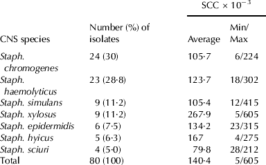
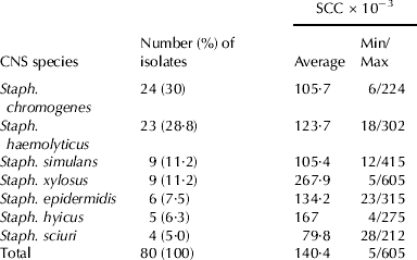
The results of somatic cell counting are presented in Table 1.
Species of coagulase negative staphylococci (CNS) isolated during routine microbiological investigation of milk samples and somatic cell count (SCC) data
Bacteriology
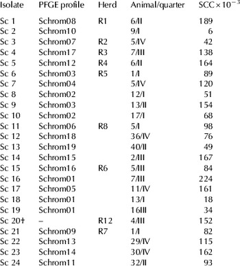
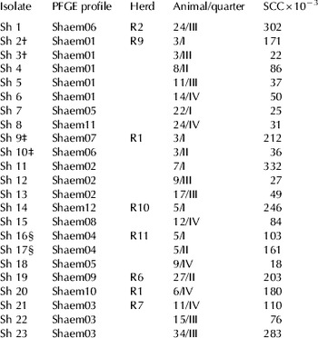
CNS were isolated from 67 (29%) out of 231 cows (80/912 udder quarters). A total of seven CNS species were identified with the most prevalent being Staph. chromogenes and Staph. haemolyticus (Table 1). Staph. chromogenes was detected in 24 animals from nine herds while Staph. haemolyticus was found in 20 animals from seven herds (Tables 2 and 3). The presence of both Staph. chromogenes and Staph. haemolyticus was confirmed in four herds, mostly in different animals, with the exception of one cow from which both species were isolated (from different quarters).
Staphylococcus chromogenes isolates: origin, somatic cell count (SCC) and PFGE results

† isolate was not typed owing to failed digestion
Staphylococcus haemolyticus isolates: origin, somatic cell count (SCC) and PFGE results

†, ‡, § two isolates from different udder quarters of the same animal
Antibiotic susceptibility testing
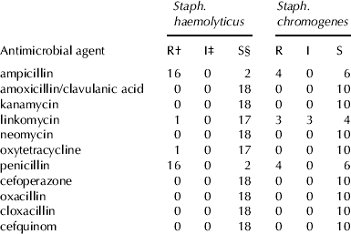
A total of 18 Staph. haemolyticus isolates were subjected to antibiotic susceptibility testing. A high percentage (88·9%) of isolates resistant to ampicillin and penicillin was detected. Among ten tested Staph. chromogenes isolates, four (40·0%) were resistant to penicillin and ampicillin and three (30·0%) to linkomycin. The results of antibiotic susceptibility testing are summarized in Table 4.
Results of susceptibility testing of 18 Staphylococcus haemolyticus and 10 Staphylococcus chromogenes isolates from bovine non-mastitic milk samples
† Number of resistant isolates
‡ Number of intermediate isolates
§ Number of susceptible isolates
PFGE
PFGE analysis of 23 Staph. chromogenes isolates collected from 23 animals in eight herds (enzymic digestion failed in one isolate) resulted in 19 PFGE profiles. Diverse profiles were seen in all herds (n=5) from which multiple isolates (2–6) were obtained, with up to four different profiles identified within a herd (Table 2). Within two herds (R5 and R6), some of the isolates shared identical PFGE profiles (Table 2). The similarity levels of the isolates within individual herds ranged from 54% to 100% (data not shown). No common PFGE types were observed among the herds.
Among 23 Staph. haemolyticus isolates collected from 20 animals originating from seven herds, a total of 12 PFGE profiles were identified. In three cases, milk samples from two udder quarters of individual animals were investigated. In two animals, Staph. haemolyticus isolates from different udder quarters showed identical PFGE profiles (Shaem01 and Shaem04) while in one animal the profiles differed (Shaem06 and Shaem07, similarity level of 69%) (Table 3). Within the herds (n=5) in which multiple Staph. haemolyticus isolates (2–6 per herd) were confirmed, distinct PFGE profiles were identified in all herds. However, within three herds some of the isolates from different animals shared a common PFGE profile (Table 3). The similarity levels of the isolates within individual herds thus ranged from 55% to 100% (data not shown). Identical PFGE profiles shared by different herds were detected twice (Shaem05 and Shaem06, respectively) (Table 3).
Discussion
The prevalence of CNS, which has gradually become the predominant pathogen isolated from dairy cows with subclinical mastitis in many countries (Myllys et al. Reference Myllys, Asplund, Brofeldt, Hirvela-Koski, Honkanen-Buzalski, Junttila, Kulkas, Myllykangas, Niskanen, Saloniemi, Sandholm and Saranpaa1998), varies among herds. The proportion of CNS-affected animals (29%) found in our study is higher than that reported by Schukken et al. (Reference Schukken, González, Tikofsky, Schulte, Santisteban, Welcome, Bennett, Zurakowski and Zadoks2009) who isolated CNS in 14·8% of cows. Sampimon et al. (2009) found similar proportions of CNS; 14·6% and 10·4% for high-SCC herds and low-SCC herds, respectively. At the quarter level, the number of infected udder quarters investigated in the present study, differed among the herds and ranged from 3·9% to 14·8% (data not shown). This concurs with data published previously (Gillespie et al. Reference Gillespie, Headrick, Boonyayatra and Oliver2009).
Among seven CNS species detected in this study, Staph. chromogenes and Staph. haemolyticus were most prevalent, representing 30% and 28·8% of all CNS isolates, respectively. These values fall within the range of previously reported data on prevalence of these two CNS species. Despite being considered as an uncommon udder pathogen with a varying prevalence from 1% to more than 50% (Matthews et al. Reference Matthews, Oliver and King1990; Taponen et al. Reference Taponen, Simojoki, Harveri, Larsen and Pyörälä2006; Jemeljanovs et al. Reference Jemeljanovs, Konosonoka and Bluzmanis2007; Gillespie et al. Reference Gillespie, Headrick, Boonyayatra and Oliver2009, Fessler et al. Reference Fessler, Billerbeck, Kadlec and Schwarz2010), Staph. haemolyticus was reported recently as one of the most prevalent CNS isolates detected in milk (Jemeljanovs et al. Reference Jemeljanovs, Konosonoka and Bluzmanis2007; Bal et al. Reference Bal, Bayar and Bal2010; Nam et al. Reference Nam, Lim, Kim, Kang, Moon, Jang, Kim, Wee, Joo and Jung2010). On the other hand, Staph. chromogenes, with a prevalence of 10–61% (Matthews et al. Reference Matthews, Oliver and King1990; Taponen et al. Reference Taponen, Simojoki, Harveri, Larsen and Pyörälä2006, Reference Taponen, Björkroth and Pyörälä2008; Sampimon et al. Reference Sampimon, Barkema, Berends, Sol and Lam2009; Sawant et al. Reference Sawant, Gillespie and Oliver2009) is regarded as one of the most important and most frequently isolated agents of subclinical mastitis with a significant impact on SCC (Devriese et al. Reference Devriese, Baele, Vaneechoutte, Martel and Haesebrouck2002; De Vliegher, Reference De Vliegher2004). The latter was not observed in our study (Table 2); however, it should be stressed that our isolates were obtained during routine microbiological examinations of milk samples from dairy cows without clinical signs of mastitis.
Antibiotic susceptibility testing of Staph. chromogenes and Staph. haemolyticus isolates revealed a high level of resistance to penicillins (Table 4). The latter is widespread among CNS as the most common resistance mechanism in staphylococci is β-lactamase production (Taponen & Pyörälä, Reference Pyörälä and Taponen2009). The reported penicillin resistance for CNS isolated from mastitis in some Northern European countries is between 25% and 61% (Taponen & Pyörälä, Reference Pyörälä and Taponen2009). Compared with earlier data (Devriese et al. Reference Devriese, Baele, Vaneechoutte, Martel and Haesebrouck2002; Taponen et al. Reference Taponen, Simojoki, Harveri, Larsen and Pyörälä2006), describing 19–38% of CNS resistant to penicillins, mostly from cases of subclinical and chronic mastitis, the resistance to penicillins has increased in recent years (Bal et al. Reference Bal, Bayar and Bal2010; Nam et al. Reference Nam, Lim, Kim, Kang, Moon, Jang, Kim, Wee, Joo and Jung2010). Recently, methicillin-resistant Staph. haemolyticus (Fessler et al. Reference Fessler, Billerbeck, Kadlec and Schwarz2010) and Staph. chromogenes (Sampimon et al. Reference Sampimon, Lam, Mevius, Schukken and Zadoks2011) has also been described in cattle.
A vast majority of Staph. haemolyticus isolates (88·9%) in the present study were resistant to penicillin and ampicillin. Resistance of Staph. haemolyticus to these two antimicrobials (48·4% and 47·3% for penicillin and ampicillin, respectively) was reported also by Nam et al. (Reference Nam, Lim, Kim, Kang, Moon, Jang, Kim, Wee, Joo and Jung2010), along with a relatively higher resistance of CNS against most antimicrobials tested than in most of the previous studies from other countries. However, owing to the low number of tested isolates in our study, it is hard to make relevant comparisons with the data collected on a larger panel of strains. As in Staph. haemolyticus isolates, resistance to penicillin and ampicillin was observed also in Staph. chromogenes isolates from this study. Data on antimicrobial susceptibility of Staph. chromogenes are scarce. In the past, the antimicrobial resistance situation of Staph. chromogenes appeared to be relatively favourable. All Staph. chromogenes strains tested in a study by Devriese et al. (Reference Devriese, Baele, Vaneechoutte, Martel and Haesebrouck2002) were susceptible to all antimicrobials, except to penicillinase-susceptible penicillins (penicillin G, ampicillin and amoxicillin). Ampicillin-resistant Staph. chromogenes isolates (18%) have also been described in a recent study by Sawant et al. (Reference Sawant, Gillespie and Oliver2009) while Sampimon et al. (Reference Sampimon, Lam, Mevius, Schukken and Zadoks2011) reported relatively rare phenotypic resistance and detection of resistance genes other than blaZ in Staph. chromogenes.
The epidemiology of CNS mastitis is still unclear, although a number of studies have been conducted to identify reservoirs of CNS. CNS strains from infected mammary glands and other sources were compared in two studies: using ribotyping and PFGE, Thorberg et al. (Reference Thorberg, Kuhn, Aarestrup, Brandstrom, Jonsson and Nielsson-Tham2006) compared Staph. epidermidis isolates from bovine milk and milker's hands while Taponen et al. (Reference Taponen, Björkroth and Pyörälä2008) compared CNS species and strains isolated from mastitic milk with the species and strains isolated from various extramammary sites. While the first PFGE-based analysis of Staph. chromogenes genetic profiles was performed over a decade ago (Shimizu et al. Reference Shimizu, Kloos, Berkhoff, George and Ballard1997), PFGE has been, to the best of our knowledge, applied to genotyping Staph. haemolyticus isolates from bovine mammary gland only once. Fessler et al. (Reference Fessler, Billerbeck, Kadlec and Schwarz2010) characterized five mecA-positive Staph. haemolyticus isolates which exhibited similarities of 60–85%.
Genotyping results of Staph. haemolyticus isolates investigated in our study demonstrate that individual animals may be infected with different strains of this microbe, since isolates originating from different quarters of the same animal showed different PFGE profiles. Likewise, distinct Staph. haemolyticus strains found within the herds reveal heterogeneous population of Staph. haemolyticus in individual herds. However, in some herds from which multiple isolates were obtained, the occurrence of possibly predominant strains was observed (herds R9, R1 and R7, Table 3). These strains could successfully spread within the quarters of one animal and among the animals. An elevation of SCC was expected as a response of the mammary gland to the infection; however, SCC in the majority of milk samples from these quarters was relatively low (Table 3). Regarding the fact that Staph. haemolyticus is usually found on teat skin and apex, the results may reflect either colonization (rather than infection of the mammary gland) or contamination of the sample during milking. The finding of identical PFGE profiles in different herds indicates a possible persistence of certain clones in the environment as no trade between the farms was recorded.
Staph. chromogenes isolates investigated in our study exhibited a marked diversity as 19 PFGE profiles were detected among 23 isolates. Despite general heterogeneity, isolates with identical or highly similar profiles were seen within some of the herds (R5 and R6, Table 2). Identical PFGE profiles found in some herds indicate a common source of infection or transmission of the infection between the animals. The fact that no common PFGE profile was found among the herds could be attributed to the diversity of Staph. chromogenes isolates which seems higher compared with the results reported previously: Taponen et al. (Reference Taponen, Björkroth and Pyörälä2008) detected ten PFGE profiles among 46 Staph. chromogenes isolates while Gillespie et al. (Reference Gillespie, Headrick, Boonyayatra and Oliver2009) analysed 66 isolates which exhibited 33 distinct profiles. However, it has to be noted that the isolates included in these studies originated from a lesser number of herds (one and three, respectively). As relative homogeneity of Staph. chromogenes within a herd was seen in our study, investigation of isolates from a small number of herds may result in lower degree of diversity. In addition, being aware of the fact that API Staph kit has been shown to be sometimes unreliable for identification of certain CNS of bovine origin (Taponen et al. Reference Taponen, Simojoki, Harveri, Larsen and Pyörälä2006), it is possible that part of the heterogeneity, observed especially in isolates identified as Staph. chromogenes, might be caused by the fact that some isolates belong to other CNS species.
In conclusion, among seven CNS species detected in milk samples from cows, Staph. chromogenes and Staph. haemolyticus were predominant. Both species exhibited a relatively high resistance to penicillin and ampicillin. PFGE enabled an insight into the diversity of these organisms. Isolates with identical or highly similar PFGE profiles were detected at the herd level despite a marked heterogeneity seen for both species. On the basis of SCC, absence of clinical signs of inflammation and heterogeneity of genotypes, we assume that CNS isolated in this study could not be considered as important causative agents of bovine mammary gland inflammation.