Patent ductus arteriosus is a common problem in preterm infants, with rates of 40–55% in those born <1000 g.Reference Koch, Hensley and Roy 1 , Reference Trus, Winthrop and Pipe 2 Haemodynamically significant ductus arteriosus in preterm infants can have important clinical consequences, particularly during the period of recovery from respiratory distress syndrome.Reference Hamrick and Hansmann 3 As pulmonary vascular resistance decreases, the increase in pulmonary blood flow can lead to pulmonary oedema and pulmonary haemorrhage. In addition, ductal steal can cause hypoperfusion of the kidneys, brain, intestines and other organs. The major effects of patent ductus arteriosus tend to occur in the first week of life and are associated with neonatal morbidities including chronic lung disease,Reference Rojas, Gonzalez and Bancalari 4 pulmonary haemorrhage,Reference Garland, Buck and Weinberg 5 intraventricular haemorrhageReference Clyman and Chrone 6 , Reference Evans and Kluckow 7 and necrotising enterocolitis.Reference Dollberg, Lusky and Reichman 8
Generally, combinations of clinical and echocardiographic assessments are used to assign haemodynamic significance to a ductus.Reference McNamara and Sehgal 9 Echocardiographic assessment of patent ductus arteriosus in preterm infants is traditionally performed by measuring ductal size and assessing the degree of pulmonary and systemic perfusion related to the transductal shunt using Doppler studies of ductal flow and calculation of the left atrium to aortic (LA:Ao) ratio. These methods give useful information about the effect of patent ductus arteriosus on pulmonary circulation but little about the effect on the myocardium. Monitoring left ventricular diastolic function using pulse wave Doppler has been shown to add significant information in preterm infants with a patent ductus arteriosus.Reference Schmitz, Stiller and Koch 10 The additional assessment of myocardial function using tissue Doppler imaging may give further information about the effect of haemodynamically significant ductus arteriosus on the myocardium. Myocardial velocity waveforms acquired using tissue Doppler imaging allow measurement of peak velocities and calculation of myocardial performance index, allowing assessment of myocardial function. Myocardial velocities are measured in systole (S′), early diastole (E′) and late diastole (A′). In general, the higher the myocardial velocities and lower the myocardial performance index, the better the myocardial function.
Our group has recently published the first study examining the role of biventricular tissue Doppler imaging assessment in preterm infants.Reference Negrine, Chikermane, Wright and Ewer 11 This study demonstrated the normal range of myocardial peak velocity values using tissue Doppler imaging and highlighted the potential application of tissue Doppler imaging as an additional tool in the assessment of neonatal cardiac function.Reference Negrine, Chikermane, Wright and Ewer 11 Tissue Doppler imaging may complement the previously described assessments of a patent ductus arteriosus, with myocardial velocities and myocardial performance index providing important information relating directly to myocardial function. Tissue Doppler imaging is also thought to be less preload dependent than blood pool pulse wave Doppler-derived data. This is the first study to investigate biventricular myocardial velocities and myocardial performance index in preterm infants with varying degrees of ductal patency.
Methods
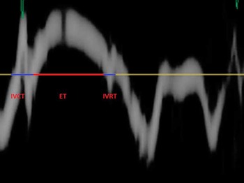
Ethical approval was obtained by the West Midlands Research Ethics Committee and infants <30 weeks of gestation were recruited prospectively following written consent. Babies with congenital abnormalities were excluded. A total of 72 infants were recruited in the study. The recruited infants underwent echocardiography by one investigator (R.P.), on day 3 of life using a Philips HD11xe system (S12-4 Hz transducer) (Philips healthcare, Best, The Netherlands). Babies were then categorised into three groups depending on the degree of ductal patency (measured at the narrowest dimension), which was determined using a previously published classification system.Reference McNamara and Sehgal 9 The groups were defined as follows: (i) haemodynamically significant ductus arteriosus (ductal diameter ≥2.5 mm and left atrium to aorta (LA:Ao) ratio ≥1.5, (ii) patent ductus arteriosus (ductal diameter <2.5 mm or LA:Ao ratio <1.5 and (iii) no patent ductus arteriosus. Babies with haemodynamically significant ductus arteriosus were treated with indometacin according to local unit guidelines (0.2 mg/kg 12 hourly for three doses). All babies underwent standard and tissue Doppler imaging echocardiography. Echocardiography was repeated between 48–72 hours after completion of the indometacin therapy. Tissue Dopper myocardial velocities were acquired from an apical four-chamber view. A pulse-wave Doppler sample gate of 0.12 cm was positioned at the lateral tricuspid and mitral annuli. Peak velocities in systole (S′), early diastole (E′) and late diastole (A′) were obtained from each site. An angle of <20 degrees was maintained using two-dimensional hold and the Doppler signal was not corrected. Average readings were taken from three to five cardiac cycles during off-line analysis. Left and right myocardial performance index was calculated. Mitral and tricuspid inflow was obtained from the standard apical four-chamber view analysing the flow of blood through the mitral valve and tricuspid valve, respectively. The myocardial performance index was also calculated. The myocardial performance index is a ratio of time intervals derived from the tissue Doppler waveform (Fig 1). The myocardial performance index evaluates both the systolic and diastolic function of a ventricle and provides a non-invasive, quantitative assessment of ventricular function. The myocardial performance index is a ratio in which the numerator is the sum of isovolumetric contraction and relaxation times and denominator is the ejection time with all the parameters measured from the same ventricle (Fig 1).
Tissue Doppler waveform showing measurements for calculation of myocardial performance index (see text).

In the haemodynamically significant ductus arteriosus group, a further analysis was undertaken following indometacin treatment to examine the following: (i) whether myocardial function improved following closure of haemodynamically significant ductus arteriosus (ii) whether differences in myocardial function could be used to predict success or failure of treatment. Statistical analysis was by GraphPad Instat v 3.05. For data comparison, one-way analysis of variance and t-test were used. Reproducibility of this method has been studied previously and has demonstrated acceptable interobserver and intraobserver bias.Reference Negrine, Chikermane, Wright and Ewer 11
Results
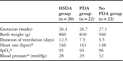
A total of 72 preterm infants born <30 weeks of gestation were recruited and a total of 64 babies were investigated – four babies died before day 3, one baby had coarctation of the aorta and three babies were considered clinically too unstable to undergo further echocardiography. In all, 20 babies were recruited into the haemodynamically significant ductus arteriosus group and 22 babies into each of the patent ductus arteriosus and no patent ductus arteriosus groups. The demographic details of the three groups are shown in Table 1. Table 2 shows mean myocardial velocities in each group.
Demographic data.

HSDA=haemodynamically significant ductus arteriosus; PDA=patent ductus arteriosus
All data expressed as mean
* At the time of initial echocardiogram
Myocardial peak velocities and myocardial performance index.

Mit=mitral; MPI=myocardial performance index; Tri=tricuspid
Values are mean (SD)
There were significantly lower velocities in the haemodynamically significant ductus arteriosus group compared with the two other groups (p<0.0001). There was a trend to lower velocities in the patent ductus arteriosus group compared with the no patent ductus arteriosus group, but this did not reach statistical significance. Myocardial performance index was higher in the haemodynamically significant ductus arteriosus group compared with the other two groups reaching statistical significance for the right ventricle (p=0.013) (Table 2). Biventricular myocardial performance index was significantly higher in the patent ductus arteriosus group compared with the no patent ductus arteriosus group (p<0.0001).
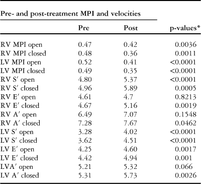
In the haemodynamically significant ductus arteriosus group, a further comparison was undertaken following indometacin therapy to examine the effect of treatment on myocardial function and to identify whether tissue Doppler imaging could be used to predict likelihood of treatment success. In the haemodynamically significant ductus arteriosus group, all received treatment with indometacin, with successful closure of patent ductus arteriosus in eight out of 20 babies. For the haemodynamically significant ductus arteriosus group, post-treatment myocardial velocities were significantly higher and myocardial performance index significantly lower than pre-treatment except right ventricular early and late diastolic and left ventricular late diastolic myocardial velocities. (Table 3)
Pre-treatment to post-treatment changes to velocities and myocardial performance index.

A′=peak late diastolic velocity; E′=peak early diastolic velocity; LV=left ventricle; MPI=myocardial performance index; RV=right ventricle; S′=peak systolic velocity
* Values are mean (paired t-test)
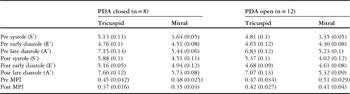
On further analysis, the pre-treatment myocardial velocities were significantly lower (p<0.001) and myocardial performance index was higher in babies whose patent ductus arteriosus remained open (n=12) compared with babies whose patent ductus arteriosus closed (n=8) with indometacin (Tables 3 and 4). These myocardial performance index differences were statistically significant for the left ventricle (p=0.014).
Pre- and post-treatment myocardial peak velocities and MPI.

MPI=myocardial performance index
Values are mean (SD)
Discussion
This study has demonstrated significantly lower biventricular myocardial velocities and higher right ventricular myocardial performance index in babies with haemodynamically significant ductus arteriosus compared with those with smaller or absent ducts, suggesting relative myocardial dysfunction in these babies. In babies with non-significant patent ductus arteriosus, there was a trend to higher myocardial velocities and significantly lower myocardial performance index compared with babies with no patent ductus arteriosus. These results correlate with data from studies utilising pulse wave Doppler to measure left myocardial ventricular function.Reference Schmitz, Stiller and Koch 10
Tissue Doppler imaging has been studied extensively in adults, less so in children, and there have been relatively few studies in neonates. All the studies involving children have concluded that tissue Doppler imaging is a valuable tool for assessing ventricular function and that further studies establishing normative data in the paediatric group should be undertaken. There are two previous studies establishing the role of tissue Doppler imaging in both term and premature babies that have been published.Reference Negrine, Chikermane, Wright and Ewer 11 , Reference Ciccone, Schcchitano and Zito 12 A study examining the acute changes in myocardial systolic function by tissue Doppler and myocardial deformation in preterm infants undergoing patent ductus arteriosus ligation has been recently published;Reference El-Khuffash, Jain and Dragulescu 13 however, this is the first study to examine the role of tissue Doppler imaging in the assessment of biventricular function in preterm infants with patent ductus arteriosus in the first week of life. Pharmacological closure is a more gradual transition rather than an acute surgical ligation and therefore the myocardium may undergo a slower period of adaptation than in the study by El-Khuffash et al. The post-intervention echocardiogram in our study was performed after a longer period of time (48–72 hours) than in the El-Khuffash study, and this may reflect the apparent differences in the results. It is clear from their data that the acute changes seen reverted to baseline by 18 hours and our measurements were taken much later than this.
A patent ductus arteriosus produces a left-to-right shunt, potentially causing volume overload of the left side of heart. If the patent ductus arteriosus is haemodynamically significant, this may result in left ventricular dysfunction. Our study has shown that in babies with haemodynamically significant ductus arteriosus there was impairment of biventricular myocardial function when compared with other groups with non-significant or absent patent ductus arteriosus.
We also examined myocardial function in babies with haemodynamically significant ductus arteriosus before and after indometacin therapy. Although the numbers studied were small, comparison of pre- and post-treatment myocardial function of babies whose patent ductus arteriosus closed (successful treatment) with those babies whose patent ductus arteriosus remained open (unsuccessful treatment) demonstrated higher pre-treatment myocardial velocities and lower myocardial performance index in the successful treatment group. This observation suggests that myocardial function is impaired by haemodynamically significant ductus arteriosus and improves significantly if the patent ductus arteriosus closes following treatment. In our study, treatment failures had worse pre-treatment biventricular myocardial function and persisting relative biventricular diastolic dysfunction after indometacin compared with treatment successes. Our data suggest that tissue Doppler imaging assessment of myocardial function in babies with haemodynamically significant ductus arteriosus may be useful in predicting those ducts that may be more resistant to medical intervention but further research is necessary. The persistence of the patent ductus arteriosus despite indometacin is associated with continuing biventricular diastolic dysfunction.
Our study has shown that tissue Doppler imaging can be used to measure myocardial velocities and myocardial performance index in preterm infants with different degrees of ductal patency. These data may help to improve our understanding of the effect of patent ductus arteriosus on myocardial function and the likelihood of response to pharmacological treatment.
There are limitations to our study. Intrinsic myocardial immaturity and hypoxia can cause myocardial dysfunction in premature babies. In all three groups, the mean gestational age was comparable. Birth weight was slightly higher in the no patent ductus arteriosus group, whereas mean ventilation days were higher in the haemodynamically significant ductus arteriosus group as might be expected. However, we have clearly demonstrated that ductal closure in this group was associated with improvement in myocardial function.
Conclusion
Preterm infants with haemodynamically significant ductus arteriosus had lower myocardial velocities and higher myocardial performance index, suggesting relative systolic and diastolic myocardial dysfunction in this group. Babies whose patent ductus arteriosus remained open despite indometacin had lower pre-treatment myocardial velocities and higher myocardial performance index than those babies whose patent ductus arteriosus closed successfully with treatment, suggesting relatively worse myocardial function in this group. Post-treatment myocardial velocities improved in babies whose patent ductus arteriosus remained open, but were still lower and myocardial performance index higher suggesting a persistence of relative biventricular diastolic dysfunction. Our study demonstrates that measurement of myocardial function in premature babies with patent ductus arteriosus is feasible and may be a helpful adjunct in the management of these babies, although larger studies are required to define the precise role of tissue Doppler imaging in this respect.
Acknowledgements
None.
Financial Support
This research received no specific grant from any funding agency, commercial or not-for-profit sectors.
Conflicts of Interest
None.
Ethical Standards
The authors assert that all procedures contributing to this work comply with ethical standards of National Research Ethics Standards (NRES), UK on human experimentation and with the Helsinki Declaration of 1975, as revised in 2008, and has been approved by the West Midlands Research and Ethics Committee.