Implications
Although this research was conducted on avalanche dogs, it highlights the possibility of using real-time PCR to measure metabolism- and oxidative stress-related gene expression to assess the impact of management practices, such as handling, transport and pre-slaughter procedures, in farmed species. Current scientific knowledge underscores the need to adopt new approaches to non-invasively monitoring of the relationships between stress, animal welfare and product quality. Real-time PCR investigation of metabolism- and oxidative stress-related genes could be a fascinating subject of future studies on acute stress in experimental models of farmed animals.
Introduction
In addition to behavioural and clinical measurements, blood sampling has been widely used to monitor animal responses to stress (Haverbeke et al., Reference Haverbeke, Diederich, Depiereux and Giffroy2008). However, in some cases, the restraint and handling procedures required for blood sampling have been shown to be themselves stressful (Kobelt et al., Reference Kobelt, Hemsworth, Bernett and Bulter2003). To avoid introducing confounding variables, new, non-invasive techniques are needed to measure responses to stress.
Exercise induces multiple biochemical changes and homoeostatic recovery events that may affect gene expression. For example, it can increase mRNA expression levels of genes involved in mitochondrial biogenesis and metabolism, such as glucose transporter 4 (GLUT4; Ren et al., Reference Ren, Semenkovich, Gulve, Gao and Holloszy1994). Muscular activity also results in an increased production of radicals and other forms of reactive oxygen species (ROS), which can disturb muscle homoeostasis (Powers and Lennon, Reference Powers and Lennon1999). The functions of superoxide dismutase 1 (SOD1), cyclooxygenase 2 (COX2) and heat shock protein 70 (HSP70) have been associated with cellular protection during exercise and cellular stress, including increased temperature, hypoxia, glucose deprivation and cellular damage (Guo et al., Reference Guo, Wharton, Moseley and Honglian2007; Golbidi and Laher, Reference Golbidi and Laher2011). Oxidative stress and antioxidants have been shown to regulate HSP70 expression (McLaughlin et al., Reference McLaughlin, Hartnett, Erhardt, Legos, White, Barone and Aizenman2003), and Catoire et al. (Reference Catoire, Mensink, Boekschoten, Hangelbroek, Muller, Schoauwen and Kersten2012) observed an effect of endurance exercise on gene expression levels in human muscle.
Despite recent advances, many important aspects of the complex molecular and cellular events that underlie the physiological adaptations triggered by exercise remain elusive. Although it is generally accepted that gene transcription regulation is involved, the gene networks or pathways that translate acute exercise effects into exercise adaptation are still unknown. Because exercise stress is a reversible event, gene expression information can be obtained before, during and after exercise activity.
In the present work, real-time PCR was employed to evaluate the expression of metabolism- and oxidative stress-related genes in saliva samples as indicators of acute exercise stress in a group of avalanche search and rescue military dogs. These animals are an incredibly important resource for searching for disaster victims. Avalanche search and rescue work requires a high level of fitness because the dogs have to cover large areas in difficult climatic conditions. Before starting their search, avalanche rescue dogs are strongly encouraged by their handlers to do their best. It is widely recognized that there is a deep relationship between military handlers and their dogs, and the quality of this relationship has been shown to influence dog efficiency and welfare (Lefebvre et al., Reference Lefebvre, Diederich, Delcourt and Giffroy2007). Sport competitions have been reported to induce a temporary stress response in dogs (Pastore et al., Reference Pastore, Pirrone, Balzarotti, Faustini, Pierantoni and Albertini2011). We previously found that training for search and rescue missions in a simulated avalanche area (SAA) induces physiological stress responses in military dogs (Diverio et al., Reference Diverio, Barbato, Guelfi, Zasso, Cavallina, Massimo, Di Mari, Murari, Santoro, Levis and Knowles2013). Specifically, we recorded significant increases of cortisol, creatine kinase (CK), aspartate aminotransferase (AST), lactate dehydrogenase (LDH), non-esterified fatty acids, heart rate and body temperature. However, these changes were moderate and limited over time, with most of the parameters progressively decreasing to completely recovery within 2 h of the end of training. These changes may be dependent on physical exercise levels. Additional factors, such as competition context, handler’s tension and environmental disturbance, may also play roles.
Here, we studied the expression of metabolism- and oxidative stress-related genes on a subsample of this group of avalanche search and rescue dogs. This group of animals is homogeneous in terms of physical variables and their ability to perform a planned exercise, thus enhancing experimental reproducibility.
To the best of our knowledge, no previous studies have been carried out to assess the effects of exercise on the mRNA transcript profiles of saliva. We chose to evaluate the expression of GLUT4, COX2, SOD1 and HSP70 mRNAs in the saliva. Such studies are non-invasive and useful for accumulating scientific rationale (Lee et al., Reference Lee, Zhou, Reiss, Yan, Zhang, Chia and David2011). As an acute exercise stress indicator, we chose body rectal temperature (RT) because it is a minimally invasive measurement that shows a tendency to rapidly recover after changes (Bouwknecht et al., Reference Bouwknecht, Oliver and Paylor2007). Transient rises in body temperature have been observed after the application of stressors in other species (Marchei et al., Reference Marchei, Diverio, Falocci, Fatjo, Ruiz-De-La-Torre and Manteca2009) and after physical exercise in dog (Matwichuk et al., Reference Matwichuk, Taylor, Shmon, Kass and Shelton1999).
Material and methods
All experimental procedures in the present study were in agreement with the Ethical Committee of the Perugia University and were conducted in accordance with the laws of the Italian Ministry of Health. There is a standing agreement between the Italian Military Force Guardia di Finanza (GdF) and the Faculty of Veterinary Medicine of Perugia allowing ethical testing of GdF working dogs.
Experimental conditions
This study was carried out during the Second Retraining Course (February 2012) for the Avalanche Search and Rescue Military Dog-Handler Units (SAGF Units) at the GdF Alpine School of Passo Rolle (Trento, Italy). During this period, all the SAGF Units were trained with two avalanche search training sessions per day, with a snow-buried operator simulating an avalanche victim. A limited experimental area was established on a field of about 1 ha, at 2170-m altitude, to resemble an avalanche environment (SAA). The snow was moved around this area and then compressed by a vehicle designed to move on snow. Three identical pits (1.5-m deep, 50×100-cm wide) were dug to avoid olfactory clues. In each of the trials, the simulated victim was entirely buried at random in one of the three pits and covered with compressed snow. A hole for air turnover was left in the pits for safety reasons, and the buried operator was never left inside for more than 60 min.
During the simulation, a team of experts of the Agenzia Regionale per la Prevenzione e Protezione Ambientale del Veneto continuously monitored meteorological conditions by collecting data through a small Oregon Scientific portable station (Oregon Scientific Wireless Weather Station, Tualatin, OR, USA) installed at a campsite near the SAA. According to the recorded data, the environmental air temperatures ranged from −8.5°C to −10.4°C with 28% humidity and a wind-chill effect of −29.0°C.
Subjects
We used a sample of 12 male dogs belonging to SAGF Units (SAGF dogs) aged from 2.8 to 5.6 years and belonging to various breeds (three Border Collies, six Belgian Shepherd Malinois, three German Shepherds). All dogs were physically (i.e. X-ray negative for hip dysplasia and considered in good health after veterinary inspection) and behaviourally tested (i.e. absence of behavioural problems or pathologies during veterinary behavioural consultancy) to certify their suitability for training work. These animals came from different locations in Italy and lived with their handlers all year round. The dogs were individually kennelled in indoor pens (2.9×2.1×2.3 m) during the experiment at a location next to the GdF Alpine School of Passo Rolle. All the animals had the same feeding regimen (standard commercial dry adult dog diet) and had been operational as search and rescue working dogs for a minimum of 1 year. They were always trained with their own handler, who had at least 2 years of working experience with dogs before the beginning of this experiment.
Experimental design
Each SAGF Unit was individually monitored during a specific training exercise, also called a simulated avalanche search and rescue (SASR) operation. According to a specific protocol, the dogs and their handlers for each SAGF Unit, equipped with pulley harnesses, were loaded into a helicopter and transported in about 8 min to the area above the SAA. Next, the SAGF Unit was lowered from the hovering helicopter to the ground. Upon arrival, the SAGF Unit moved to an adjacent sampling point, where an operator subjected the dogs to the first saliva and RT sample collection (T1).
Then, the SAGF Unit began the SASR to search for the simulated victim, who was buried in one of the three pits. During the SASR, the handler could vocally encourage the dog, covering the SAA with the dog at a variable distance. When the dog located the site where the operator was hidden, the handler rewarded the dog by playing with it for a maximum of 3 min. Then, the SAGF Unit returned to the sampling point where the operator collected a second saliva sample (T2). A maximum of 10 min of SASR time was allowed for the SAGF Unit to find the simulated victim. Considering the 3-min reward time and the 1–2 min required to reach the sampling point, the T1 to T2 sampling interval ranged from a minimum of 6 to a maximum of 15 min. After T2 sampling, each SAGF dog was led by its handler to its living pen to rest. The third saliva sample (T3) was collected 2 h after the end of the SASR (T2), just outside the SAGF dog pen. Baseline samples (T0) were also collected from the SAGF dogs the day before SASR between 0800 and 0900 h.
The procedures to collect saliva and RT at T0, T1, T2 and T3 sampling times from the SAGF dogs are described in detail as follows. For saliva sample collection, the handler asked the dog to sit and then gently manipulated the dog to remain still for a few seconds with its mouth open. The operator gentle rubbed the inside of the dog’s cheeks with two cotton swabs. The handler then asked the dog to stand and gently petted and distracted it for 1 min while the GdF veterinarian measured RT with a digital thermometer (MB TERMO 7126500, Reckitt Benckiser SpA, Milano, Italia).
Saliva and RT sampling time ranged from 2 to 3 min. During this time, the SAGF dogs did not show any behavioural signs of stress or anxiety; they remained calm and focused on their handlers.
Saliva collection, RNA extraction and real-time PCR
Each buccal swab collected from SAGF dogs at the four sampling times (T0, T1, T2 and T3) was immediately placed into 500 µl phosphate-buffered saline and 1.3 ml RNA Later (Ambion, Austin, Texas, USA). RNA Later stabilizes and protects cellular RNA, eliminating the need to immediately process or freeze samples. Afterwards, all specimens were kept at −20°C until attempts at isolation were made. The saliva was removed from the cotton swabs by centrifugation (3000 r.p.m., 15 min). Total RNA purification kits (Promega, Madison, WI, USA) were used to extract RNA according to the manufacturer’s instructions. RNA was quantified with the Qubit RNA assay (Life Technologies, Carlsbad, CA, USA) and stored at −20°C until use.
Next, 20 ng total RNA was reverse transcribed in 20 μl iSCRIPT cDNA (Bio-Rad, Hercules, CA, USA) using random hexamers according to the manufacturer’s suggestions. Controls without reverse transcriptase were included to check for genomic DNA contamination. mRNA expression in saliva samples was measured by real-time PCR according to Guelfi et al. (Reference Guelfi, Zerani, Brecchia, Parillo, Dall’Aglio, Maranesi and Boiti2011). The optimized PCR assay of a 20-µl PCR reaction volume contained 10 μl TaqMan Gene Expression Master Mix (Applied Biosystems, Foster City, CA, USA), 1 µl TaqMan Gene Expression Assays (Table 1) and water to 20 µl. All reagents were mixed and distributed into a 96-well PCR plate before adding 4 µl cDNA (1–100 ng). Sample amplification fidelity was verified by agarose gel electrophoresis. PCR was performed on iCycler iQ (Bio-Rad) with an initial incubation at 95°C for 10 min, followed by 45 cycles at 95°C for 15 s and 60°C for 60 s, during which time fluorescence data were collected. The cycle threshold (Ct) was automatically computed for each trace. PCR products were purified and sequenced by QIAquick PCR Purification Kit (Qiagen Inc., Valencia, CA, USA) according to the manufacturer’s protocol. PCR amplification efficiency was determined by using the slope of the standard curve: efficiency=10−1/slope−1. The slope was then utilized to determine the amplification efficiency. PCR conditions were optimized to generate >95% PCR efficiency, and only reactions between 95% and 100% efficiency were included in subsequent analyses.
TaqMan Gene Expression assay used in the studies are listed by assay numbers

MGB=minor groove binder; NFQ=non-fluorescent quencher.
TaqMan Gene Expression assay consist of a pair of unlabelled PCR primers and a probe with a FAM dye label on the 5' end, and MGB NFQ on the 3' end.
The Ct value corresponding to the PCR cycle number at which fluorescence emission in real time reached a threshold above the baseline emission was determined. The Ct value for each sample was then exported and analysed in Microsoft Excel (Redmond, WA, USA). The relative expressions of GLUT4, COX2, SOD1 and HSP70 genes were normalized to beta-actin (ACTB) housekeeping gene levels. For the comparative Ct method, the difference in Ct between the target gene and ACTB was calculated for each sample. The expression level in any sample was determined by the
 $${\rm 2}^{{{\minus}\Delta \Delta C_{t} }} $$
method (Livak and Schmittgen, Reference Livak and Schmittgen2001). All reactions were prepared in triplicate.
Statistical analysis
The results of reverse transcription, real-time PCR and RT were analysed by repeated measures ANOVA. Significance was set at P<0.05. All post hoc analyses were performed using Tukey’s method for multiple comparisons (GraphPad Prism 5.04, La Jolla, CA, USA).
Results
RT
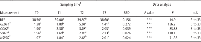
The effects of SASR on RT are reported in Table 2. Soon after the descent from the helicopter (T1), the RT of the SAGF dogs was 38.96±0.15°C (mean±s.e.), which was not significantly different from the mean baseline value (T0=38.55±0.14°C). RT peaked at T2 (39.49±0.12°C), which was significantly higher than T0 (P<0.001) and T1 (P<0.05). Complete RT recovery was observed after a 2-h rest (T3=38.57±0.08°C), significantly decreasing from T2 (P<0.001), reaching values similar to those measured at baseline (T0).
Sampling time differences in RT and saliva mRNA expression of GLUT4, COX2, SOD1 and HSP70 genes in the 12 dogs belonging to Avalanche Search and Rescue Military Dog-Handler Units (SAGF dogs)

RT=rectal temperature; GLUT4=glucose transporter 4; COX2=cyclooxygenase 2; SOD1=superoxide dismutase 1; HSP70=heat shock protein 70; RSD=residual standard deviation of ANOVA; d.f.=degree of freedom of effect of interest and of denominator; SASR=Simulated Avalanche Search and Rescue; SAA=simulated avalanche area.
a,b,cValues within a row significantly differ at P<0.05.
1 T0: baseline samples collected from 0800 to 0900 h, the day before the trial –(SASR operation); T1: at arrival on the SAA after helicopter transport, when began to search the simulated disperse victim; T2: at the end of the SASR, when the buried victim was discovered; T3: 2 h after the T2.
2 Measured by a digital thermometer.
3
Real-time data expressed by a means of
 $${\rm 2}^{{{\minus}\Delta \Delta C_{t} }}$$
.
***Difference (P<0.001) due to sampling time.
GLUT4, COX2, SOD1 and HSP70 gene expression
Approximately 200 µl of saliva was obtained from each cotton swab, with no significant differences among subjects. The total RNA yield was 30 ng/100 μl of saliva. The effects of SASR on all metabolism- and oxidative stress-related genes expressions are listed in Table 2. All metabolism- and oxidative stress-related genes under investigation showed a peak in expression at T2 and fell back to initial values at T3. Baseline values of GLUT4 (T0=1.37±0.03, mean±s.e.) significantly (P<0.001) increased at T1 (1.89±0.02). There was a more than twofold increase in GLUT4 gene expression at T2 (5.04±0.08), which was significantly (P<0.001) higher than T0 and T1. Following a 2-h rest period, the GLUT4 values (T3=1.41±0.02) were similar to those measured in saliva samples collected at the baseline (T0) (Table 2). The trend of COX2 gene expression was similar to that of GLUT4. The mean COX2 expression values progressively and significantly increased from T0 to T1 (P<0.001; T0=1.90±0.03; T1=2.30±0.06), up to T2 (P<0.001; 3.08±0.10). A 2-h rest allowed complete recovery of COX2 gene expression in the SAGF dogs (T3=2.03±0.04). Similarly, SOD1 levels reached a significant (P<0.001) peak at T2 (2.85±0.06) with respect to T1 (1.69±0.07). Then, a significant decrease (P<0.001) in SOD1 expression was observed at T3 (2.12±0.06), although the values were still higher (P<0.001) than those measured at T1 (Table 2). No significant differences were recorded between SOD1 at T0 (1.96±0.05) and T3, indicating a re-establishment of baseline levels (Table 2). A temporary increase in HSP70 gene expression was also observed. The trend was comparable with that recorded for RT. HSP70 gene expression values peaked at T2 (2.68±0.12), and were significantly different (P<0.001) from those measured at T0 (1.97±0.04), T1 (1.84±0.12) and T3 (2.01±0.12). No significant differences were observed between T0 and T3 HSP70 gene expression values (Table 2).
Discussion
In the present study, we presented a novel characterization of salivary mRNAs coded by metabolism- and oxidative stress-related genes before, during and after dogs completed a simulated search and rescue operation. Overall, the changes recorded in GLUT4, COX2, SOD1 and HSP70 gene expression levels most likely reflect their role in the control of sugar metabolism and oxidative pathways. The peak in GLUT4 expression at the end of the SASR (T2) could be due to a proportional increase in insulin responsiveness of the glucose transport process in the skeletal muscle, as described in humans (Cortright and Dohm, Reference Cortright and Dohm1997). Insulin and contraction signalling pathways both result in GLUT4 translocation (Cortright and Dohm, Reference Cortright and Dohm1997). In rat, increases in mitochondrial biogenesis and GLUT4 glucose transporter expression have been found to be the major adaptive responses of skeletal muscle to endurance exercise (Kazuhiko et al., Reference Kazuhiko, Sang, Mitsuru, John and Dong-Ho2011). Given that the biological role of GLUT4 is to transport glucose into muscle cells and considering that glycogen re-synthesis has a high metabolic priority during the post exercise recovery period, transcriptional activation of GLUT4 after a single bout of exercise is likely to be involved in re-establishing glycogen homoeostasis in skeletal muscle (Mahoney et al., Reference Mahoney, Parise and Melov2005). In human skeletal muscle, GLUT4 gene expression increased immediately after exercise, but remained significantly higher than the baseline for the following 3 h (Kraniou et al., Reference Kraniou, Cameron-Smith, Misso, Collier and Hargreaves2000). In our study, GLUT4 mRNA expression exhibited a fast homoeostatic recovery after the dogs performed an avalanche search and rescue activity. This trend suggests that GLUT4 gene expression could be a useful indicator of carbohydrate (CHO) metabolism and exercise stress in dogs.
The peak in salivary expression of SOD1 mRNA recorded at T2 might be a consequence of the generation of superoxide radicals in response to exercise. Superoxide radicals are involved in erythrocyte conversion of haemoglobin to methemoglobin during physical exercise. These are ultimately converted into hydrogen peroxide (H2O2) by SOD1. Notably, SOD1 also acts as one of the primary enzymatic antioxidant defences of the cell against damage caused by superoxide radicals. Skeletal muscles contain other endogenous protective mechanisms, such as COX2, that reduce the harmful effects of ROS in cells. The transient rise of COX2 gene expression recorded at T2 confirms the importance of this protective mechanism. COX2 converts arachidonic acid to prostaglandin H2 (PGH2), the precursor of series-2 prostanoids. The beneficial actions of COX2 derive from PGE2 and/or PGI2 production. In fact, elevated prostanoid production by COX2 protects cardiomyocytes against oxidative stress and exerts anti-apoptotic effects in several cell types (Adderley and Fitzgerald, Reference Adderley and Fitzgerald1999). Similarly to SOD1, COX2 mRNA expression returned to baseline values after a 2-h rest (T3). This homoeostatic recovery of SOD1 and COX2 gene expressions suggest that they play specific roles in the oxidative pathways activated by exercise.
The HSP70 gene plays a specific role in cytoprotection (Santoro, Reference Santoro2000). The peak of HSP70 mRNA expression recorded at T2 could reflect its protective role against a variety of cellular reactions triggered by acute exercise stress during the SASR. Previous human studies (Powers and Lennon, Reference Powers and Lennon1999; Guo et al., Reference Guo, Wharton, Moseley and Honglian2007) have shown that HSP70 synthesis is enhanced to protect cells against various stress conditions (Golbidi and Laher, Reference Golbidi and Laher2011). Guo et al. (Reference Guo, Wharton, Moseley and Honglian2007) hypothesized that HSP70 might protect cells from ischaemic injuries by regulating cellular redox status. This is primarily regulated by the balance between cellular oxidant and reductant levels. The cellular redox environment plays an important role in several cellular functions, including the regulation of proliferation, differentiation and cell death (Schafer and Buettner, Reference Schafer and Buettner2001). According to Schafer et al., the upregulation of HSP70 and SOD is one mechanism by which cells manage potential cytotoxicity induced by exercise-related stress. A variety of stressors associated with exercise, such as decreased intracellular pH, reactive oxygen and nitrogen species production, depletion of glucose and glycogen stores, increased cytosolic calcium levels and cardiomyocyte stretching, can contribute to HSP70 elevation during physical exercise (Powers et al., Reference Powers, Locke and Demirel2001). Similarly, we observed a temporary increase in HSP70 mRNA expression after exercise stress. The quick return to baseline values after rest might reflect the ability of the SAGF dogs to recover their energy metabolism.
The transient rise and subsequent recovery of SAGF dog RT at T3 is similar to those reported in other working dogs after strenuous exercise (Matwichuk et al., Reference Matwichuk, Taylor, Shmon, Kass and Shelton1999). Stress- and exercise-induced body temperature increase is a universal phenomenon that occurs in many species (Bouwknecht et al., Reference Bouwknecht, Oliver and Paylor2007). Actually, in relation to stress, body temperature regulation is an integrative response to different stimuli at the level of the hypothalamus, which is involved in the regulation of a variety of responses occurring in stress and anxiety (Dunn and Berridge, Reference Dunn and Berridge1990). In addition, body temperature increases after exercise because a portion of the nutrient energy is converted to heat during cellular metabolism (Matwichuk et al., Reference Matwichuk, Taylor, Shmon, Kass and Shelton1999). The similar temporal trends in body temperature and the expression of genes controlling sugar metabolism and oxidative pathways may be explained by their concurrent involvement in the response to exercise.
As suggested in humans (Pilegaard et al., Reference Pilegaard, Ordway, Saltin and Neufer2000), our findings seem to indicate that the mechanisms involved in substrate utilization during exercise and/or restoration of muscle glycogen after exercise are linked to the transcriptional activation of selected metabolic genes in dogs as well. Overall, the exercise involved in the SASR proved to alter mRNA gene expression, but the changes were acute. A similar trend of change over time was also observed for RT. These findings suggest that the military dogs only exhibited a transient response to the rescue operation. According to a previous study (Diverio et al., Reference Diverio, Barbato, Guelfi, Zasso, Cavallina, Massimo, Di Mari, Murari, Santoro, Levis and Knowles2013), military dogs performing a rescue operation may show a significant but short-lasting rise in cortisol, glucose, AST, LDH and CK blood concentrations. Most changes were moderate and progressively decreased over time, with complete recovery after 2 h, demonstrating that military dogs were able to rapidly recover from acute exercise stress despite extreme working conditions (Diverio et al., Reference Diverio, Barbato, Guelfi, Zasso, Cavallina, Massimo, Di Mari, Murari, Santoro, Levis and Knowles2013).
Thus, the T2 to T3 period could be considered as the dog recovery time, and the variation in the expression pattern of the metabolism- and oxidative stress-related genes recorded in this period could be used for estimating resilience in dogs. However, more studies are required that compare salivary mRNA expression levels with measures of resilience. The recovery of the expression of metabolism- and oxidative stress-related genes at T3 might indicate that the previous physical training of these military dogs, which is designed to prepare them for avalanche searching, was able to enhance their performance and their capacity to repeat high-intensity efforts. High performance levels and an ability to recover from hard exercise are essential for these working dogs, and they can make the difference between life and death for avalanche victims. However, more studies are required to compare the responses of trained and untrained dogs in the same test.
It remains to be elucidated if the ability to quickly re-establish energy homoeostasis may be attributable to epigenetic regulation, as was shown in humans. Further studies are needed to investigate how training can modulate epigenetic mechanisms, thereby increasing the current body of knowledge in this field.
Our study aimed to assess gene expression responses of dogs to an acute exercise stress and to develop a non-invasive monitoring method. The present findings are encouraging for the creation of a robust and streamlined clinical assay for detecting exercise stress in dogs. This non-invasive gene expression method may be exploited and validated in future studies because of its potential value as an acute exercise stress assessment tool in dogs and other species.
Acknowledgements
The authors wish to thank the Guardia di Finanza General Command for supporting this project and Col. Alciati Secondo, Col. Stefano Murari, Mar. Valter Levis, Dr Anselmo Cagnati and Renato Zasso for their valuable collaboration. The authors also thank the SAGF Unit handlers for their invaluable collaboration and, of course, their wonderful SAGF dogs. We are grateful to Sara Ricchetti for English revising our manuscript. They are also grateful to the two anonymous referees who preciously helped to improve the quality of this manuscript.