Introduction
18F-fluoro-deoxy-glucose (18F-FDG) positron emission tomography computed tomography (PET-CT) is an established functional imaging technique in the diagnostic investigation of several oncological conditions, including non-small cell lung cancer.Reference Bar-Shalom, Valdivia and Blaufox1 All organs are examined in a single scanning episode for the presence of a primary or metastatic malignancy. Incidental findings within other organs or anatomical regions are not uncommon; it has been reported that 5–12 per cent of all PET-CT scans highlight areas of concern regarding possible synchronous second malignancies.Reference Ozkol, Alper, Aydin, Ozkol, Topal and Akpinar2, Reference Beatty, Williams, Aldridge, Hughes, Vasudeva and Gucwa3
PET-CT scans are performed regularly within thoracic units to screen and monitor for both new and recurrent lung carcinomas. The considerable overlap in aetiological risk factors for both lung cancer and head and neck cancer presents a definite risk of synchronous malignant pathologies in these patients. It has been suggested that 4–15 per cent of patients presenting with new head and neck cancer will have a synchronous pulmonary tumour.Reference Ghosh, Roland, Kumar, Tandon, Lancaster and Jackson4, Reference Houghton, Hughes, Garvey, Beasley, Hamilton and Gerlinger5
This is the first study to specifically review incidental PET-CT head and neck findings in lung carcinoma patients – a high-risk population for synchronous head and neck cancer.
Materials and methods
Hospital records of all patients with lung cancer who underwent PET-CT imaging were identified from the Liverpool thoracic multidisciplinary team database over a five-year period (January 2008 – December 2012) and retrospectively reviewed. Incidental clinically significant PET-CT findings in the head and neck area were identified from the contemporaneous imaging report approved by a single consultant radiologist. The investigation and subsequent management of these patients was recorded.
This study was approved by our institution's Clinical Information and Audit Department; all data were anonymised and encrypted throughout. Data collation and the calculation of all descriptive statistics were performed using Microsoft Excel for Mac 2011 (Redmond, Washington, USA).
Results
Between January 2008 and December 2012, 609 patients underwent PET-CT imaging as a diagnostic investigation for lung carcinoma. Seven patients with a known history of head and neck cancer were excluded from the analysis. Seventy-six of the patients (12.5 per cent) were found to have incidental abnormal 18F-fluoro-deoxy-glucose (18F-FDG) uptake in the head and neck region. Only 28 of these patients went on to be formally reviewed by otolaryngology and/or maxillofacial specialists.
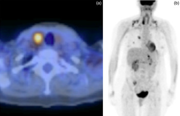
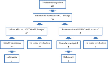
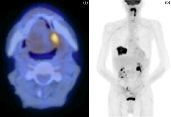
One patient was found to have synchronous primary malignant pathology in the head and neck region (papillary carcinoma of the thyroid; Figure 1). In this patient, PET-CT demonstrated focally increased 18F-FDG activity within the right lobe of the thyroid. The lesion was histologically defined as a papillary carcinoma. Another patient was diagnosed with non-Hodgkin's lymphoma after open biopsy of an 18F-FDG avid neck node. Cytological analysis of fine needle aspiration samples from two further patients undergoing investigation for 18F-FDG avid neck nodes showed secondary metastatic disease from the known lung carcinoma. Figure 2 shows all patients included in the study, the presence of incidental PET-CT findings in each case and subsequent investigations.
Positron emission tomography computed tomography hybrid images of a 54-year-old woman with an incidental 18F-fluoro-deoxy-glucose avid ‘hot spot’ in the head and neck region.

Incidental findings in positron emission tomography computed tomography scans investigating lung carcinoma from January 2008 to December 2012.

Forty-three patients with incidental findings had no formal investigation arranged by the thoracic medical team and were not referred for otolaryngology and/or maxillofacial review. This group included patients who were not referred following simple clinical examination of the incidental region and those who were not investigated because of the advanced nature of their lung carcinoma.
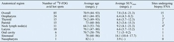
For some patients, PET-CT scans revealed more than one 18F-FDG avid ‘hot spot’ in the head and neck region. Overall, 85 such hot spots were found in all 76 patients. Table I depicts these hot spots by anatomical region and shows the average level of 18F-FDG uptake (defined in terms of the maximum standardised uptake value) at each site.
Incidental 18F-FDG avid ‘hot spots’ found on PET-CT in the head and neck region by anatomical region*

* 85 hot spots in 76 patients. †Both cases had follicular thyroid cytology and diagnostic hemi-thyroidectomy was performed. One case found to be papillary carcinoma. ‡One case found to be non-Hodgkin's lymphoma following open biopsy. Two cases found to present secondary metastases from chest primary after cytological analysis. PET-CT = positron emission tomography computed tomography; FDG = fluoro-deoxy-glucose; y = years; SUVmax = maximum standardised uptake value; FNA = fine needle aspiration
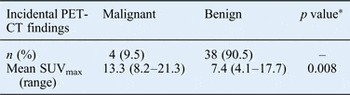
Table II shows the incidental findings by hot spot (not by patient). For the 42 incidental hot spots that went on to be investigated, the maximum standardised uptake value for findings with a benign or malignant diagnosis are compared.
Subgroup analysis of incidental PET-CT findings formally investigated or referred to head and neck services

*Student's t-test. PET-CT = positron emission tomography computed tomography; SUVmax = maximum standardised uptake value
Discussion
Positron emission tomography is a non-invasive diagnostic tool that provides information about metabolic events in tissues. Several radiopharmaceuticals are used in PET imaging, but 18F-fluoro-deoxy-glucose (18F-FDG), a glucose analogue, remains by far the most common. It is well known that malignant cells utilise glucose at greater rates than normal tissue, mainly via glycolysis for energy production regardless of the oxygen availability; this phenomenon is known as the Warburg effect.6 Fluoro-deoxy-glucose is taken up by normal and tumour cells in the first stages of the normal glucose pathway; however, unlike glucose, FDG cannot undergo further metabolism after its initial phosphorylation to form FDG-6-phosphate and is thus trapped in metabolically active tumour cells. This trapping phenomenon is exploited for FDG-PET imaging, which can often detect early-stage malignant disease before any structural abnormality is evident. It can also help to exclude the presence of malignancy in an anatomically altered structure. These added benefits have been well documented in a variety of malignancies.Reference Zimmer, Snyderman, Fukui, Blodgett, McCook and Townsend7 With the introduction of hybrid molecular imaging, in which PET and CT are combined in a single system (PET-CT), the accuracy in localising areas of increased glucose metabolism to their correct anatomical sites has significantly increased. The sensitivity and specificity of this novel imaging technique has subsequently improved.
Positron emission tomography computed tomography in the head and neck region
18F-fluoro-deoxy-glucose PET-CT imaging is utilised in the investigation of head and neck cancer; it is used for both assessing suspected recurrence and evaluating patients with metastatic lymph nodes and unknown primary tumours.Reference Roland and Paleri8 However, since 18F-FDG uptake reflects metabolic activity, it may also be absorbed in certain non-neoplastic pathologies and can accumulate in particular organs under normal physiological conditions (Figure 3).Reference Schoder, Carlson, Kraus, Stambuk, Gonen and Erdi9 In this patient, PET-CT demonstrated focally increased 18F-FDG activity at the left tongue base. However, biopsy samples revealed no dysplasia or malignancy.
(a) Coronal and (b) axial positron emission tomography computed tomography hybrid images of a 70-year-old man with an incidental 18F-fluoro-deoxy-glucose avid ‘hot spot’ in the head and neck region.

On PET-CT, normal physiological activity can be observed at multiple sites in the body, including the palatine tonsils, tongue base and salivary glands. Asymmetrical uptake within the salivary glands can be either positional or reflect inflammatory changes following surgical or radiation therapy. Physiological 18F-FDG activity is seen in the anterior floor of the mouth because the genioglossus muscle actively prevents the tongue from falling back when patients are supine in the CT scanner. If patients grind their teeth or chew, the muscles of mastication may appear prominently. Excessive talking after injection can cause prominent 18F-FDG uptake within the larynx; this is why a ‘silence protocol’ should be initiated during the scanning procedure. Such laryngeal uptake is normally very subtle, appearing in the form of a symmetrical inverted ‘V’ shape, but focal unilateral uptake within the larynx can suggest pathology and/or muscle overuse.Reference Cook, Fogelman and Maisey6
Hence, reporting PET-CT in the head and neck region often causes interpretation difficulties. Retrospective studies of head and neck patients undergoing PET-CT as part of their primary investigation show excellent sensitivity but relatively poor specificity, owing to physiological uptake at sites such as the larynx and tongue (95 per cent and 60 per cent, respectively, in one such study).Reference Zimmer, Snyderman, Fukui, Blodgett, McCook and Townsend7 The use of PET-CT to detect nodal metastasis in oral carcinoma patients has been limited by reports of a large proportion of false positives, mostly attributed to benign lymphangitis.Reference Schoder, Carlson, Kraus, Stambuk, Gonen and Erdi9
Maximum standardised uptake value as a predictor of malignancy
Histopathological analysis has revealed that regions of particularly high 18F-FDG focal uptake have a high probability of cellular transformation.Reference Agress and Cooper10 The level of 18F-FDG uptake is defined in terms of maximum standardised uptake value (maximal standardised uptake value). In the setting of incidental colorectal findings on PET-CT, the presence of foci with a maximum standardised uptake value of at least 4.35 has been reported to increase the likelihood of an underlying advanced colorectal neoplasm.Reference Cho, Kim, Kim, Park, Yoo and Kim11 In 2011, a study of 38 incidental head and neck findings from PET-CT scans also found that a high maximum standardised uptake value was a strong predictor of malignancy.Reference Al-Hakami, Makis, Anand, Mlynarek, Black and Stern12 Of the 38 incidental PET-CT findings in this study, 21 per cent represented new primary head and neck malignancies.
Of the 76 patients with incidental PET-CT findings in our case series, 4 (5.3 per cent) had findings which represented a demonstrable malignant process. When viewed in terms of individual hot spots, the 76 patients had a total of 85 hot spots, 4.7 per cent of which represented malignancy.
One obvious limitation of our study is the large number of patients who were not formally investigated by the thoracic medical team or referred for otolaryngology and/or maxillofacial review. A large proportion of this group were not further investigated because of the advanced nature of their pulmonary disease; however, the incidental PET-CT findings in these patients could represent synchronous, undetected head and neck malignancies. For this reason, if this patient group were included when assessing the predictive value of PET-CT, the incidental findings of this case series would probably represent a gross underestimate.
• Reports indicate that 4–15 per cent of new head and neck cancer patients have a synchronous pulmonary tumour
• In this study, 12 per cent of lung cancer patients had incidental positron emission tomography computed tomography findings in the head and neck region
• In all, 5.3 per cent had a demonstrable malignant process
• Head and neck region findings with a high maximum standardised uptake value probably represent malignancy
Four of the 28 patients (14.3 per cent) who had their incidental findings fully investigated (formal investigation or specialist review) had demonstrable malignancy. When comparing the uptake figures for the individual hot spots in this subgroup, the maximum standardised uptake value of malignant incidental findings was significantly higher than that of the cases in which head and neck findings were ultimately found to be benign (p = 0.008, Table II).
Conclusion
This study shows that a malignant process should be suspected when focal 18F-fluoro-deoxy-glucose uptake is detected in the head and neck region by PET-CT. Such incidental findings can be clinically significant in a considerable proportion of cases, e.g. 14.3 per cent of patients who underwent thorough investigation in this series. Our study also suggests that PET-CT findings in the head and neck region with a high maximum standardised uptake value are significantly more likely to represent malignant pathology. This supports the existing body of evidence regarding incidental findings in other regions of the body.Reference Agress and Cooper10, Reference Cho, Kim, Kim, Park, Yoo and Kim11