Introduction
During fetal life, gas exchange occurs across the placenta and the future airways are filled with liquid, which is critical for lung growth and structural development.Reference Harding and Hooper1 At term, the lungs need to have developed appropriately in order for them to perform their postnatal function of gas exchange. Animal studies have shown that a number of intrauterine perturbations disrupt fetal lung development and compromise postnatal gas exchange. In sheep, hypoxia reduces lung liquid secretion and decreases lung growth and development.Reference Harding and Hooper1 Placental insufficiency decreases alveolar number and increases alveolar wall thickness in lambsReference Maritz, Cock and Louey2 and these alterations persist into adulthood.Reference Maritz, Cock, Louey, Suzuki and Harding3 Such alterations in the lung architecture reduce the respiratory surface area and increase the diffusion barrier for gas exchange. In humans, intrauterine growth restriction is associated with an increased risk of respiratory insufficiency in neonatesReference Maritz, Morley and Harding4 and decrease forced expired flow rate in children aged 7–15 years,Reference Nikolajev, Heinonen, Hakulinen and Lansimies5 and gestational undernutrition has been associated with increased prevalence of obstructive airway diseases in adulthood.Reference Lopuhaa, Roseboom and Osmond6 As maternal alcohol consumption can also be associated with intrauterine growth restriction,Reference de Sanctis, Memo, Pichini, Tarani and Vagnarelli7 it is likely that prenatal ethanol exposure may also compromise postnatal respiratory health.
Prenatal alcohol [ethanol (EtOH)] exposure is prevalent in society, despite many countries recommending that women abstain from alcohol consumption during pregnancy.Reference Giglia and Binns8 Although it is well known that maternal consumption of large amounts of alcohol induce fetal alcohol spectrum disorder, with fetal alcohol syndrome being at the most severe end of the spectrum,Reference de Sanctis, Memo, Pichini, Tarani and Vagnarelli7 the effects of low or moderate amounts of gestational alcohol consumption on fetal growth and development are not well known. Even less information is available regarding the effects of alcohol/EtOH exposure to the fetal lung, despite the suggestion in the 1960s and 1970s when low doses of EtOH were used clinically to arrest preterm labour, that maternal EtOH treatment may increase the incidence of neonatal respiratory distress syndrome.Reference Zervoudakis, Krauss and Fuchs9
Studies of neonatal mice and guinea pigs have shown that prenatal EtOH exposure [maternal blood alcohol level (BAC) ∼0.05–0.08% in pregnancy] decreases alveolar macrophage numberReference Gauthier, Ping, Gabelaia and Brown10 and function and increases macrophage oxidative stress,Reference Ping, Harris, Brown and Gauthier11, Reference Gauthier, Young and Gabelaia12 thereby indicating poor pulmonary immune defence. Rodent fetal studies have demonstrated that high amounts of maternal EtOH consumption in pregnancy decreases lung growth (rats; maternal BAC 0.17%)Reference Inselman, Fisher, Spencer and Atkinson13 and delays lung development (mice; 3.75 g/kg EtOH administered, maternal BAC not reported),Reference Wang, Gomutputra, Wolgemuth and Baxi14 whereas more moderate amounts of maternal EtOH consumption by sheep (fetal BAC of 0.11%) decreases pulmonary surfactant protein mRNA levels and increases pulmonary collagen deposition.Reference Sozo, O'Day and Maritz15 Although these few animal studies provide vital information on the effects of maternal alcohol consumption on the fetal and neonatal lung, the EtOH exposure induced in these models began in either mid-gestation, when branching morphogenesis of the lung was almost complete, or late-gestation, during the period of pulmonary alveolarization. Here we have utilized our model of low-dose EtOH exposure throughout pregnancy, as this pattern of alcohol consumption in pregnancy is likely to be more common in society than high-dose alcohol abuse. We hypothesized that maternal gestational EtOH consumption, from conception to birth, would detrimentally affect lung development. The aim of this study was to utilize our recently developed rodent model of maternal EtOH consumptionReference Probyn, Zanini, Ward, Bertram and Moritz16 to determine the effects of chronic low-dose prenatal EtOH exposure on structural and non-respiratory aspects of the fetal and postnatal lung.
Methods
Animals
Control and prenatal EtOH exposed fetuses and offspring used in this study were a subset of those described previously.Reference Probyn, Zanini, Ward, Bertram and Moritz16 Briefly, time-mated virgin Sprague–Dawley rats were fed a liquid diet either lacking (control, n = 34) or containing 6% (v/v) ethanol (EtOH-fed, n = 33) from embryonic day (E) 1 until either the dam was killed at E20 for the collection of the fetal lung or her offspring were born. This amount of EtOH produced a plasma EtOH concentration of 0.03%.Reference Probyn, Zanini, Ward, Bertram and Moritz16 For dams that delivered their pups, the liquid diet was removed once the pups were born, [designated postnatal day (PN) 0], and the dams provided an ad libitum standard laboratory rat chow diet and water. All dams nursed their own pups until weaning at PN28, at which time male and female offspring were separated and housed with same-sex littermates until post-mortem examination and tissue collection.
Pulmonary tissue collection
Fetal tissue
A subset of dams (control, n = 9; EtOH-fed, n = 11) were killed at E20Reference Probyn, Zanini, Ward, Bertram and Moritz16 for the collection of the fetal lung. Fetal sex was determined via the ano-genital distance. After the fetal head was removed, an incision was made from the abdomen to the neck opening, taking care not to cut the lungs, and the chest wall removed. The heart and aorta were ‘pinched’ off from the lungs using forceps and the lungs removed from the thoracic cavity. The oesophagus and superior vena cava were freed from the lungs and the lungs weighed and snap-frozen in liquid nitrogen for gene expression studies (n = 10–12 per sex per treatment from 9 to 11 litters).
Postnatal tissue
Control and EtOH-exposed rats were killed at PN1, 8–10 months or 19 ± 1 months of age. At PN1, after decapitation, the lungs of 9–10 male and female pups of 8–10 litters were pressure fixed in situ via the trachea at a distending pressure of 20 cmH2O with 4% paraformaldehyde in 0.2% phosphate-buffered saline (PBS). Adult offspring were weighed before being heavily anaesthetized with 1 ml/kg i.p. ketamine/xylazine. The abdominal cavity was opened, the diaphragm was pierced at the sternum and the thoracic cavity opened. For 8-month-old (n = 9–12 per sex per treatment from 5 to 6 litters) and 19-month-old (n = 13–17 per sex per treatment from 10 to 13 litters) offspring the lungs were removed, weighed and snap-frozen in small sections, making sure to exclude obvious airways. For 10-month-old offspring, the whole animal was perfused with 0.2% PBS and the lungs fixed with 4% paraformaldehyde in 0.2% PBS. Fixation was performed through the simultaneous combination of perfusion fixation through the abdominal aorta and pressure fixation through the trachea at the distending pressure of 20 cmH2O. Lung volume was determined at 10 months using volume displacementReference Scherle17 (n = 9 to 10 per sex per treatment from 7 to 9 litters).
Frozen tissues (E20, 8 and 19 months) were used to determine mRNA and protein levels, whereas fixed tissues were used for stereological analyses (PN1), volume displacement measurements (10 months) and for the semi-quantification of collagen content (10 months). In addition, frozen pulmonary tissue from 8-month-old offspring was dried at 37°C to determine dry weight.
Gene expression
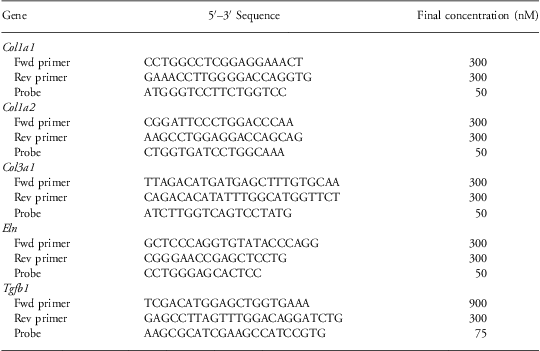
Following the extraction of total RNA using an RNeasy Mini Kit (QIAGEN, Doncaster, ViC., Australia), 1 μg of RNA was reversed transcribed into cDNA using the Taqman reverse transcription reagents kit (Applied Biosystems, Scoresby, ViC., Australia). Real-time PCR was performed in the StepOne™ Real-Time PCR system (Applied Biosystems) and relative mRNA levels [in arbitrary units (AU)] analysed using the cycle of threshold fluorescence (C t) method as reported previously.Reference Singh, Cullen-McEwen and Kett18 Genes of interest were either purchased as assay on demand (AOD; Applied Biosystems; FAM-labelled probe) or custom designed. Genes purchased as AOD include surfactant protein (SP)-A, (Sftpa1, assay ID: Rn00824545_m1), SP-B (Sftpb, assay ID: Rn00684778_m1), SP-C (Sftpc, assay ID: Rn00569225_m1), SP-D (Sftpd, assay ID: Rn00563557_m1), B-cell CLL/lymphoma 2 (Bcl2, assay ID: Rn99999125_m1) and BCL-2-associated protein X (Bax; assay ID: Rn01480159_g1), aquaporin 1 (Aqp1; assay ID: Rn00562834_m1), Aqp3 (assay ID: Rn00581754_m1), Aqp5 (assay ID: Rn00562837_m1), epithelial sodium channel (ENaC)-α (Scnn1a, assay ID: Rn00580652_m1), ENaC-β (Scnn1b, assay ID: Rn00561892_m1) and ENaC-γ (Scnn1 g, assay ID: Rn00566891_m1). Custom-made genes of interest [elastin (Eln), collagen type I α1 (Col1a1) and α2 (Col1a2), collagen type III α1 (Col3a1), transforming growth factor (TGF)-β1 (Tgfb1)], are shown in Table 1. Each gene of interest was multiplexed with the house-keeping gene, 18 s rRNA (VIC-labelled probe; TaqMan® Ribosomal RNA Control Reagents kit, Applied Biosystems). For each reaction, mRNA levels of the gene of interest were expressed relative to 18 s rRNA, as gene expression of 18 s rRNA did not differ between treatment groups. Reactions contained 50ng of cDNA, 1× mastermix (2× Taqman Universal PCR Mastermix; Applied Biosystems), gene of interest (final concentration of 0.5× AOD or as shown in Table 1), 20 nM 18 s forward primer, 80 nM 18 s reverse primer and 50nM 18 s probe (final volume of 20 μl); for Sftpa1, Sftpb, Sftpc and Sftpd 2ng cDNA and 0.1× AOD were used.
Custom-designed primers and probes used in gene expression studies

Protein expression
Total protein was extracted from 25 to 50 mg of lung taken from 8- and 19-month-old control and EtOH-exposed males. A volume of standard RIPA lysis buffer equal to 10× the volume of tissue was added and tissues homogenized with an IKA Ultra-Turrax T8 homogenizer (Labtek, Brendale, Australia). Using a temperature of 4°C, the homogenates were shaken for 50 min and centrifuged at 11,000 r.p.m. for 20 min. The supernatant was collected and assayed for total protein content using the Bicinchoninic Acid Protein assay kit (Sigma-Aldrich Pty. Ltd., Sydney, Australia). Thirty μg of protein per sample (n = 5–7 from each treatment group at each age) were subjected to SDS-PAGE (4% stacking, 12% resolving) and the proteins transferred to an Immobilon® FL PVDF membrane (Merck Millipore, Kilsyth, Australia). The membrane was blocked for 1 h at 4°C with 3% fish gelatin in 1× PBS (Sigma-Aldrich Pty. Ltd.) before 4°C incubations with primary antibodies (overnight with rabbit polyclonal anti-SFTPB (1:2,500, Abcam, Cambridge, UK, Cat # ab40876) or rabbit polyclonal anti-SFTPD (1:1,000, Abcam, Cat # ab101703), plus 1 h with mouse polyclonal anti-β-actin antibody (endogenous control), 1:10,000 (Sigma-Aldrich Pty. Ltd., Cat # A1978)). Following washes with 1× PBS containing 0.1% Tween-20, the membrane was incubated at room temperature for 1 h with the secondary antibodies goat anti-rabbit (LI-COR IRDye 800; Millennium Science, Surrey Hills, Australia) and goat anti-mouse (LI-COR IRDye 680; Millennium Science). The levels of SFTPB (42 kDa), SFTPD (43 kDa) and β-actin (42 kDa) were analysed via densitometry using the LI-COR Odyssey infrared imaging system (Millennium Science) using the 800 nm (SFTPB and SFTPD) and 700 nm (β-actin) wavelengths. After accounting for background, the ratios of SFTPB and SFTPD to β-actin were calculated to determine protein levels.
Histological analyses
The trachea was removed from fixed lungs and the lungs processed to paraffin. The PN1 lung was embedded whole, whereas the 10-month-old lung was separated into the left and right lung and sectioned into 1 cm thick blocks. One block from the upper, middle and lower section of the 10-month-old right lung was processed to and embedded in paraffin. Lungs of both ages were sectioned at 5 μm.
Stereological analyses
At PN1, five sections ∼130 μm apart were mounted onto Superfrost Plus slides (Thermo Fisher Scientific Inc., ViC., Australia), stained with H&E and photographed using the Aperio ScanScope XT (Aperio Technologies Inc., CA, USA). Resulting images were viewed using ImageScope software (version 10.2.2.2319; Aperio Technologies Inc.) and morphological measurements of airspace volume and distal airspace diameter made with the aid of test gridsReference Weibel19 superimposed over scanned images of the lungs at a magnification of ×100. The calculation for airway diameter was modified by multiplying it by the airspace fraction because the calculation assumes a minimal and consistent contribution of airway wall thickness to the linear intercept measurement. These measurements were made from six fields of view per tissue section, one from each of the upper, middle and lower section of the left and right lung (total of 30 fields of view/animal).
Pulmonary collagen deposition
At 10 months, one 5 μm section per block was mounted onto a Superfrost Plus slide (Thermo Fisher Scientific Inc.) and stained with the Masson's Trichrome stain to view total collagen deposition. Three blocks (one each from the upper, mid and lower region of the right lung) were examined from each animal. The slides were then photographed using the Aperio ScanScope XT (Aperio Technologies Inc.) and the resulting images viewed using ImageScope software (version 10.2.2.2319; Aperio Technologies Inc.). Deposition of collagen was assessed using a scoring system based on the degree of Trichrome (blue) staining using a protocol modified from La Saux et al.Reference Le Saux, Teeters and Miyasato20 An investigator blinded to the coding of the samples scored 10 fields of view per section at ×200 magnification per region of lung (control, n = 9; EtOH, n = 6) using a grading scale. The grading scale for collagen deposition was: 1 – low basal level of staining across some of the lung section; 2 – low level staining across most/all of the lung section; 3 – high level of staining across some of the lung section; and 4 – high level of stain across most/all of the lung section. A final score for each section was determined using the average value of each of the 10 fields of view from each lung region.
Statistical analyses
Data obtained from E20 and PN1 offspring were analysed using two-way ANOVA with sex and treatment as factors. Where P < 0.05, a Tukey post-hoc analysis was performed. Data obtained from adult offspring were analysed with two-tailed Student's t-test comparing treatments with each sex; in the case of a failed normality test, a Mann–Whitney rank sum test was used. Analysis of Trichrome staining was performed using a Mann–Whitney rank sum test. Statistical analyses were performed using the statistical package SigmaStat (Systat Software, San Jose, CA, USA). Values are expressed as mean ± s.e.m. and the level of significance taken as P ⩽ 0.05.
Results
Maternal parameters
The effect of daily ad libitum 6% (v/v) EtOH consumption by the pregnant dam has been described in detail previously.Reference Probyn, Zanini, Ward, Bertram and Moritz16 Briefly, this type of EtOH consumption did not alter maternal weight gain, dietary consumption, caloric intake or gestational outcome. On average, gestational length was 22.5 ± 0.5 days for both groups, litter size was 10 ± 1 pups for control dams and 11 ± 1 for EtOH-fed dams and the ratio of male to female offspring was ∼1:1. Of the 39 litters born to control dams, there were two in utero deaths. For EtOH-fed dams, there were three in utero deaths from 38 litters.
Body and lung weight and volume
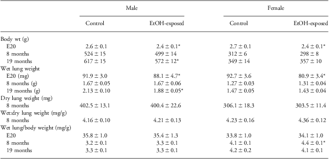
At E20, male and female EtOH-exposed fetuses were significantly lighter than controls (P = 0.002; Table 2). At 8 months of age, there was no difference in body weight between male and female groups, although both male and female EtOH-exposed offspring were 5% lighter than controls (Table 2). At 19 months of age, male, but not female, EtOH-exposed offspring were significantly lighter than same-sex controls (P = 0.027).
Body and lung weight of Control and ethanol (EtOH)-exposed offspring
E, embryonic day.
Data is mean ± s.e.m.
*P < 0.05 compared with same-sex control.
Although EtOH-exposed fetuses had significantly lighter lungs than controls (P = 0.048), lung weight at E20 was appropriate for body size (Table 2). For the 8-month-old offspring, neither absolute nor relative wet lung weights were different between male groups. Similarly, dry lung and wet-to-dry lung weights were similar between male EtOH-exposed and control offspring. For 8-month-old female offspring, although there was no difference in absolute lung weight, the relative lung weight of EtOH-exposed offspring was heavier than that of controls (P = 0.014; Table 2). However, dry lung and wet-to-dry lung weights were similar between EtOH-exposed and control female offspring. At 10 months, lung volume at 20 cmH2O was not different between male or female groups (male control, 12.84 ± 1.18 ml; male EtOH-exposed, 12.47 ± 0.42 ml; female control, 10.75 ±0.62 ml; female EtOH-exposed, 10.84 ± 0.53 ml). For 19-month-old male EtOH-exposed offspring, absolute, but not relative, lung weight was lighter than controls (P = 0.023); there was no difference in either absolute or relative lung weight between 19-month-old female EtOH-exposed and control groups.
Early pulmonary development and maturation
E20 mRNA levels
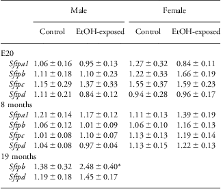
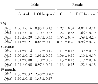
Surfactant protein mRNA levels were similar between groups of each sex (Table 3). However, the Aqp1, but not Aqp3 or Aqp5, mRNA level was downregulated (P = 0.042) in EtOH-exposed fetuses (Aqp1 – Fig. 1a; Aqp3 and Aqp5 – Supplemental Table S1). Although not significant, the Scnn1a mRNA level also tended to be reduced (P = 0.106) by EtOH exposure (Fig. 1b). There were no differences in Eln, Col1a1, Bcl2 and Bax mRNA levels between EtOH-exposed and control offspring of either sex (Supplemental Table S1). Although not reaching statistical significance, Tgfb1 mRNA levels of EtOH-exposed fetuses were decreased for both sexes (male EtOH-exposed 0.99 ± 0.16 AU v. control 1.21 ± 0.17 AU and female EtOH-exposed 0.80 ± 0.11 AU v. control 1.16 ± 0.21; P treatment = 0.086).
Surfactant protein mRNA levels in control and ethanol (EtOH)-exposed fetuses and offspring
E, embryonic day.
Data are expressed in arbitrary units as mean ± s.e.m.
*P ⩽ 0.05.
mRNA levels of pulmonary (a) aquaporin 1 (Aqp1) and (b) epithelial sodium channel alpha (Scnn1a) for control and ethanol (EtOH)-exposed fetuses at Embryonic day (E) 20. Data are expressed as mean ± s.e.m. *P⩽0.05.
PN1 airspace volume and airway diameter
At the level of the light microscope, the histological appearance of the lung did not appear different between EtOH-exposed and control groups (Fig. 2). The percentage of airspace volume was similar in male control (79.4 ± 0.5%), male EtOH-exposed (79.4 ± 0.8%), female control (80.2 ±0.6%) and female EtOH-exposed (80.3 ± 0.6%) offspring. Airway diameter was also similar between male control (128.1 ± 3.5 μm), male EtOH-exposed (125.7 ± 2.4 μm), female control (131.2 ± 4.6 μm) and female EtOH-exposed (131.7 ± 4.1 μm) offspring.
Histological representations of the lung taken from male offspring at postnatal day 1. The development of the lung was not different between (a) control and (b) ethanol-exposed offspring. Sections are stained with H&E; scale bar shows 100 μm.
Adult pulmonary structure and non-respiratory function
Pulmonary gene expression was measured in male and female offspring at 8 months of age. However, as no changes were found between female groups, only data generated from male offspring are shown below. Results from female offspring are provided in Supplementary Table S2. From 8 months onwards, only male offspring were studied.
Gene expression
At 8 months, there was no difference in surfactant protein mRNA levels between male control and EtOH-exposed offspring (Table 3 and Fig. 4a). Of the pulmonary structural genes analysed at 8 months (Eln, Col1a1, Col1a2, Col3a1, Bcl2, Bax, Tgfb1), only Col3a1 was altered by chronic 6% prenatal EtOH exposure, being increased by almost 50% (P = 0.022; Fig. 3a). The mRNA levels of the ENaCs (Scnn1a, Scnn1b and Scnn1 g) and Aqp5 were not different between groups (Supplementary Table S2).
mRNa levels of pulmonary surfactant protein B (Sftpb) in lung tissue from (a) 8-month-old and (b) 19-month-old control and ethanol (EtOH)-exposed offspring. Protein levels of pulmonary surfactant protein B (SFTPB) (normalized to the housekeeping protein, β-actin) in lung tissue from (c and e) 8-month-old and (d and ƒ) 19-month-old control and EtOH-exposed offspring. Data are expressed as mean ± s.e.m. *P ⩽ 0.05.
mRNA levels of (a) Collagen 3a1 (Col3a1) in the lung taken from 8-month-old male offspring. Pulmonary collagen content at 10 months of age was visualized using trichrome staining of lung sections taken from male (b) control and (c) ethanol (EtOH)-exposed offspring, and the intensity of trichrome staining semi-quantified (d) to determine the degree of collagen deposition. Scalebar represents 100 μm. Bars for all graphs are mean ± s.e.m. *P ⩽ 0.05.
At 19 months, although the Sftpd mRNA level was not altered by prenatal EtOH exposure, the Sftpb mRNA level was increased by 80% (P = 0.050; Table 3 and Fig. 4b). The mRNA levels for Eln, Col1a1, Col1a2, Col3a1 and Aqp5 were not different between male groups (Supplementary Table S3).
Protein levels
Pulmonary SFTPD protein levels were not different between control and EtOH-exposed offspring at 8 or 19 months of age (data not shown). In contrast, with prenatal EtOH exposure SFTPB protein levels tended to be elevated (P = 0.09; Fig. 4c and e) at 8 months and were significantly decreased (−24%) at 19 months (P = 0.010; Fig. 4d and 4f).
Lung histology
Lung sections taken from male EtOH-exposed animals appeared to contain more collagen staining than those from controls. Representative sections are shown in Fig. 3b and 3c. A semi-quantification analysis of the distal pulmonary tissue showed that EtOH-exposed males had increased collagen deposition compared with controls (P = 0.04; Fig. 3d).
Discussion
Few studies have explored effects of low-moderate amounts of prenatal alcohol exposure on the lungs. For this study, we have used a rat model of chronic, low-moderate dose (6% v/v) prenatal EtOH exposure to examine whether it alters structural and non-respiratory aspects of the pulmonary system. We found that maternal consumption of EtOH to produce a plasma EtOH concentration of ∼0.03% (30 mg/dl) had little impact on the structure of the developing lung. However, at E20, prenatal alcohol treatment downregulated Aqp1 and caused a non-significant trend for decreased Scnn1a mRNA levels, both of which are genes involved in lung liquid clearance. Postnatally, the pulmonary system of female offspring were unaffected by prenatal EtOH exposure. For adult male EtOH-exposed rats, the mRNA level of Col3a1 was upregulated at 8 months, and by 10 months the lungs contained increased collagen deposition. By 19 months, SFTPB protein levels were decreased by almost 25% despite an 80% increase in Sftpb mRNA. These results suggest that daily fetal exposure to even low-moderate amounts of EtOH can subtly affect the pulmonary system, potentially compromising the respiratory function of adult male offspring.
We have previously reported that chronic low-moderate prenatal EtOH exposure is associated with fetal growth restriction at E20 and slowed postnatal growth in male, but not female, offspring between 7 and 12 months of age.Reference Probyn, Zanini, Ward, Bertram and Moritz16 We now show that these male EtOH-exposed offspring were also lighter than male controls at 19 months of age, demonstrating that the decreased growth of male EtOH-exposed offspring continues throughout adulthood. Previously we demonstrated that although chronic low-dose prenatal EtOH exposure did not alter wet lung weight at 8 months, lung weight relative to body weight of both male and female EtOH-exposed offspring was increased.Reference Probyn, Zanini, Ward, Bertram and Moritz16 In this present study, only 8-month-old EtOH-exposed female offspring had a heavier relative lung weight, however, only a subset of the animals reported in the previous study were included in the present study. As chronic low-moderate dose prenatal EtOH exposure did not alter dry lung weight or ex vivo lung volume, these findings suggested fluid retention within the lungs at post-mortem for 8-month-old female EtOH-exposed rats. As a short (3–6-week) period of alcohol consumption by adult rats has previously been shown to cause pulmonary oedemaReference Guidot, Modelska and Lois21 and reduce the ability of the lung to clear liquid from the airways,Reference Holguin, Moss, Brown and Guidot22 we measured pulmonary gene expression levels of the epithelial sodium channels and aquaporin 5 of 8-month-old offspring. In contrast to the post-mortem finding of increased wet lung weight, the mRNA levels of Aqp5, Scnn1a, Scnn1b and Scnn1 g were unaltered at 8 months by our prenatal treatment, and therefore we must conclude that chronic low-moderate dose prenatal EtOH-exposure does not affect pulmonary fluid homeostasis in adulthood.
Surfactant production is first detected during the cannalicular stage of lung development,Reference Flecknoe, Wallace, Cock, Harding and Hooper23 which begins at ∼E18.5 in rats.Reference Yamada, Suzuki, Gejyo and Ushiki24 We found that at E20 surfactant protein mRNA levels were not different between control and EtOH-exposed fetuses, indicating appropriate biochemical maturation of the lung. In contrast, Sozo et al. Reference Sozo, O'Day and Maritz15 found that Sftpa1, Sftpb and, to some extent, Sftpd mRNA levels were downregulated in the 134-day ovine fetus that had experienced almost 40 days of late gestational exposure to moderate levels of EtOH (ovine gestation ∼140 days; fetal plasma EtOH concentration of 0.11%). In both models, prenatal EtOH exposure began before the onset of surfactant production, and therefore differences are most likely due to the dose of EtOH and blood alcohol concentration reached. For our model, the pattern of maternal EtOH consumption indicates fetuses were exposed 0.03% EtOH,Reference Probyn, Zanini, Ward, Bertram and Moritz16 whereas Sozo et al. measured the fetal plasma EtOH concentration to be 0.11%. Although in the present study surfactant protein mRNA expression levels were unaltered at E20, Aqp1 and Scnn1a mRNA expression were downregulated or tended to be decreased at this age. As all EtOH-exposed pups survived the transition from the liquid-filled fetal lung to the air-filled postnatal lung, decreased expression of these genes at E20 did not compromise lung liquid reabsorption at birth.
Perinatally, we found that pulmonary Eln, Col1a1, Tgfb1, Bax and Bcl2 mRNA levels and the airspace volume and airway diameter of the lung was unchanged by our regime of prenatal EtOH exposure. These findings suggest that low-moderate dose prenatal EtOH exposure from conception to term (maternal plasma EtOH concentration of 0.03% produced through a 15% EtOH-derived calorie diet) does not alter pulmonary organogenesis. In contrast, two acute mid-gestational exposures of the fetal mouse lung to high doses of EtOH (produced through a 25% EtOH derived calorie diet) has been shown to delay the progression of the lung from the canalicular to the saccular stage and increase protein levels of the branching morphogenesis-associated protein, HOXB5, at E18.5;Reference Wang, Gomutputra, Wolgemuth and Baxi14 at this age-branching morphogenesis should be complete.Reference Volpe, Martin, Vosatka, Mazzoni and Nielsen25 In addition, daily exposure of the ovine fetal lung to moderate amounts of EtOH during the last trimester of gestation (producing a fetal plasma EtOH concentration of 0.11%) upregulates pulmonary Col1a1 and Col1a2 mRNA levels and increases the percentage of collagen within the lung.Reference Sozo, O'Day and Maritz15 Together, these three studies suggest that EtOH effects pulmonary organogenesis in a dose- and/or time-dependent manner.
In healthy individuals, pulmonary senescence is associated with decreased lung function and altered lung structure.Reference Meyer26 In humans, decreased birth weight accelerates pulmonary senescenceReference Maritz, Morley and Harding4 and leads to adult onset respiratory diseases such as chronic obstructive airway disease.Reference Lopuhaa, Roseboom and Osmond6, Reference Lucas, Inskip and Godfrey27 Animal studies have demonstrated that prenatal insults such as nicotine exposure (reviewed in Maritz and HardingReference Maritz and Harding28) and placental insufficiencyReference Maritz, Cock, Louey, Suzuki and Harding3 also retard fetal growth and decrease adult respiratory health. As alcohol abuse in adulthood is a contributing factor for acute respiratory distress syndrome,Reference Moss, Bucher, Moore, Moore and Parsons29 we thought it possible that prenatal EtOH exposure would accelerate pulmonary senescence. For male EtOH-exposed offspring, Col3a1 mRNA was upregulated at 8 months of age, leading to significantly increased collagen deposition at 10 months of age. These findings contrast those of Sozo et al. who reported upregulated Col1a1 and Col1a2 mRNA expressions and an increased percentage of collagen in the ovine fetal,Reference Sozo, O'Day and Maritz15 but not postnatal,Reference Sozo, Vela and Stokes30 lung following daily late-gestational moderate dose EtOH exposure. Whereas the authors of those studies in young lambs suggest that pulmonary repair occurs upon the removal of EtOH, our findings from adult rats suggest that prenatal EtOH exposure contributes to increased lung collagen deposition in the adult. The functional consequences of this are unknown and warrant further investigation; however, we speculate respiratory function would have been compromised for these male EtOH-exposed rats. Lung function studies are necessary to determine whether chronic low-moderate prenatal EtOH exposure does in fact decrease adult respiratory health or accelerate pulmonary senescence. Furthermore, as cigarette smoking causes pulmonary fibrosis in humans,Reference Katzenstein31 it would be interesting to determine whether cigarette smoke/nicotine exposure of the adult rat lung that was prenatally exposed to EtOH provides a ‘second hit’ that exacerbates the alterations in pulmonary collagen observed in the current study, and induces an emphysema-like phenotype.
The bronchoalveolar lavage fluid of adults suffering from acute respiratory distress syndrome is SP-B deficient.Reference Greene, Wright and Steinberg32, Reference Gregory, Longmore and Moxley33 Using adult compound SP-B−/− mice, Melton et al. demonstrated that a 75% reduction in SP-B is lethal, increasing the pulmonary surface tension and decreasing hysteresis.Reference Melton, Nesslein and Ikegami34 In the current study, we found that 19-month-old male rats exposed to low-moderate amounts of EtOH throughout gestation had a 25% reduction in pulmonary tissue SP-B protein levels. As SP-B, with SP-C, increases phospholipid uptake into type II alveolar epithelial cells (reviewed in GrieseReference Griese35), it is likely that male EtOH-exposed offspring of the current study had decreased pulmonary surfactant within the airways. SP-B is also responsible for the rapid absorption of surfactant at the air-liquid interface.Reference Griese35 Thus, our finding of decreased SP-B in pulmonary tissue taken from aged (19 month) male EtOH-exposed offspring suggests a compromised surfactant system and this, together with the increased collagen deposition seen at 10 months, suggests that pulmonary function becomes compromised with age.
In conclusion, we have demonstrated that although daily fetal exposure to low-dose EtOH results in fetal growth restriction it does not alter pulmonary development. However, for male but not female offspring, chronic low-moderate dose prenatal EtOH exposure increases pulmonary collagen deposition in adulthood and decreases pulmonary tissue surfactant protein B levels in old age. We suggest that such alterations may lead to pulmonary fibrosis and decrease adult respiratory function. Along with our recent findings that low-dose prenatal ethanol in the rat alters glucose homeostasisReference Probyn, Parsonson and Gardebjer36 and induces an anxiety-like phenotypeReference Cullen, Burne, Lavidis and Moritz37 in the adult, this work highlights that even low-moderate maternal alcohol consumption during pregnancy can result in long-term detrimental outcomes for offspring.
Acknowledgements
The authors are grateful for the assistance of Harry Davis, Chelsea Stewart, Emma-Kate Lock, Lauren Wilson, Karrona Tep and Carlie Cullen in these studies.
Financial support
This work was supported by the National Health and Medical Research Council of Australia (grant ID511162) and The University of Queensland Early Career Researcher Grant (author M.E. Probyn).
Conflicts of Interest
None.
Ethical standard
The authors assert that all procedures contributing to this work comply with the Australian Code of Practice for the Care and Use of Animals for Scientific Purposes (7th edition, National Health and Medical Research Council, 2004) and use of animals has been approved by the Animal Welfare Unit of The University of Queensland.
Supplementary materials
To view supplementary material for this article, please visit http://dx.doi.org/10.1017/S2040174413000305