The healthcare environment is a reservoir for multidrug-resistant bacterial organisms (MDROs) that contribute to risk for healthcare-associated infections (HAIs). Prior studies have highlighted specific sites in the healthcare environment as reservoirs for MDROs. Surface contamination with MDROs frequently occurs on high-touch objects (HTOs) and within bathroom and wastewater sites in hospitals. Reference Buchan, Graham and Lindmair-Snell1–Reference Regev-Yochay, Smollan and Tal8 In the case of Clostridioides difficile, this contamination has been shown to contribute directly to infection risk: patients occupying rooms where the previous inhabitant was infected with C. difficile are at increased risk of this infection, and surface contamination has been shown to precede acquisition. Reference Chen, Knelson and Gergen9–Reference Shaughnessy, Micielli and DePestel11
The persistence of MDROs and HAI-associated bacteria in the healthcare environment depends on the deposition of bacteria from patients and healthcare workers, environmental features that sustain bacterial growth, and on environmental cleaning practices. Reference Weber, Anderson and Rutala12 Studies of the hospital microbiome have demonstrated bidirectional exchange between environmental surfaces and patient skin, with increased similarity over time. Reference Lax, Sangwan and Smith13 However, the hospital environment is not uniform. Because deposition, environmental surface selection, and cleaning practices vary across sites within a single hospital room, the environmental microbiome may likewise vary across close sites. We hypothesized that the competing distance between the source patient and the wastewater reservoirs may serve as an effective means to model bacterial colonization of the hospital room environment.
To define the impact of distance from patient and wastewater sources (ie, sinks and toilets) on the specific MDROs colonizing the healthcare environment, we performed a prospective cohort study with systematic sampling and MDRO culture of rooms occupied by patients from whom an MDRO had been isolated. To further discern the relationship between culturable MDROs and bacterial DNA deposition in the healthcare environment, we complemented our systematic, bacterial culture with 16S rRNA gene sequencing of the environmental samples.
Materials and methods
Study design, setting, and population
We performed a prospective cohort study at the Hospital of the University of Pennsylvania (HUP), an academic medical center in Philadelphia, Pennsylvania. We enrolled hospital rooms of newly admitted patients who had had a positive clinical or surveillance culture for an eligible MDRO within the prior 30 days (see Table 1 for eligible MDROs) between November 4, 2019, and March 12, 2020. We performed longitudinal, comprehensive bacterial culture to detect MDRO colonization in the environment (eligible for sampling on days 1, 2, 4, 6, 14, 21, and 28 unless discharged or transferred from their room). We sampled 3 environmental zones in each room corresponding to distances from the patient’s bed: (1) near patient, comprising the patient bed railing, the television remote control, the nurse call button, and the overbed tray table; (2) intermediate distance, comprising the intravenous fluid pump control pad, the supply cart keypad, the in-room computer keyboard and mouse; and (3) sites distant from the patient and in close proximity to wastewater sites, comprising the light switch, doorknob, toilet handle and seat, as well as the handles and basin of the bath and/or in-room sinks. In total, 51 participants were enrolled, and 136 sampling events performed, generating 408 samples. Approval was granted by the University of Pennsylvania Institutional Review Board (IRB protocol no. 833724).
Recovery of MDROs from the Hospital Environment a

Note. MDRO, multidrug-resistant organism; CR, carbapenem resistant; ESBL/CR, extended-spectrum β-lactamase (ESBL)–producing Enterobacterales, carbapenem-resistant (CR) Enterobacterales; VRE, vancomycin-resistant Enterococcus; MRSA, methicillin-resistant Staphylococcus aureus.
a A comprehensive environmental sampling and selective culture strategy captured 11 MDROs. For each MDRO, the table presents the total number of times they were recovered from the healthcare environment, and the frequency with which they were detected from cultured environmental specimens.
Causal model
We hypothesized that the physical distance from patient and wastewater sources of MDROs defines MDRO colonization of the healthcare environment. We further hypothesized that the bacterial DNA deposition in the environment reflects environmental contamination with bacteria and risk for persistent MDRO colonization.
Specimen collection and MDRO culture
Upon subject enrollment, environmental surfaces were sampled longitudinally until discharge from the enrolling ICU room, or day 28. Two composite samples were obtained from each in-room area (near, intermediate, and far), with a sponge stick (3M, New Ulm, MN) used for bacterial culture and a flocked swab (Copan Diagnostics, Carlsbad, CA) for microbiome analysis. Sampling sites and sampling procedures were standardized with an approximate surface area of < 350 cm2 per site for each sponge stick and 50 linear cm per site for each flocked swab. Sponge-stick specimens for bacterial culture were collected in stomacher bags to which phosphate-buffered saline with 0.02% Tween (PBST) was added. This mixture was processed using a Seward Stomacher mechanical homogenizer (Seward, West Sussex UK). After centrifugation, the cell pellet was plated on 6 selective media, including BD MRSA Chromagar (Becton Dickinson, Franklin Lakes, NJ), Hardy Chromagar CRE (Hardy Diagnostics, Santa Maria, CA), CHROMagar KPC (CHROMagar, Paris, France), Hardy Chromagar ESBL, Thermo SPECTRA VRE (Thermo Fisher Scientific, Waltham, MA), and Anaerobe Systems CCFA-HT (Anaerobe Systems, San Martin, CA). Cultures were incubated aerobically for 18–24 hours at 35–37°C, with the exception of Anaerobe Systems CCFA-HT which was incubated in anaerobic conditions for 18–48 hours. All bacterial colonies with appropriate morphology and chromogenic features were subcultured to blood agar and confirmed by MALDI-TOF with the exception of methicillin-resistant Staphylococcus aureus (MRSA). Due to the inability to recover isolates after a laboratory shutdown during the COVID-19 pandemic, we were unable to assess recovery of 1 MDRO from 3 specimens, and we were unable to assess recovery of 6 MDROs from 3 specimens. The presented data and models account for these missing cultures.
16S rRNA gene sequencing, quantitative PCR, and bioinformatics
A subset of samples (123 samples from 16 participants) were included for microbiome analysis, representing the earliest samples collected and processed prior to the COVID-19 pandemic. Parallel surface samples were collected with flocked nylon swabs and were then subjected to 16S rRNA gene sequencing and microbiome analysis. Swabs were frozen at −80°C prior to DNA extraction and sequencing. DNA extraction was performed using MoBio PowerSoil DNA isolation kit (Qiagen, Hilden, Germany). The V1–V2 hypervariable region of the 16S rRNA gene was amplified using barcoded primers 27F (5′AATGATACGGCGACCACCGAGATCTACACTATGGTAATTGTAGAGTTTGATCCTGGCTCAG-3′) and 338R (5′CAAGCAGAAGACGGCATACGAGATNNNNNNNNNNNNAGTCAGTCAGCCTGCTGCCTCCCGTAGGAGT-3′). Thermocycler conditions were as follows: 5 minutes at 95°C, 30×(30 seconds at 95°C, 30 seconds at 56°C, 90 seconds at 72°C), and 8 minutes at 72°C. The 16S rRNA gene sequencing was performed with the Illumina MiSeq platform (Illumina, San Diego, CA) with 250-bp paired-end reads, permitting high-quality coverage of the 316-bp V1–V2 amplicon. Amplicon sequence variants (ASVs) were identified using DADA2 within the Quantitative Insights Into Microbial Ecology 2 (QIIME2) pipeline. Reference Callahan, Wong and Heiner14,Reference Bolyen, Rideout and Dillon15 Genus-level taxonomic assignment was performed via comparison to the Greengenes reference database, Reference McDonald, Price and Goodrich16 using the naive Bayes classifier implemented in scikit-bio. Reference Bokulich, Kaehler and Rideout17 To account for the compositional nature of microbiome measures, which may confound the analysis of relationships between bacterial community members, the centered log-ratio transform was applied to genus-level abundances prior to modeling. Reference Gloor, Macklaim, Pawlowsky-Glahn and Egozcue18 The 16S rRNA gene quantitative PCR (qPCR) also targeted the V1-V2 hypervariable region, with primers BSF8 (5’-AGAGTTTGATCCTGGCTCAG-3’) and BSR357 (5’-CTGCTGCCTYCCGTA-3’) and probe 5’-TAACAC ATGCAAGTCGA-3’. The standard curve was generated with a plasmid containing the 16S rRNA gene derived from Streptococcus.
Distance data collection
Distances from the environmental sampling site to the patient bed were recorded using a laser measuring device (Bosch GLM 20 laser measure, Bosch, Los Angeles, CA). Researchers positioned the laser measure at the head of the bed within the room and recorded distances to the in-room computer workstation, the medication cart, IV pole, and bathroom door. The intermediate surfaces were 2.04 m (interquartile range [IQR], 1.88–2.19 m) from the patient, and the far surfaces were 2.81 m (IQR, 2.37–3.04 m) from the patient. Surfaces in the near group were approximately 0.5 m from the head of the patient’s bed.
Definition of exposures and outcomes
The primary exposure of interest was the distance from the patient (measured toward the wastewater sites). The primary outcome of interest was the detection of a culturable MDRO, including MRSA, C. difficile, vancomycin-resistant Enterococcus (VRE) spp, extended-spectrum β-lactamase (ESBL)–producing Enterobacterales, carbapenem-resistant (CR) Enterobacterales, Acinetobacter spp, or Pseudomonas spp. Secondary outcomes of interest included the detection of MDRO genera by 16S rRNA gene sequencing. Clostridioides difficile was included within the primary outcome of MDRO detection due to its importance as an HAI and prior evidence supporting environmental transmission.
Statistical analysis
Data were organized using R statistical software version 3.6.1, 19 and plots were generated using the ggplot2 package. Reference Wickham20 We examined the relationship between distance from the patient (toward the wastewater sites) and the probability of MDRO detection using a Gaussian process model (exponentiated-quadratic kernel) for distance, with models fit separately for each of 11 detected MDROs. The same Gaussian process model evaluated the relationship between distance and the detection of MDRO genera. We also evaluated a mixed-effects model incorporating a random effect for participants to account for clustering of longitudinal data. To understand the relationship between bacterial DNA contamination and risk for MDRO detection, we used a mixed-effects model with random effects for genus and sampling site. Models were fit using Stan Hamiltonian Monte Carlo (HMC) version 2.21, via the brms package with default, weakly informative priors. Reference Carpenter, Gelman and Hoffman21,Reference Bürkner22 Prior predictive modeling was performed, and models were fit with 4 chains of 1,000 iterations, confirmed with HMC diagnostics (no divergent iterations, Rhat statistic <1.1 for all parameters, and E-BFMI >0.2). Reference McElreath23–Reference Gelman, Vehtari and Simpson25 We examined parameter distributions at 50%, 80%, and 95% posterior credible intervals to understand the relationship between exposure and outcome variables.
Power and sample size
We anticipated that the enrollment of 50 participants would permit 85% power to detect a difference of 20% in MDRO culture recovery between near and far specimen collection sites, with type 1 error <0.05.
Availability of data
Sequence data have been uploaded to the National Center for Biotechnology Information’s Sequence Read Archive (NCBI SRA): PRJNA689547. Data, analysis scripts, and model code are available at https://github.com/bjklab.
Results
Detection of MDROs in the proximity to patient-associated MDROs
We enrolled 51 patient rooms at the time of admitting a patient who had been recently diagnosed with MDRO colonization or infection by clinical culture and sampled the rooms repeatedly, obtaining a median of 3 sampling events per patient room (IQR, 2–4). Prior to patient entry, 75% of rooms (n = 38) underwent terminal cleaning with ultraviolet germicidal irradiation (UVGI). All rooms received daily cleaning using Oxivir Plus, a hydrogen-peroxide cleaning product, with the exception of patients in C. difficile isolation whose rooms were cleaned with bleach. The most common enrolling isolate for patients included in the study was C. difficile (n = 16, 31%), followed by ESBL-producing Enterobacterales (ESBL) (n = 15, 29%), MRSA (n = 13, 25%), VRE spp (n = 7, 14%), and CR gram-negative organisms (n = 4, 8%). Across 51 participants, we performed 136 sampling events. At each event, we collected composite samples from 3 in-room sites (near, intermediate, and far), and each composite sample was plated on 6 MDRO-selective media. Table 1 shows the 11 MDROs identified across all enrolled participants, the total number of times they were recovered from the healthcare environment, and the frequency with which they were detected from cultured environmental specimens.
Impact of distance from patient on MDRO detection by bacterial culture
We related the probability of MDRO detection by selective culture to the distance from the patient and toward the wastewater sites, using mean distances from each of the 3 in-room sites sampled at each sampling event. Figure 1 depicts the impact of distance from patient on each of the 11 MDROs encountered in across our cohort. The probability of detecting ESBL and CR gram-negative organisms, including Enterobacterales, Acinetobacter spp, and Pseudomonas spp, increased with distance from the patient. In contrast, C. difficile and MRSA were more likely to be detected in proximity to the patient and less likely to be detected in the wastewater sites. Notably, VRE were distributed more evenly across all areas. Given the patterns of MDRO detection observed at the genus level, we also examined the relationship between aggregate gram-negative or gram-positive MDROs and the distance from patient (Supplementary Fig. 1 online). This analysis confirmed the substantial increase in gram-negative MDRO detection with increasing distance from the patient (and toward the wastewater site), with the opposite pattern observed among gram-positive MDROs.
Impact of distance from the patient on detection of multidrug-resistant organisms (MDROs) in the healthcare environment. Distance from the head of the patient’s bed and towards the wastewater sites is shown on the horizontal axis. The vertical axis depicts the probability of MDRO detection by selective culture for each of 11 detected MDROs according to a logistic regression model incorporating a Gaussian process prior for distance. The black line shows the best estimate, and shades of red indicate 50%, 80%, and 95% posterior credible intervals. Note. ESBL, extended-spectrum beta-lactamase; CR, carbapenem-resistant; VRE, vancomycin-resistant Enterococcus; MRSA, methicillin-resistant Staphylococcus aureus.

Impact of distance from patient on bacterial DNA deposition
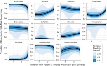
We were able to complement comprehensive, culture-based surveillance with 16S rRNA gene sequencing to detect bacterial DNA deposition in the healthcare environment in a subset of 123 specimens from 16 study participants. In Figure 2, we compare the impact of distance from patient on the bacterial DNA reads assigned to the 11 genera from which MDROs were cultured. We found that Enterobacteriaceae, including Citrobacter spp, Enterobacter spp, Klebsiella spp, and Raoultella spp, exhibited a similar pattern as bacterial cultures, increasing with distance from the patient and in proximity to the wastewater sites. In contrast, bacterial DNA assigned to Acinetobacter spp and Pseudomonas spp were distributed more evenly across all areas, though bacterial culture was more likely to detect these genera in the wastewater sites. Bacterial DNA reads assigned to Staphylococcus spp were also distributed evenly across all areas, though bacterial culture was more likely to detect MRSA in close proximity to patients.
Impact of distance from the patient on bacterial DNA deposition in the healthcare environment. Distance from the head of the patient’s bed and toward the wastewater sites is shown on the horizontal axis. The vertical axis depicts the probability of bacterial genus detection by 16S rRNA gene sequencing for genera corresponding to each of 11 detected multidrug-resistant organisms (MDROs) according to a logistic regression model that incorporates a Gaussian process prior for distance. The black line shows the best estimate, and shades of blue indicate 50%, 80%, and 95% posterior credible intervals.

Relation between bacterial DNA deposition and MDRO detection in the healthcare environment
To evaluate the relation between bacterial DNA contamination detected by untargeted 16S rRNA gene sequencing and environmental MDRO risk, we performed mixed-effects regression to determine the association between genus-level bacterial DNA abundance and the probability of MDRO detection across all observed MDRO genera and sampling sites. A positive association was observed across multiple genera and sites, but the only association that achieved high posterior certainty was Pseudomonas spp in the far-from-patient site, finding that as the probability of detecting Pseudomonas DNA increased, so too did the probability of culturing CR Pseudomonas spp at the site (odds ratio [OR], 1.47; 95% confidence interval [CI], 1.09–1.99). We further examined how bacterial community features, defined by 16S rRNA gene sequencing and qPCR, changed with distance from the patient and wastewater sites, as well as the association between bacterial community features and MDRO detection. We found that total bacterial abundance (16S rRNA gene copies per cm2 of surface area measured by qPCR) increased with proximity to wastewater sites, but the increase was small (1,466 copies per cm2; 95% CI, 670–2454 copies per cm2). Bacterial community Shannon diversity and richness (number of ASVs) did not change significantly with distance (Fig. 3). The only association between a bacterial community feature (ie, total bacterial abundance, diversity or richness) and probability of MDRO detection that achieved high posterior certainty was the association between total 16S rRNA gene copies and Pseudomonas detection (OR, 1.19; 95% CI, 1.03–1.41).
Impact of distance from the patient on total bacterial abundance, bacterial community diversity, and richness. Distance from the head of the patient’s bed and towards the wastewater sites is shown on the horizontal axis. The vertical axis depicts (A) the total bacterial abundance measured by 16S rRNA gene qPCR, (B) the bacterial community diversity measured by the Shannon index, or (C) the bacterial community richness measured by number of ASVs, according to a linear regression model that incorporates a Gaussian process prior for distance. The black line shows the best estimate, and shades of blue indicate 50%, 80%, and 95% posterior credible intervals.

Discussion
We sought to define how bacterial deposition and persistence in the healthcare environment is affected by proximity to patient and wastewater sources, and we found clear distinctions between near-patient and far-from-patient/near-wastewater sites in patient rooms. The probability of detecting ESBL-producing and CR gram-negative organisms, including Enterobacterales, Acinetobacter spp, and Pseudomonas spp, increased with distance from the patient. In contrast, C. difficile and MRSA were more likely to be detected close to the patient and less likely to be detected in the wastewater sites.
We compared culturable MDROs to bacterial DNA deposition to identify areas where bacteria are deposited but are not viable due to either environmental features (eg, microenvironments that do not support bacterial growth or persistence) or cleaning practices. In some cases, the pattern of culturable MDROs matched bacterial DNA deposition: detection of Enterobacterales increased with distance from the patient and in proximity to the wastewater sites, mirroring the overall detection of 16S copies or bacterial abundance. But in other cases, the discrepancy between bacterial DNA detection and culturable MDROs suggested environmental selection. Although bacterial DNA assigned to Pseudomonas and Staphylococcus were distributed evenly across all areas, bacterial culture identified distinct hot spots for CR Pseudomonas spp far from patients and MRSA near patients. Notably, both bacterial DNA assigned to Enterococcus spp and culturable VRE were detected across distances, suggesting that bacterial deposition from stool is extensive. The decreasing probability of C. difficile and decreasing Clostridioides DNA deposition as sampling became closer to the wastewater sites may be attributable to more effective cleaning practices specific to these pathogens within wastewater sites.
Because deposition of bacteria from patients and healthcare workers, environmental features that sustain bacterial growth, and environmental cleaning practices vary within patient rooms, we identified microenvironments as we collected samples along an axis from the patient’s bed toward the wastewater sites. By simultaneously measuring viable MDROs by selective bacterial culture and bacterial DNA deposition by 16S rRNA gene sequencing, and by incorporating distance from patient and wastewater reservoirs, we defined hot spots specific to each MDRO, and we linked those hot spots to specific features of the healthcare environment. Our approach yields new insight into the contamination of surfaces within the hospital rooms and may be used to guide future studies focused on preventing transmission of MDROs through targeted environmental cleaning approaches.
Our study has several limitations. First, sampling sites were divided into 3 composite in-room categories rather than multiple individual specimens. Although this approach limits the specificity of surface-specific contamination, it decreased the resources needed for sampling a greater number of surfaces in total. Second, bacterial culture was limited to only multidrug-resistant pathogens and C. difficile. This approach was taken to limit detection to pathogens of greatest clinical significance within healthcare settings, but it is possible that culturable, non-MDRO bacteria persist in different distributions. Third, culture was nonquantitative to limit complexity of specimen processing. These limitations decrease the specificity of site-specific features but should not reduce the effectiveness of this methodology for modeling contamination of the healthcare space. Fourth, 16S sequencing was used to assign genus-level taxonomic assignment rather than species-level identification, and only a subset of specimens underwent 16S sequencing. Finally, our study was restricted to a single academic medical center. We expect that the environmental MDROs observed would differ at other medical facilities, according to local prevalence. However, we believe that modeling MDRO deposition and persistence using our distance-based approach will yield similar insights.
In conclusion, using distance modeling between the patient bed and wastewater sites is an effective method to identify organism-specific hot spots for bacterial contamination. Although further studies are needed to validate this method, this technique may be useful in identifying a patient-specific approach to room design and environmental cleaning to prevent persistence of contamination and transmission of pathogens to subsequent room occupants.
Supplementary material
To view supplementary material for this article, please visit https://doi.org/10.1017/ice.2021.323
Acknowledgments
We acknowledge 16S rRNA gene sequencing support provided by the PennCHOP Microbiome Center.
Financial support
This research, B.J.K., M.J.Z., and E.L. are supported in part by the CDC Epicenters for the Prevention of Healthcare Associated Infections (cooperative agreement FOA no. CK16-004). B.J.K. is supported by the National Institute for Allergy and Infectious Diseases (grant nos. K23 AI121485 and L30 AI120149). M.J.Z. is supported by the National Institute for Allergy and Infectious Diseases (grant no. K23 AI143925).
Conflicts of interest
J.H.H.’s contributions were made at the listed affiliations, but she is currently employed by, and holds shares in, the GSK group of companies.