Implications
The present study aimed to determine numerous blood variables in slaughter horses in the lairage, immediatelly before stunning and during exsanguination. Horses are social herd animals with the nature of prey animals. When entering the slaughterhouse, they have to cope with several stressors. All blood variables remained unchanged between the lairage and the stunning box, indicating no physiological stress responses to novelty of the pre-slaughter environment and handling. Significant increase of selected blood variables at exsanguination may partially be attributed to stimulation of sympathetic nervous system caused by stunning and bleeding.
Introduction
Stress is a broad term that is not easy to define. On the basis of neuroendocrine response to stressors, it has simply been defined as any event resulting in increased activity of the hypothalamo–pituitary–adrenal axis. The final stage of this activation is the secretion of glucocorticoids (Harbuz and Lightman, Reference Harbuz and Lightman1992). In contrast, stress has been defined as a condition in an animal that results from the action of one or more stressors that may be of either external or internal origin and can be classified as physical, physiological or interoceptive in nature (von Borell, Reference von Borell2001). The stress response is a pattern of behavioural, endocrine, neural, immune, haematologic and metabolic changes directed towards the restoration of homoeostasis (Muir, Reference Muir2004). Consequently, stress can be measured and monitored in terms of physiological and behavioural alterations that indicate the animal's state of well-being or otherwise (Harbuz and Lightman, Reference Harbuz and Lightman1992; von Borell, Reference von Borell2001; Muir, Reference Muir2004).
Before slaughter, including transportation, lairage and handling immediately before slaughter, production animals are exposed to various stressors that can trigger behavioural changes and several physiological responses, such as increased heart rate and concentrations of stress-related hormones (glucocorticoids, catecholamines and beta-endorphins; Grandin, Reference Grandin1997; von Borell, Reference von Borell2001; Terlouw et al., Reference Terlouw, Arnould, Auperin, Berri, Le Bihan-Duval, Deiss, Lefevre, Lensink and Mounier2008). The latter might lead to changes of biochemical, haematological and oxidative stress variables (Mitchell et al., Reference Mitchell, Hattingh and Ganhao1988; Wernicki et al., Reference Wernicki, Urban-Chimel, Kankofer, Mikucki, Puchalski and Tokarzewski2006; Miranda-de la Lama et al., Reference Miranda-de la Lama, Rivero, Chacon, Garcia-Belenguer, Villarroel and Maria2010). Being fear and flight animals, horses can respond to different stressful conditions by displaying behavioural changes that might include strenuous physical activity with muscle damage and oxidative stress (Ishida et al., Reference Ishida, Hobo, Takahashi, Nanbo, Sato, Hasegawa and Mukoyama1999; Winther Christensen et al., Reference Winther Christensen, Kelling and Lindestrøm Nielsen2005; Werner and Gallo, Reference Werner and Gallo2008).
The effects of transport and pre-slaughter handling on meat quality and fluctuations of blood constituents indicative of stress have been studied mostly in cattle (Mitchell et al., Reference Mitchell, Hattingh and Ganhao1988; Cockram and Corley, Reference Cockram and Corley1991; Tadich et al., Reference Tadich, Gallo, Bustamante, Schwerter and van Schaik2005), pigs (Young et al., Reference Young, Rosenvold, Stagsted, Steffensen, Nielsen and Andersen2003; Hambrecht et al., Reference Hambrecht, Eissen, Newman, Smits, den Hartog and Verstegen2005) and sheep (Jarvis and Cockram Reference Jarvis and Cockram1994; Jarvis et al., Reference Jarvis, Cockram and McGilp1996; Miranda-de la Lama et al., Reference Miranda-de la Lama, Rivero, Chacon, Garcia-Belenguer, Villarroel and Maria2010) but less frequently in horses (Stull, Reference Stull1999; Werner and Gallo, Reference Werner and Gallo2008; Micera et al., Reference Micera, Albrizio, Surdo, Moramarco and Zarrilli2010).
Horses are social herd animals with the nature of prey animals. Emotional stimuli such as isolation from the herd and exposure to a novel environment facilitate the release of adrenocorticotropic hormone, cortisol and catecholamines (Alexander and Irvine, Reference Alexander and Irvine1998; Hada et al., Reference Hada, Onaka, Kusunose and Yagi2001). A slaughterhouse presents a multi-factorial stressor for horses. They are separated from the co-specific group, placed into the new, not hierarchical, social structure, and in new environmental conditions that include novel visual, olfactory and auditory stimuli, which all present potential danger for a horse and thus a stressful situation. Stress-related hormones, such as catecholamines and cortisol, and lactate, glucose, creatine kinase and packed cell volume (PCV) have already been shown to increase after stunning in comparison with pre-stunning values (Werner and Gallo, Reference Werner and Gallo2008; Micera et al., Reference Micera, Albrizio, Surdo, Moramarco and Zarrilli2010). However, no information is available on the biochemical, haematological and antioxidant variables in horses during lairage, in the stunning box immediately before stunning and during exsanguination. We have therefore studied changes of serum cortisol and biochemical, haematological and blood antioxidant variables during lairage, in the stunning box immediately before stunning and during exsanguination.
Material and methods
Animals and management
The study was conducted from February to April 2007. A total of 24 Slovenian warm-blooded horses (13 F/11 M), from 2 to 10 years old, with a mean (±s.d.) body weight of 389.7 ± 126.1 kg, were included in the study. Horses were further divided into two groups, a group of young horses that consisted of 13 horses (7 F/6 M, 2 to 5 years old) and a group of old horses that consisted of 11 horses (6 F/5 M, 6 to 10 years old).
The horses included in the study were farm horses. They were taken from their individual stalls, loaded alone into one-horse trailers and transported for between 45 min and 2 h from various parts of Slovenia to a commercial slaughterhouse. Distance between individual farms and slaughterhouse was between 10 and 50 km. The selection criteria for inclusion in this study were that the horses were healthy and in a good body condition, without any lameness, injury or infectious diseases. Sick or injured animals were excluded from the study. During the period of lairage (from 2 to 5 h), horses had free access to water but not to feed. Horses were handled in such a way as to avoid harm or injury. Each horse had enough room to stand up, lie down or turn around. They were in visual contact with slaughterhouse personnel and other horses.
After the lairage, horses were moved to the stunning box, without use of goad, and stunned using a penetrating captive bolt pistol impelled by air (type EFA VB 215, 12 bar, ∅ = 10 mm; Schmid & Wezel, Maulbronn, Germany) and bled by jugular vein sticking. Horses spent maximum 5 min in the stunning box. All horses were stunned with one shot. After stunning, the horses showed paddling movements with their legs. Maximum stun-to-stick interval was 90 s. The following signs of correct stunning were checked: the animal collapses immediately and does not attempt to stand up, respiratory arrest, the body and muscles of the animal become rigid immediately after the shot and the eyelid is open with the eyeball facing straight ahead and is not rotated.
All procedures were conducted in accordance with the European law (EU Directive 93/119/EC) on the protection of animals at the time of slaughter or killing and complied with applicable Slovenian governmental regulations (Animal Protection Act, Official Gazette of the Republic of Slovenia, No. 43/2007).
Collection and preparation of blood samples
Blood samples for determination of serum cortisol and haematological, biochemical and blood antioxidant enzyme variables were collected by venipuncture of the left jugular vein in the lairage pen, 60 min before stunning (lairage), and immediately before stunning (stunning box). At exsanguination, blood samples were collected from the wound at the time of jugular vein sticking. During blood collection in the lairage pen and in the stunning box, horses were gently restrained with a halter. Blood samples collected in the stunning box were collected within 5 min after leaving the lairage.
Blood samples for the determination of serum cortisol and biochemical profile, with the exception of lactate and glucose, were collected into serum separator tubes (Vacuette; Greiner Bio-One, Kremsmunster, Austria) and stood for 30 min to clot before centrifugation at 1300 × g at 4°C for 10 min. Serum was separated and aliquoted into two cryotubes. Serum samples for the determination of cortisol concentration were kept frozen at −20 ± 2°C until analysed. Biochemical profiles were determined on the day of blood sample collection.
Blood samples for plasma glucose and lactate determination were collected into tubes containing lithium iodoacetate and heparin (Vacuette; Greiner Bio-One, Kremsmuenster, Austria). Tubes were centrifuged at 1500 × g for 15 min at 4°C. Plasma was separated and analysed on the day of blood sample collection.
Tubes with K3EDTA anticoagulant (Vacuette; Greiner Bio-One, Kremsmunster, Austria) were used for collection of blood samples for the determination of haematological parameters. Ethylenediamine tetraacetic acid blood samples were stored at room temperature and analysed between 1 and 5 h after sampling.
Blood samples for the determination of glutathione peroxidase (GSH-Px) and superoxide dismutase activities were collected into tubes containing anticoagulant lithium heparin (Vacuette; Greiner Bio-One, Kremsmunster, Austria) and immediately frozen at −80°C until analysis.
Biochemical analyses and serum cortisol determination
Biochemical profiles included serum electrolytes (sodium (Na), potassium (K), chloride (Cl), magnesium (Mg), inorganic phosphate (iP), calcium (Ca)), creatinine, urea, total proteins, albumin, alkaline phosphatase (AP), creatine kinase (CK), alanine aminotransferase (ALT) and aspartate aminotransferase (AST), as well as plasma glucose and lactate. Electrolyte concentrations were determined by electrolyte analyser Ilyte Na/K/Cl (Instrumentation Laboratory, Lexington, MA, USA). Other biochemical variables were determined using an automated biochemistry analyser RX-Daytona (Randox, Crumlin, UK).
Serum cortisol concentrations were determined using commercial enzyme immunoassay (Active Cortisol EIA, DPC, Los Angeles, USA). Absorbancies of calibrators and samples were measured spectrophotometrically, using a microplate reader Anthos (Anthos Labtech Instruments GmbH, Salzburg, Austria). Cortisol concentrations were calculated by a computer programme WinRead, which is a functional part of the reading system. All samples were analysed within a single run, with variability coefficients of 2.05% and 5.29% for low ( = 116.75 nmol/l) and high (
 = 694.42 nmol/l) values, respectively.
Haematological analyses
Haematological analyses were performed using an automated laser haematology analyser Technicon H*1 (Siemens, Munich, Germany) with species-specific software (H*1 Multi-Species V30 Software, Tarrytown, New York, USA). The haematological variables measured were red blood cell count (RBC), haematocrit (HCT) and haemoglobin concentration (HGB).
Measurement of GSH-Px activity
Activity of GSH-Px in whole blood haemolysates was determined spectrophotometrically with an automated biochemical analyser RX-Daytona (Randox) using the commercial Ransel kit (Randox Laboratories, Crumlin, UK), which is based on the method of Paglia and Valentine (Reference Paglia and Valentine1967). Activity of GSH-Px was expressed as Units/g of haemoglobin (U/g HGB).
Measurement of SOD activity
SOD activity in whole blood haemolysates was determined spectrophotometrically with an automatic biochemical analyser RX-Daytona (Randox), using commercially available Ransod kit (Randox Laboratories, Crumlin, UK), which is based on the original method of McCord and Fridovich (Reference McCord and Fridovich1969). Activity was expressed as U/g HGB.
Statistical evaluation
Data were analysed by the use of R programming language and environment for statistical computing and graphic. Means and standard errors of the mean (s.e.) were calculated for serum cortisol and biochemical, haematological and antioxidant enzyme variables. To test whether the data were normally distributed, the histograms were inspected visually and Shapiro–Wilk tests performed. The effects of age, time of sampling and age by time of sampling interactions were evaluated by repeated measures ANOVA. When the time of sampling effect was significant, means were compared by Tukey's HSD (honestly significant difference) test with 95% family-wise confidence level. A value of P < 0.05 was considered significant.
Results
According to ante-mortem inspection, horses included in the study were healthy and in a normal body condition. During lairage and in the race before entering the stunning box, horses were restless. They were walking around the lairage with their tail swishing, ears twitching, shifting from one foot to the other and turning around. In the race, horses were shifting from one foot to the other with their tail swishing and ears twitching.
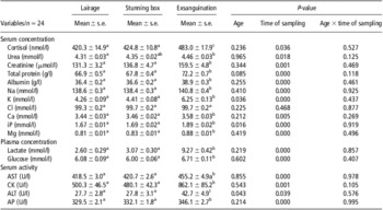
Significant age effects were found for serum activity of ALT and serum concentrations of potassium and inorganic phosphate (Table 1); however, there were no significant age and time of sampling interactions for any of the blood variables measured (Tables 1, 2 and 3). With the exception of serum concentration of chloride and the activity of blood antioxidant enzymes, significant time of sampling effect was found for all other blood variables (Tables 1, 2 and 3).
Effects of age and sampling time on blood chemistry of slaughter horses

AST = aspartate aminotransferase; CK = creatine kinase; ALT = alanine aminotransferase; AP = alkaline phosphatase; Na = sodium; K = potassium; Cl = chloride; Ca = calcium; iP = inorganic phosphate; Mg = magnesium.
a,bMean values with different superscripts in the same row differ significantly (P < 0.05). Mean values with superscript t tend to differ from the lairage and the stunning box values (P < 0.07).
n = number of samples analysed.
Effects of age and sampling time on haematological variables in slaughter horses

RBC = red blood cell count; HCT = haematocrit; HGB = haemoglobin concentration.
a,bMean values with different superscripts in the same row differ significantly (P < 0.05).
n = number of samples analysed.
Effects of age and sampling time on the activity of antioxidant enzymes in the blood of slaughter horses

SOD = superoxide dismutase; GSH-Px = glutathione peroxidase.
n = number of samples analysed.
In horses, the plasma lactate and glucose concentrations, serum potassium concentration, the activities of the serum muscle enzymes AST and CK, and values of most of the other biochemical (Table 1) and haematological variables (Table 2), were significantly (P < 0.05) higher at exsanguination, than in blood sampled while they were in the lairage and in the stunning box. The most prominent was the increase of plasma lactate and serum potassium concentrations and serum activity of CK. In contrast, the serum concentrations of cortisol and chloride and the activities of ALT and blood antioxidant enzymes (Table 3) were not significantly different between the pre-slaughter period and exsanguination. However, there was a tendency for an increase in serum cortisol concentration and the activity of ALT (P < 0.07) at exsanguination.
There were no significant differences for any of the biochemical, haematological and blood antioxidant enzyme variables between the lairage and the stunning box sampling time.
Discussion
The present study demonstrated no physiological stress responses of investigated horses to stressors, such as novelty of the pre-slaughter environment and handling, present in the slaughterhouse between the lairage and the stunning box as serum cortisol and all other blood variables did not change significantly between the lairage and the stunning box sampling times. However, at exsanguination, significantly higher levels of plasma lactate and glucose, serum muscle enzymes and haematological variables, than at the pre-slaughter period, might partially be attributed to stimulation of the sympathetic nervous system, caused by stunning and bleeding (Mitchell et al., Reference Mitchell, Hattingh and Ganhao1988; Shaw and Tume, Reference Shaw and Tume1992; Werner and Gallo, Reference Werner and Gallo2008). Plasma catecholamine concentrations have not been measured in the present study; however, their increased concentration after stunning have been found in horses, cattle and lambs (Mitchell et al., Reference Mitchell, Hattingh and Ganhao1988; Linares et al., Reference Linares, Bornez and Vergara2008; Micera et al., Reference Micera, Albrizio, Surdo, Moramarco and Zarrilli2010).
Cortisol is one of the most used indicators of stress in slaughter animals, despite its variable excretion (Mitchell et al., Reference Mitchell, Hattingh and Ganhao1988; Werner and Gallo, Reference Werner and Gallo2008; Micera et al., Reference Micera, Albrizio, Surdo, Moramarco and Zarrilli2010). On the basis of previous studies, the plasma cortisol concentration, in blood collected at exsanguination, was unlikely to have been affected by stunning and exsanguination and might provide information on pre-slaughter handling stress (Shaw and Tume, Reference Shaw and Tume1992; Werner and Gallo, Reference Werner and Gallo2008; Micera et al., Reference Micera, Albrizio, Surdo, Moramarco and Zarrilli2010). Cortisol concentration determined in blood samples collected post slaughter has been used to evaluate stress reactions of animals undergoing slaughter (Cockram and Corley, Reference Cockram and Corley1991; Werner and Gallo, Reference Werner and Gallo2008; Micera et al., Reference Micera, Albrizio, Surdo, Moramarco and Zarrilli2010). In the present study, serum cortisol concentration slightly exceeded the upper value of normal range (Eades and Bounous, Reference Eades and Bounous1997) at all sampling times. The slightly higher than normal serum cortisol concentration in the blood sampled in the lairage might have been due to transport stress (Fazio et al., Reference Fazio, Medica, Aronica, Grasso and Ferlazzo2008; Werner and Gallo, Reference Werner and Gallo2008). There was a tendency for an increase in serum cortisol concentration at exsanguination; however, serum cortisol concentrations did not differ significantly among sampling times, which is in agreement with the study of Werner and Gallo (Reference Werner and Gallo2008). Our result contrasts with the previous report on horses that reported significantly higher serum cortisol concentration at exsanguination than in the lairage, 45 min before exsanguination (Micera et al., Reference Micera, Albrizio, Surdo, Moramarco and Zarrilli2010). The difference might be because of variations between the horses studied, the slaughterhouse environments, pre-slaughter handling and the time of blood collection. Because serum cortisol concentration did not change significantly between the lairage and the stunning box sampling times, short pre-slaughter handling that included the collection of the first blood sample in the lairage and movement of horses from lairage to stunning box, and the stressors present in the slaughterhouse such as exposure to new social structure, unknown sounds, sights, smells and other slaughter animals did not cause a stress response (as assessed by the serum cortisol concentration).
Pre-slaugher stress and exsanguination have been reported to significantly increase plasma lactate in cattle and horses (Mitchell et al., Reference Mitchell, Hattingh and Ganhao1988; Werner and Gallo, Reference Werner and Gallo2008). In the present study, the plasma lactate concentration in the lairage and stunning box slightly exceeded the upper value of the normal range (Kaneko et al., Reference Kaneko, Harvey and Bruss1997a) and was significantly higher at exsanguination, when plasma lactate concentration was more than four times higher than the upper value of the normal range for horses. The result is in agreement with previous reports on cattle and horses (Mitchell et al., Reference Mitchell, Hattingh and Ganhao1988; Werner and Gallo, Reference Werner and Gallo2008). Hyperlactatemia observed at exsanguination was most probably a consequence of rapid glycogenolysis, stimulated by muscle movements and catecholamines released by stunning and bleeding (Shaw and Tume, Reference Shaw and Tume1992; McCorry, Reference McCorry2007; Micera et al., Reference Micera, Albrizio, Surdo, Moramarco and Zarrilli2010). The increase in catecholamines reported by Micera et al. (Reference Micera, Albrizio, Surdo, Moramarco and Zarrilli2010) may explain the significant increase in plasma lactate concentration, as well as the significant increase of plasma glucose concentration at exsanguination observed in investigated horses.
Reports on physiological responses of slaughter animals to transport, pre-slaughter handling and exsanguination often include measurement of activities of serum muscle enzymes – CK and AST (Tadich et al., Reference Tadich, Gallo, Bustamante, Schwerter and van Schaik2005; Werner and Gallo, Reference Werner and Gallo2008; Miranda-de la Lama et al., Reference Miranda-de la Lama, Rivero, Chacon, Garcia-Belenguer, Villarroel and Maria2010). Horses may respond to stress situations by displaying behavioural changes, which can include physical activity such as struggling, kicking, pawing, jumping, rearing and bucking. The latter may lead to muscle exertion with muscle damage and consequently to increased activities of muscle enzymes (Boissy, Reference Boissy1995; Winther Christensen et al., Reference Winther Christensen, Kelling and Lindestrøm Nielsen2005; Werner and Gallo, Reference Werner and Gallo2008). In the present study, the activities of CK and AST exceeded the upper value of normal ranges at all sampling times (Eades and Bounous, Reference Eades and Bounous1997). Transport of animals is physically demanding and can result in muscle damage and consequently increased activity of serum muscle enzymes (Stull and Rodiek, Reference Stull and Rodiek2000; Tadich et al., Reference Tadich, Gallo, Bustamante, Schwerter and van Schaik2005). Exsanguination resulted in a significant increase of serum CK and AST activities, which is in agreement with the study of Werner and Gallo (Reference Werner and Gallo2008). The significant increase of serum CK and AST activities could be partially ascribed to enzyme leakage from skeletal muscle cells because of increased membrane permeability resulting from catecholamine-induced rapid glycogenolysis or even because of muscle damage during exsanguination and muscle activity after stunning. Serum CK and AST activities at lairage and stunning box, higher than the upper value of the reference range, could be partially attributed to transport stress and physical activity of horses such as walking, turning around and shifting from one foot to the other, during pre-slaughter procedures, respectively.
Serum albumin and total protein concentrations, as well as haematocrit, are often used as indicators of dehydration (Eades and Bounous, Reference Eades and Bounous1997; Kaneko et al., Reference Kaneko1997b). In contrast, in horses, splenic contraction triggered by catecholamines results in the release of a large mass of erythrocytes into the circulation, thereby increasing the haematocrit/PCV, with little or no change in serum protein concentration (Eades and Bounous, Reference Eades and Bounous1997; Kaneko et al., Reference Kaneko1997b). In the present study, serum albumin and total protein concentrations and values of haematological variables did not exceed the normal range at any of the sampling times (Eades and Bounous, Reference Eades and Bounous1997; Colahan et al., Reference Colahan, Merritt, Moore and Mayhew1999). At exsanguination, serum total protein and albumin concentrations were significantly higher than at the lairage and stunning box stages, but negligible compared with increase of haematological parameters, which indicates that splenic contraction triggered by increased concentration of catecholamines may be responsible for these changes instead of dehydration. The results are in agreement with those of similar studies conducted on horses (Werner and Gallo, Reference Werner and Gallo2008) and cattle (Mitchell et al., Reference Mitchell, Hattingh and Ganhao1988; Cockram and Corley, Reference Cockram and Corley1991).
In the present study, most of the serum electrolytes had higher concentrations at exsanguination than at lairage and stunning box stages, although the values, with the exception of potassium, remained within the normal range (Kaneko et al., Reference Kaneko, Harvey and Bruss1997a). Hyperkalaemia present at exsanguination could be ascribed to haemolysis (Frank et al., Reference Frank, Bermes, Bickel and Watkins1978); however, the serum samples of the investigated horses were not haemolysed, according to visual inspection. Hyperkalaemia is a result of imbalance of its distribution between intracellular and extracellular space. The potassium intracellular concentration is more than 23 times that in the extracellular fluid (Frank et al., Reference Frank, Bermes, Bickel and Watkins1978). Thus, a minor shift of potassium (K+) between intracellular and extracellular fluid leads to a very large increase in plasma potassium concentration. Hyperkalaemia determined at exsanguination may be because of increased passive fluxes promoted by increased release of catecholamines, released from adrenal medulla caused by stunning and bleeding (Shaw and Tume, Reference Shaw and Tume1992), metabolic acidosis and muscle activity after stunning. Catecholamines released promote the release of potassium via alpha 1 and 2 adrenergic receptors (Moratinos and Reverte, Reference Moratinos and Reverte1993). The increase in concentrations of other electrolytes and serum concentrations of creatinine and total proteins probably reflects mostly splenic contraction induced by increased concentrations of circulating catecholamines and some degree of dehydration.
Although activities of antioxidant enzymes were determined in whole blood haemolysates rather than in meat, these enzymes could potentially have provided useful information on meat quality in horses (Dotan et al., Reference Dotan, Lichtenberg and Pinchuk2004; Kirschvink et al., Reference Kirschvink, de Moffarts and Lekeux2008). For example, exercise is associated with increased production of reactive oxygen species (ROS), which may lead to oxidative stress and eventually to damage of biological molecules and key cellular components. Both increased and decreased activity of antioxidant enzymes have been reported after different types of exercise as a consequence of enhanced ROS production, either by upregulation of enzyme activity or utilisation of the antioxidant enzymes to counter the ROS (Deaton and Marlin, Reference Deaton and Marlin2003; Kirschvink et al., Reference Kirschvink, de Moffarts and Lekeux2008). In slaughter horses, oxidative stress might be associated with a stress response, when the latter includes the types of physical activity observed in the horses in this study. During stunning and bleeding, tissue hypoxia can also increase the production of ROS, thus leading to disturbance of antioxidant–oxidant equilibrium. Although the horses included in the study were physically active in the lairage pen and during waiting in the race to be stunned, no significant changes of GSH-Px and SOD activities in the blood indicative of oxidative stress were observed. At all sampling times, the blood activities of SOD and GSH-Px were in agreement with published data for the enzymes in healthy horses (Kaneko et al., Reference Kaneko, Harvey and Bruss1997a; Gorecka et al., Reference Gorecka, Sitarska and Klucinski2002).
Conclusion
The present study showed no physiological stress responses of the investigated horses to stressors, such as novelty of the pre-slaughter environment and handling, present in the slaughterhouse between the lairage pen and the stunning box. However, the significantly higher values, at exsanguination, for the plasma lactate and glucose concentrations, serum muscle enzyme activities and haematological variables, than during the pre-slaughter period, might partially be attributed to stimulation of the sympathetic nervous system, caused by stunning and bleeding.
Acknowledgements
The authors acknowledge the financial support of the Slovenian Research Agency (research programme P4-0053). The authors thank Professor Andrej Blejec for statistical evaluation of the data, Roger Pain for review of English and Matej Lončar for technical assistance.