





Introduction
Electroconvulsive therapy (ECT) is highly efficient in treating severe depression (Kolar, Reference Kolar2017). Being superior to pharmacotherapy in its antidepressant effect (UK ECT Review, 2003), it remains a very important treatment option. Alas, the intervention appears to be accompanied by anterograde and retrograde amnesia in a number of cases (Fraser, O’Carroll, & Ebmeier, Reference Fraser, O’Carroll and Ebmeier2008; Mathiassen et al., Reference Mathiassen, Semkovska, Lundsgaard, Gbyl and Videbech2026; Semkovska & McLoughlin, Reference Semkovska and McLoughlin2010), ultimately limiting the application of ECT.
When considering the amnesic side effects of ECT, it is important to distinguish between subtypes of amnesia, as differential effects are suggested in the literature. A meta-analysis by Semkovska and McLoughlin of the cognitive side effects, including verbal and visual anterograde amnesia, indicates that both types of anterograde amnesia are evident shortly after ECT, but they are resolved within 2 weeks (Semkovska & McLoughlin, Reference Semkovska and McLoughlin2010). A more recent meta-analysis by Landry et al. found that only verbal, and not visual, anterograde amnesia was evident within a month after ECT, and the amnesia was also resolved at follow-ups beyond 1 month after ECT (Landry et al., Reference Landry, Moreno, Patry, Potvin and Lemasson2021). This meta-analysis also investigated autobiographical amnesia and showed short-term, but not long-term, impairment (Landry et al., Reference Landry, Moreno, Patry, Potvin and Lemasson2021). This result contrasts with an earlier systematic review by Fraser, O’Carroll, and Ebmeier (Reference Fraser, O’Carroll and Ebmeier2008) and our more recent meta-analysis (Mathiassen et al., Reference Mathiassen, Semkovska, Lundsgaard, Gbyl and Videbech2026), both of which suggest long-term autobiographical amnesia. The latter meta-analysis highlights the need for longitudinal studies conducting long-term follow-up assessments of autobiographical amnesia, while applying an appropriate control group, to control for the confounding effect of time and depression in the appraisal of the duration of the amnesic side effect, as only a few studies addressed these issues (Bjoerke-Bertheussen et al., Reference Bjoerke-Bertheussen, Schoeyen, Andreassen, Malt, Oedegaard, Morken, Sundet, Vaaler, Auestad and Kessler2018; Kessler et al., Reference Kessler, Schoeyen, Andreassen, Eide, Malt, Oedegaard, Morken, Sundet and Vaaler2014; Semkovska & O’Grady, Reference Semkovska and O’Grady2017).
We performed such a longitudinal study, assessing autobiographical memory as well as verbal and visual anterograde memory both before, shortly after, and 6 months after ECT, in patients with depression. Assessments were performed at corresponding time points in control groups consisting of patients with depression receiving pharmacotherapy and/or psychotherapy and healthy controls, respectively.
Our primary aim was to investigate the effect of ECT on different aspects of memory function. Based on the literature presented above, we hypothesized the following:
-
1) Autobiographical amnesia will be evident in the ECT group at both short- and long-term follow-up.
-
2) Verbal anterograde amnesia will be evident in the ECT group at short- but not long-term follow-up.
-
3) Visual anterograde amnesia will be evident in the ECT group at short- but not long-term follow-up.
Given the clinical value of predicting ECT-associated amnesia, our secondary aim was to explore potential predictors of the amnesia. Based on the literature (Legendre et al., Reference Legendre, Stern, Solomon, Furman and Smith2003; Martin et al., Reference Martin, Katalinic, Ingram, Schweitzer, Smith, Hadzi-Pavlovic and Loo2013; Sackeim et al., Reference Sackeim, Prudic, Fuller, Keilp, Lavori and Olfson2007), we chose a priori to investigate whether age and cognitive reserve were associated with the degree of post-ECT amnesia.
Method
This study was part of The Danish Neuropsychological Study of the Adverse Effects of ECT (DANSECT). The preparation of this manuscript was performed in compliance with the Strengthening the Reporting of Observational Studies in Epidemiology (STROBE) guideline.
Study design
This longitudinal, prospective, and observational study shares methodology with the Danish ECT/MRI Project (Gbyl et al., Reference Gbyl, Rostrup, Raghava, Carlsen, Schmidt, Lindberg, Ashraf, Jørgensen, Larsson, Rosenberg and Videbech2019, Reference Gbyl, Støttrup, Mitta Raghava, Xue Jie and Videbech2021, Reference Gbyl, Lindberg, Wiberg Larsson, Rostrup and Videbech2022, Gbyl et al., Reference Gbyl, Labanauskas, Lundsgaard, Mathiassen, Ryszczuk, Siebner, Rostrup, Madsen and Videbech2024; Gbyl, Jørgensen, & Videbech, Reference Gbyl, Jørgensen and Videbech2022), of which DANSECT (Lundsgaard et al., Reference Lundsgaard, Mathiassen, Wiberg Larsson, Videbech, Gbyl and Vestergaard2025; Mathiassen et al., Reference Mathiassen, Lundsgaard, Gbyl, Miskowiak, Fagerlund, Larsson, Lindberg and Videbech2025) was an extension. We consecutively included inpatients with moderate to severe depression, referred to ECT, from mental health centers in the Capital Region of Denmark from December 2020 to July 2023. Assessments were performed at pre-ECT (baseline/T1): 1–2 days prior to ECT, post-ECT (T2): 5 (± 2) days after ECT (median = 5, range = 2–25), and 6 (± 2) months after the post-ECT (T3).
We included patients referred to ECT for either unipolar or bipolar depression cf. ICD-10 (WHO, 1993), between the ages of 18 and 95 years, and proficient in Danish. Exclusion criteria were as follows: (1) patients who had received ECT during the last 6 months; (2) patients receiving maintenance ECT; (3) patients receiving compulsory treatment; (4) psychotic disorders, except psychotic depression; (5) dependency syndrome; (6) history of unconsciousness for more than 5 minutes following head trauma; and (7) neurological disease or other physical illness affecting the brain.
The diagnosis of depression was initially made by a psychiatrist in the clinic. After referral to our project, the diagnosis was confirmed or rejected by the project neuropsychologist or the project medical doctor using the Mini-International Neuropsychiatric Interview (MINI) (Sheehan et al., Reference Sheehan, Lecrubier, Sheehan, Amorim, Janavs, Weiller, Hergueta, Baker and Dunbar1998). To rule out organic etiology giving rise to the depressive symptoms, a physical examination, electrocardiogram, blood tests, and a urine-based drug test were performed.
We recruited study participants for two control groups, depression controls (DC) and healthy controls (HC). The DC group consisted of patients with depression receiving other treatments than ECT (antidepressant medication and/or psychotherapy) and was recruited from the same psychiatric wards, in addition to an outpatient clinic. Additionally, they met the same inclusion criteria as the ECT group. The HC group was recruited from the local blood bank in the Capital Region of Denmark. If they did not fulfil the criteria for a depressive episode or any of the remaining exclusion criteria, they were eligible for inclusion. The DC group was recruited within a week after admission (inpatients) or before initiation of the treatment program (outpatients). Both control groups were tested at time points corresponding to the ECT group: baseline (T1), 30 days after baseline (T2), and 6 (± 2) months after T2 (T3). The purpose of the HC group was to represent the normal trajectory of performance on the autobiographical memory interview, given that the instrument lacks formal psychometric validation and that the effect of time is essential to autobiographical memory consistency. Therefore, this group was only administered the autobiographical memory interview, not the anterograde memory tests.
ECT
ECT was administered by use of Thymatron IV (Somatics, LLC, USA). The treatment was provided in compliance with the standard clinical practice of the regional mental health services. ECT was delivered three times a week. The pulse width was 0.5–1.0 ms (brief pulse). In principle, the electrode placement was bitemporal, but this could be altered to right unilateral electrode placement by decision of the treating physician in order to limit disproportionate cognitive impairment. The first charge was determined using the half-age method. Thiopental (2–4 mg/kg) was used as anesthetic. Suxamethonium (0.75 mg/kg) was used as a muscle relaxant. The research group was not involved in any clinical decisions regarding treatment.
Clinical assessments
Memory and depression severity
Autobiographical memory was assessed by use of the Columbia Autobiographical Memory Interview – Short Form (CAMI-SF) (McElhiney, Moody, & Sackeim, Reference McElhiney, Moody and Sackeim2001). Verbal anterograde memory was assessed with the Verbal Learning Test – Delayed (VLT-D), a subtest of the Screen for Cognitive Impairment in Psychiatry (SCIP-D) (Jensen et al., Reference Jensen, Støttrup, Nayberg, Knorr, Ullum, Purdon, Kessing and Miskowiak2015). Parallel versions were administered at T2 and T3. Visual anterograde memory was measured with the Rey Complex Figure Test (RCFT) three-minute retention (Meyers & Meyers, Reference Meyers and Meyers1995). To measure depression severity, we used the 6-item Hamilton Rating Scale for Depression (HAMD-6) (Hamilton, Reference Hamilton1960). The 6-item version was chosen because it has proven to be as sensitive as the 17-item version and because its items are more equidistant, ultimately enhancing within- and between-group comparisons (Lecrubier & Bech, Reference Lecrubier and Bech2007).
Cognitive reserve
Cognitive reserve was operationalized through two proxy measures: level of education and level of intelligence (estimated with the Vocabulary subtest of the Wechsler Adult Intelligence Scale IV (WAIS-IV) (Wechsler, Reference Wechsler2008), assessed at post-ECT). The Vocabulary subtest was chosen as an estimator of the level of intelligence, since this is the subtest of the WAIS-IV battery correlating the strongest with the full-scale IQ (Lezak, Howieson, Bigler, & Tranel, Reference Lezak, Howieson, Bigler and Tranel2004) and since it has demonstrated particular resilience against brain detriments (Lezak et al., Reference Lezak, Howieson, Bigler and Tranel2004). Level of education was based on the International Standard Classification of Education (Eurostat & UNESCO Institute for Statistics, 2015) and was divided into seven levels. See Table S1 in the Supplementary Material for the exact division.
Statistical analysis
Primary analyses
To assess the effect of the independent variables (ECT, DC, and HC) on the dependent variables (autobiographical memory, visual anterograde memory, and verbal anterograde memory), a repeated measures mixed ANOVA was conducted for each dependent variable with three levels of the time variable (T1, T2, and T3). To explore interactions and main effects, Bonferroni-adjusted pairwise comparisons were performed. Specifically, memory scores of the ECT group were compared at the different time points (within-subjects), and the scores were subsequently compared to those of the two control groups at these time points (between-subjects).
As we suspected the ECT group to have a higher depression severity at baseline than the DC group, we conducted an independent samples t-test to compare baseline depression severity between the two groups. To explore whether such a difference in depression severity influenced our estimation of the amnesic effect of ECT, the baseline HAMD-6 score was added as a covariate in the primary analyses. To investigate whether the antidepressant effect influenced the memory decline, the change in HAMD-6 score from pre- to post-ECT was also added as a covariate in the primary analyses.
Secondary analyses
We tested the predictive value of age and cognitive reserve on memory performance at T2 and T3. To assess whether these predetermined predictors were associated with memory performance at T2 and T3, we repeated the primary analyses for the ECT subjects only, adding the predictors as covariates.
Post hoc analyses
In the verbal anterograde memory test, baseline performance differed between the two groups, with the DC group performing better than the ECT group. To dissect whether the interaction between group and time in fact reflected a difference in memory decline between groups, we added the baseline verbal anterograde memory score as a covariate to the primary analysis.
To estimate the prevalence of post-ECT amnesia, we calculated the percentage of ECT patients who exhibited a memory decline below the DC group mean by at least 2 standard deviations. This was done at both T2 and T3. A tangible estimation of the severity of the autobiographical amnesia was performed through a calculation of the mean decline in memory consistency, respectively, from T1 to T2 and T1 to T3.
As medication status differed between groups, and since an anticholinergic effect of tricyclic antidepressants and antipsychotics may affect cognition (Chandramouleeshwaran, Rajji, Khan, & Inglis, Reference Chandramouleeshwaran, Rajji, Khan and Inglis2024), the medication status for these drugs at baseline was added as a covariate in each of the primary statistical analyses to explore whether differing medication affected results.
To explore robustness of the main findings, the data were also analyzed using a Linear Mixed-Effects Model. Details are provided in the Supplementary Material.
Results
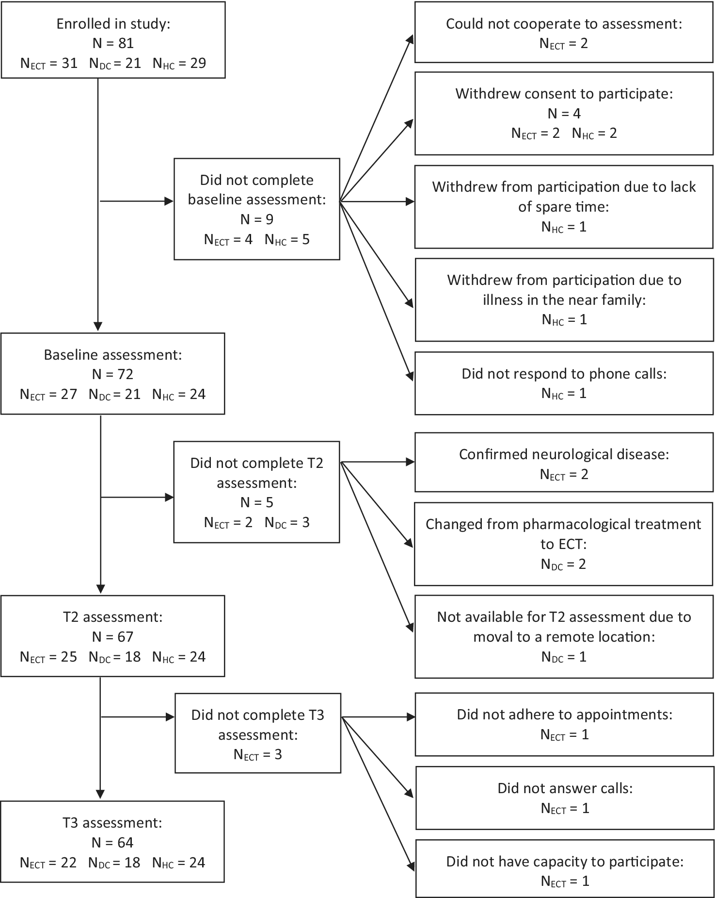
See the flow diagram in Figure 1 for an overview of the inclusion process and participation over time. Baseline demographic and clinical characteristics of the three groups, in addition to ECT parameters, are presented in Table 1.
Flowchart of inclusion process. DC, depression controls; ECT, electroconvulsive therapy group; HC, healthy controls.

Figure 1. Long description
The flowchart follows a central vertical path with three branching points to the right.
1. Enrolled in study: N = 81 (N- ECT = 31, N-DC = 21, N-HC = 29).
- Branch: Did not complete baseline assessment: N = 9 (N- ECT = 4, N-HC = 5). Reasons include: Could not cooperate to assessment (N- ECT = 2); Withdrew consent to participate (N = 4, N-ECT = 2, N-HC = 2); Withdrew due to lack of spare time (N- HC = 1); Withdrew due to illness in the near family (N- HC = 1); Did not respond to phone calls (N- HC = 1).
2. Baseline assessment: N = 72 (N- ECT = 27, N-DC = 21, N-HC = 24).
- Branch: Did not complete T2 assessment: N = 5 (N- ECT = 2, N-DC = 3). Reasons include: Confirmed neurological disease (N- ECT = 2); Changed from pharmacological treatment to ECT (N- DC = 2); Not available due to removal to a remote location (N- DC = 1).
3. T2 assessment: N = 67 (N- ECT = 25, N-DC = 18, N-HC = 24).
- Branch: Did not complete T3 assessment: N-ECT = 3. Reasons include: Did not adhere to appointments (N- ECT = 1); Did not answer calls (N- ECT = 1); Did not have capacity to participate (N- ECT = 1).
4. T3 assessment: N = 64 (N- ECT = 22, N-DC = 18, N-HC = 24).
Clinical characteristics of included subjects at baseline and ECT parameters a

Table 1. Long description
The table is organized into five main sections: Demographics, Memory performance, Depression variables, Psychopharmacology, and ECT parameters.
1. Demographics:
- Age: ECT group 45.6, DC 37.1, HC 47.8. Significant difference between ECT and DC (p = 0.048).
- Gender: Male percentage is 48.0% for EC T, 28.6% for D C, and 62.6% for H C.
- Education and Vocabulary: Significant differences found between ECT and HC (p = 0.022 and 0.020 respectively).
2. Memory performance:
- CAMI-SF: Scores are similar across all groups (51.8 to 52.4).
- VLT-D and RCFT: Significant differences between ECT and DC (p = 0.007 and 0.023).
3. Depression variables:
- HAMD-6: ECT group 14.7, DC 11.4 (p < 0.001).
- HAMD-17: ECT group 27.3.
4. Psychopharmacology:
- Significant differences between ECT and DC for TCA (24% vs 0%, p = 0.016), Lithium (24% vs 0%, p = 0.016), and Antipsychotics (64% vs 24%, p = 0.006).
5. ECT parameters (E CT group only):
- 92% received bilateral placement.
- Average of 10.9 sessions.
- Total seizure time 455.6 seconds.
- Total dose 2752.3 m C.
- 68% were responders and 32% were remitters.
Notes: BL, bilateral electrode placement; BZD, benzodiazepines; CAMI-SF, Columbia Autobiographical Memory Interview – Short Form; DC, depression controls; ECT, electroconvulsive therapy; HAMD-6, Hamilton Rating Scale for Depression-6; HAMD-17, Hamilton Rating Scale for Depression-17; HC, healthy controls; mC, millicoulombs; Mixed, a combination of BL and right unilateral electrode placement; n, number of subjects; RCFT, Rey Complex Figure Test; TCA, tricyclic antidepressants; VLT-D, Verbal Learning Test – Delayed. P values below 0.05 indicate the significant differences between the study groups and are set in bold.
a Mean (standard deviation) or frequency (percentage) are reported. P-values are reported for independent samples t-test or χ2 test in comparison of the mean values of the ECT group and control group.
b Hypnotics and medication pro re nata (as needed) not included.
c Patients who were changed from BL to right unilateral electrode placement along the treatment course.
d Defined as a post-ECT reduction in HAMD-6 score of at least 50%.
e Defined as a post-ECT HAMD-6 score below 5.
Amnesia following ECT
A repeated measures mixed ANOVA was performed for each of the three memory outcomes to investigate the impact of ECT, depression, and time on autobiographical, verbal, and visual anterograde amnesia. Approximate normal distribution was confirmed for each outcome across time points within each group. See Table 2 for mean values and standard deviations. Results of the testing of assumptions for the following analyses can be viewed in the Supplementary Material.
Memory and depression scores over time across groups

Table 2. Long description
The table presents Mean and SD values for three groups across three time points: T1, T2, and T3.
1. ECT Group (n = 22):
* CAMI-SF: T1 (Mean 51.90, SD 4.40), T2 (Mean 34.62, SD 8.48), T3 (Mean 36.33, SD 6.23).
* VLT-D: T1 (Mean 6.28, SD 2.47), T2 (Mean 4.83, SD 2.79), T3 (Mean 6.72, SD 2.68).
* RCFT: T1 (Mean 19.25, SD 6.30), T2 (Mean 18.20, SD 5.07), T3 (Mean 21.75, SD 5.39).
* HAMD-6: T1 (Mean 14.72, SD 2.35), T2 (Mean 6.56, SD 4.00), T3 (Mean 6.57, SD 3.53).
2. DC Group (n = 18):
* CAMI-SF: T1 (Mean 52.11, SD 4.31), T2 (Mean 47.22, SD 4.81), T3 (Mean 44.83, SD 5.08).
* VLT-D: T1 (Mean 8.39, SD 1.94), T2 (Mean 8.33, SD 1.94), T3 (Mean 7.83, SD 2.38).
* RCFT: T1 (Mean 24.50, SD 6.18), T2 (Mean 28.06, SD 5.49), T3 (Mean 28.82, SD 4.21).
* HAMD-6: T1 (Mean 11.38, SD 3.34), T2 (Mean 7.67, SD 4.67), T3 (Mean 5.50, SD 5.07).
3. HC Group (n = 24):
* CAMI-SF: T1 (Mean 52.25, SD 4.18), T2 (Mean 45.96, SD 4.70), T3 (Mean 44.42, SD 4.62).
Note: CAMI-SF, Columbia Autobiographical Memory Interview – Short Form; DC, depression controls; ECT, electroconvulsive therapy; HAMD-6, Hamilton Rating Scale for Depression-6; HC, healthy controls; RCFT, Rey Complex Figure Test; SD, standard deviation; VLT-D, Verbal Learning Test – Delayed.
a Number of completers.
Autobiographical amnesia
For the autobiographical memory outcome, there was a significant interaction between time and group, FHF (3.45, 103.51) = 22.261, p < 0.001, partial η 2 = 0.426. See Figure 2a.
Memory performance over time across groups. CAMI-SF, Columbia Autobiographical Memory Interview – Short Form; error bars, 95% confidence interval; RCFT, Rey Complex Figure Test; VLT-D, Verbal Learning Test – Delayed.

Figure 2. Long description
The figure consists of three panels labeled A, B, and C, and a shared legend. The legend identifies three groups: Electroconvulsive therapy (solid blue line), Depression control group (dashed maroon line), and Healthy control group (dotted teal line).
* Panel A: The y-axis is CAMI-S F, ranging from 35 to 55. At T1, all groups start near 52. By T2, the Electroconvulsive therapy group shows a sharp decline to approximately 35, while the control groups show a slight decline to 46-48. At T3, the Electroconvulsive therapy group recovers slightly to 37, while control groups continue a gradual decline to 45.
* Panel B: The y-axis is VLT D, ranging from 4 to 10. The Depression control group maintains a steady score between 8 and 8.5 across all time points. The Electroconvulsive therapy group starts at 6.3 at T1, drops to a low of 5 at T2, and recovers to 6.8 at T3.
* Panel C: The y-axis is RCF T, ranging from 15 to 35. The Depression control group shows a steady increase from 25 at T 1 to 29 at T3. The Electroconvulsive therapy group starts at 19.5 at T1, dips slightly at T2, and increases to 22 at T3.
All data points include vertical error bars representing 95 percent confidence intervals.
Bonferroni-adjusted pairwise comparisons revealed that baseline scores on the CAMI-SF were equal across groups. Within-group comparisons showed that all groups scored significantly lower on the CAMI-SF at T2 compared to T1 (p ≤ 0.002 in all cases). From T2 to T3, scores were only significantly different (reduced) within the DC group (p = 0.034). At both T2 and T3, the ECT group scored significantly lower than both control groups (p < 0.001 in all cases). At no time point did the two control groups differ in the CAMI-SF scores (p ≥ 0.519 in all cases).
The independent samples t-test confirmed that the baseline depression severity of the ECT group (mean = 14.72, SD = 2.36) was higher than that of the DC group (mean = 11.38, SD = 3.34), t (44) = 3.966, p < 0.001. Yet, adjusting for depression severity did not significantly affect the results at either T2 (p = 0.398) or T3 (p = 0.095). The same was the case for antidepressant effect (T2: p = 0.643; T3: p = 0.169).
Verbal anterograde amnesia
A significant interaction between time and group was found on the verbal anterograde memory performance, F (2, 68) = 6.138, p = 0.004, partial η 2 = 0.153. See Figure 2b.
Pairwise comparisons with Bonferroni adjustments showed that the VLT-D score in the ECT group was significantly impaired at T2 relative to T1 (p = 0.009), but returned to baseline at T3, with no difference between T3 and T1 (p = 1.0). In comparison, the DC group did not change significantly on the VLT-D score at any time point (p ≥ 0.841 in all cases). Noteworthy, a between-group comparison showed that the groups differed significantly at both baseline (p = 0.007) and T2 (p < 0.001) but not at T3 (p = 0.197).
Results did not change significantly after adjusting for either depression severity (T2: p = 0.594; T3: p = 0.784) or antidepressant effect (T2: p = 0.638; T3: p = 0.838).
Visual anterograde amnesia
There was a significant interaction between time and group on RCFT performance, F (2, 66) = 5.677, p = 0.005, partial η2 = 0.147. See Figure 2c.
Pairwise comparisons adjusted with Bonferroni showed that the RCFT scores of the ECT and DC groups differed at all time points. As for within-group changes, the DC group performed significantly better at T2 than at T1 (p = 0.012), while the ECT group remained stable (p = 1.0). At T3, the DC group remained stable (p = 1.0), while the ECT group improved significantly (p < 0.001).
Predictors of ECT-related amnesia
Parameter estimates showed that neither age, vocabulary, nor level of education was significantly associated with either the autobiographical or verbal anterograde memory performance at either T2 or T3 (p ≥ 0.090 in all cases).
Post hoc statistical analyses
Baseline memory performance
The baseline verbal anterograde memory score was a significant covariate, F (1, 33) = 31.042, p < 0.001, partial η 2 = 0.206. Nonetheless, parameter estimates revealed that the anterograde memory performance of the ECT group was still significantly lower than the DC group at T2 (B = −1.985, SE = 0.677, p = 0.006, partial η 2 = 0.485), and there were still no group differences at T3 (p = 0.669), when adjusting for the baseline performance.
Prevalence of amnesia
At post-ECT, 75% of the ECT patients showed autobiographical amnesia and 24% exhibited verbal anterograde amnesia. At the 6-month follow-up, 57% of the ECT patients still showed autobiographical amnesia.
Severity of autobiographical amnesia
The ECT group declined 31% in autobiographical memory consistency from T1 to T2. In comparison, the DC group declined 9%, and the HC group declined 12%. The difference between the ECT and both control groups was significant (p < 0.001). This difference was still present at T3 (p < 0.001), where the ECT group showed a stable total decline in consistency of 30% in contrast to 14% of the DC group and 15% of the HC group.
Medication status
Controlling for medication with either tricyclic antidepressants or antipsychotics did not significantly change the main results, and effect sizes did not change notably (CAMI-SF: η 2 = 0.366–.388, p < 0.001; VLT-D: η 2 = 0.152–162, p ≤ 0.004; RCFT: η 2 = 0.158–.189, p ≤ 0.004).
Linear mixed-effects model
Applying a Linear Mixed-Effects Model to analyze the data did not affect the main findings. See Supplementary Material for the exact results and plots.
Discussion
Overall, we found that ECT was associated with significant both short- and long-term autobiographical amnesia, supporting our first hypothesis. Results also indicated verbal anterograde amnesia immediately after ECT, resolving after 6 months, which supported hypothesis 2. ECT was not associated with significant impairments in visual anterograde memory. The evident group differences at T2 and T3 were most likely attributable to superior test–retest effects in the control group, opposing hypothesis 3. Neither age nor cognitive reserve was associated with the degree of any type of ECT-related amnesia at any time point.
Our finding that ECT was associated with both short- and long-term autobiographical amnesia is in line with our recent meta-analysis of controlled longitudinal studies (Mathiassen et al., Reference Mathiassen, Semkovska, Lundsgaard, Gbyl and Videbech2026). This meta-analysis indicated a moderate detrimental effect of ECT on autobiographical memory, which was stable, and inferior to the control groups, at long-term follow-up (6–12 months) (Mathiassen et al., Reference Mathiassen, Semkovska, Lundsgaard, Gbyl and Videbech2026). In contrast, Landry et al. (Reference Landry, Moreno, Patry, Potvin and Lemasson2021) found short- but not long-term autobiographical amnesia in their meta-analysis. Although the total sample size was larger in the latter study, the autobiographical memory scores were highly heterogeneous, and there was no demand for control groups, which introduced uncertainty about the confounding effects of time and depression. Our meta-analysis (Mathiassen et al., Reference Mathiassen, Semkovska, Lundsgaard, Gbyl and Videbech2026) was strict in only including controlled studies, and the heterogeneity of our results was low.
Our differential findings regarding subtypes of anterograde amnesia are consistent with the meta-analysis by Landry et al. (Reference Landry, Moreno, Patry, Potvin and Lemasson2021), who also found the short-term verbal anterograde amnesia to be the only anterograde memory deficit after ECT. Contrastingly, the earlier meta-analysis by Semkovska and McLoughlin (Reference Semkovska and McLoughlin2010) found short-term impairments in both verbal and visual anterograde memory. Nonetheless, the effect sizes were large and small, respectively, indicating that the verbal anterograde amnesia was more substantial than the visual.
We found that age, vocabulary, and level of education were not associated with either type of ECT-related memory loss. This contrasts with the large study by Sackeim et al. (Reference Sackeim, Prudic, Fuller, Keilp, Lavori and Olfson2007), finding more autobiographical amnesia to be predicted by a lower estimated IQ and that more anterograde amnesia was predicted by both older age and lower estimated IQ. Additionally, Martin et al. (Reference Martin, Katalinic, Ingram, Schweitzer, Smith, Hadzi-Pavlovic and Loo2013) also found estimated IQ, and not age, to predict autobiographical amnesia. The most apparent difference between these studies and ours was the substantially larger sample sizes, suggesting that suboptimal statistical power is the most likely explanation for our nil findings.
Retrograde amnesia
Our first hypothesis was supported, as the ECT group declined substantially more in autobiographical memory consistency after ECT and remained below both control groups after 6 months, as reflected by a large effect-sized interaction.
Despite the larger depression severity of the ECT group, as an inherent consequence of the observational design, depression severity was not found to be a significant covariate either short- or long-term, minimizing the risk of our finding being a type I error. The same was the case with antidepressant effect, further substantiating our finding. These results are in line with our observation that depressive and healthy controls did not differ in the trajectory of their autobiographical memory decline. A suboptimal sensitivity to episodic details of the CAMI-SF might have masked a potential group difference, and depression severity as a covariate (Mathiassen et al., Reference Mathiassen, Semkovska, Lundsgaard, Gbyl and Videbech2026), as patients with depression are particularly found to recall fewer details of their episodic autobiographical memories (Söderlund et al., Reference Söderlund, Moscovitch, Kumar, Daskalakis, Flint, Herrmann and Levine2014). Our findings may also reflect that the autobiographical memory function of patients with depression is primarily impaired due to overgeneralization and negative recollective bias, rather than an outright tendency to fail to recall otherwise accessible memories (Bergouignan et al., Reference Bergouignan, Lemogne, Foucher, Longin, Vistoli, Allilaire and Fossati2008; Nandrino et al., Reference Nandrino, Pezard, Posté, Réveillère and Beaune2002; Söderlund et al., Reference Söderlund, Moscovitch, Kumar, Daskalakis, Flint, Herrmann and Levine2014; Williams et al., Reference Williams, Barnhofer, Crane, Hermans, Raes, Watkins and Dalgleish2007). Hence, our findings challenge the assumption that the autobiographical memory recall of patients with depression in general is profoundly inferior to that of healthy people (Semkovska & McLoughlin, Reference Semkovska and McLoughlin2013). Our recent meta-analysis substantiates this view, as the study found that depression severity did not moderate the degree of autobiographical amnesia, and the depressive and healthy controls did not differ in the trajectory of their autobiographical memory decline (Mathiassen et al., Reference Mathiassen, Semkovska, Lundsgaard, Gbyl and Videbech2026). Nevertheless, certain illness factors may still separate our patient groups with respect to memory function, for example, in the number of past depressive episodes, which has been linked to cognitive functioning (MacQueen et al., Reference MacQueen, Galway, Hay, Young and Joffe2002).
Age could be thought to confound the difference in autobiographical memory decline between groups, since the patients in the ECT group were slightly older than those in the DC group. However, age was not found to be a significant covariate, which is in line with the meta-analysis by Landry et al. (Reference Landry, Moreno, Patry, Potvin and Lemasson2021), who found no influence of age on autobiographical memory after ECT. Yet, our null-finding contrasts with our recent meta-analysis, where we found a higher age to be associated with less autobiographical memory loss (Mathiassen et al., Reference Mathiassen, Semkovska, Lundsgaard, Gbyl and Videbech2026). Finally, others have found a higher age to be related to more autobiographical memory loss (Lisanby et al., Reference Lisanby, Maddox, Prudic, Devanand and Sackeim2000). The ambiguous findings as to whether age constitutes a protective factor, a risk factor, or is in fact unimportant for the development of post-ECT autobiographical amnesia may be related to the seemingly bidirectional effects associated with increasing age. First, it appears intuitive that a higher age may be associated with more pronounced autobiographical amnesia following ECT, as advancing age has been linked to greater deficits in other cognitive domains (Sackeim et al., Reference Sackeim, Prudic, Fuller, Keilp, Lavori and Olfson2007). Yet, as increasing age is associated with reduced richness of detail in autobiographical memories (Levine et al., Reference Levine, Svoboda, Hay, Winocur and Moscovitch2002), it is possible that patients of higher age report fewer details in the autobiographical memory interview at baseline. Thus, the sensitivity to memory loss at subsequent interviews may be reduced for the elderly, as a floor effect might mask memory loss to some extent. Furthermore, a superior antidepressant effect of ECT in older patients (Rhebergen et al., Reference Rhebergen, Huisman, Bouckaert, Kho, Kok, Sienaert, Spaans and Stek2015) may result in a more favorable cognitive outcome overall, thus possibly counteracting amnesic effects to some degree.
Anterograde amnesia
As temporary verbal anterograde amnesia occurred in the ECT group, and not in the DC group, our second hypothesis was supported. The group difference remained significant after controlling for baseline performance, suggesting a robust finding, consistent with earlier studies (Landry et al., Reference Landry, Moreno, Patry, Potvin and Lemasson2021; Semkovska & McLoughlin, Reference Semkovska and McLoughlin2010). As in the case of autobiographical memory, neither depression severity nor antidepressant effect was found to be a significant covariate, which further strengthens our finding.
The third hypothesis was not supported by our findings, since no within-subjects change was observed after ECT on visual anterograde memory performance. Most likely, the improved performance in the DC group at T2 reflects larger test–retest effects in this group, which is coherent with the literature suggesting only small or no impairment in this type of memory function (Landry et al., Reference Landry, Moreno, Patry, Potvin and Lemasson2021; Semkovska & McLoughlin, Reference Semkovska and McLoughlin2010). However, since we did not administer parallel versions of the complex figure at T2 and T3, a test–retest effect in the ECT group cannot be entirely ruled out, potentially concealing a small memory decline, risking a type II error. Yet, since memory consolidation is continuously disturbed during the ECT course, the benefit of practice in the ECT group appears negligible, ultimately making a type II error unlikely.
Measuring autobiographical memory over time
Although the CAMI-SF is the most frequently used instrument to assess autobiographical memory decline in the context of ECT (Semkovska, Noone, Carton, & McLoughlin, Reference Semkovska, Noone, Carton and McLoughlin2012), it has several limitations. Importantly, the instrument has not undergone formal psychometric validation, and therefore, prudent interpretation of results is generally warranted. Furthermore, repeated questioning about specific autobiographical information and events at T1 and T2 may have been more beneficial to the control groups, as the ECT patients most likely were more cognitively impaired at the time. The repeated exposure also makes the instrument susceptible to reconsolidation effects (Kroes et al., Reference Kroes, Tendolkar, Van Wingen, Van Waarde, Strange and Fernández2014), which ultimately may lead to exacerbated group differences in autobiographical memory decline to some extent.
It has been proposed that the baseline assessment on the CAMI-SF may be imprecise, as the accuracy of these answers cannot be verified (Semkovska & McLoughlin, Reference Semkovska and McLoughlin2014). This could ultimately lead to inappropriate characterization of memory loss at subsequent assessments in the case of divergent answers. This is a potential issue in all groups, but baseline memory impairment may be relatively pronounced in the ECT group due to a higher severity in various illness factors possibly affecting response accuracy. However, this potential inaccuracy most likely will consist of limited richness of detail in memory recall rather than incorrect memory retrieval or actual confabulation (Sigurdsson, Gudjonsson, Kolbeinsson, & Petursson, Reference Sigurdsson, Gudjonsson, Kolbeinsson and Petursson1994; Weiss-Cowie, Verhaeghen, & Duarte, Reference Weiss-Cowie, Verhaeghen and Duarte2023). In effect, this might in fact mask memory loss in the ECT group at subsequent assessments, as the necessary details demanded to obtain a full score in an item would be lower compared to control groups. Nevertheless, the CAMI-SF does appear to remain the most sensitive assessment tool for its purpose, even though its sensitivity to episodic autobiographical memory decline could in fact be further improved (Mathiassen et al., Reference Mathiassen, Semkovska, Lundsgaard, Gbyl and Videbech2026). Considering other tools for assessing autobiographical memory, the Kopelman Autobiographical Memory Interview (Kopelman AMI) could be a viable alternative. As this is also a structured interview, it bears a similar ecological validity while also resolving some of the issues related to reconsolidation and practice effects. However, the Kopelman AMI emphasizes semantic elements of autobiographical memories, and the episodic items comprise the entire lifespan. To employ the Kopelman AMI in a meaningful way in the context of ECT would necessitate focusing on items in ‘Recent Events’ and ‘Recent Information’ (Fraser, O’Carroll, & Ebmeier, Reference Fraser, O’Carroll and Ebmeier2008).
Strengths and limitations
The main strength of our study was the depression-controlled, longitudinal design, complemented with a long-term follow-up, a strong combination which is rarely seen in the field (Mathiassen et al., Reference Mathiassen, Semkovska, Lundsgaard, Gbyl and Videbech2026). Through this design, we were able to isolate the effect of ECT on memory decline to a considerable extent by controlling for the effect of both time and depression. Although the observational design of our study provides a high degree of ecological validity, it is also associated with risks of confounding group differences. Even though depression severity, antidepressant effect, and medication status did not seem to confound our results, other group differences (e.g. number of depressive episodes) might have.
Although the anterograde memory tests are psychometrically validated instruments with available normative data, it is still a limitation that the healthy controls were only subjected to the autobiographical memory tests. Thus, a conservative interpretation of the long-term effects on anterograde memory should be applied. As regards the short-term effects of ECT on particularly anterograde memory, these may have been ameliorated by the variability in the time of assessment at T2, since five ECT patients were assessed later than a week after the last ECT session (range = 8–25 days).
Conclusion
Our results suggest that ECT may be associated with autobiographical amnesia, possibly lasting half a year or more. This might correspond to approximately twice the amount of memory loss otherwise expected due to depression and the passage of time, although the exact clinical importance of this remains unclear. Furthermore, our results suggest that ECT may also be associated with transient verbal anterograde amnesia, while the risk of visual anterograde amnesia appears to be minimal. Controlling for depression severity, antidepressant effect, or medication status did not change the results, though we cannot rule out residual confounding. Age and cognitive reserve did not predict memory decline.
Considering our findings in the context of the existing literature, it appears reasonable to distinguish between the risk of anterograde and retrograde amnesia in the information provided to patients, as only the latter memory domain appears prone to long-term impairment. Accordingly, it may be warranted to prioritize monitoring autobiographical over anterograde memory loss in the clinical management of memory side-effects.
Supplementary material
The supplementary material for this article can be found at http://doi.org/10.1017/S0033291726106187.
Data availability statement
The data that support the findings of this study are available from the corresponding author, A.B.M., upon reasonable request.
Funding statement
This work was supported by the following contributors through unrestricted grants: The Danish Health Authority (P.V., 4-1613-417/1), Jascha Fonden (P.V., 2022-0300 and 2024-0098), Helsefonden (P.V., 18-B-0266 and 22-B-0237), Tømmerhandler Johannes Fogs Fond (P.V., 2022-0141), Grosserer L. F. Foghts Fond (P.V., 22.102], Læge Gerhard Linds Legat (A.B.M., 01-10046), and Mental Health Services CPH (P.V., 61151-05).
Competing interests
A.B.M. reports a speaker’s honorarium from Lundbeck A/S. C.C.L. reports a speaker’s honorarium from Lundbeck A/S. B.F. reports no conflicts of interest. K.M. reports honoraria from Lundbeck A/S, Gedeon Richter, and Angelini. P.V. reports no conflicts of interest.
Ethical standards
The authors assert that all procedures contributing to this work comply with the ethical standards of the relevant national and institutional committees on human experimentation and with the Helsinki Declaration of 1975 as revised in 2024. All procedures involving human subjects/patients were approved by the Danish Research Ethics Committee (H-19038861).
Patient consent
Written and verbal informed consent was obtained from all study participants prior to enrolment.
Clinical trial registration
The study protocol was registered on ClinicalTrials.gov (ID: NCT04160286) prior to recruitment of study participants.


 Open access

