





Introduction
Development of the circadian system begins in utero, as the fetus establishes biological rhythms influenced by maternal diet, hormones, and environmental conditions. Reference Ding, Weger and Liu1–Reference Astiz and Oster3 This sensitive period lays the foundation for circadian rhythm regulation, which continues to mature and adapt throughout early life. Reference Wong, Wright and Spencer4 Circadian rhythms in turn regulate the sleep–wake cycle and various physiological functions including metabolism, thermoregulation, and hormone secretion. Reference Qian and Scheer5–Reference Lee, Field and Sehgal7
As individuals transition into adolescence, they experience a delay in sleep phase where their natural sleep–wake cycle and circadian-related biological processes shift to a later time as they transition through puberty. Reference Carskadon8,Reference Roenneberg, Kuehnle and Pramstaller9 Moreover, circadian disruptions (i.e., mismatch between internal circadian rhythms and external sleep-wake behaviors) experienced during adolescence have been linked to increased risk of obesity, impaired glucose tolerance, high blood pressure, and metabolic syndrome. Reference Milagro, Gómez-Abellán, Campión, Martínez, Ordovás and Garaulet10–Reference Peng, Zhu, Goldberg, Vaccarino and Zhao12 One of the emerging pathways is via epigenetic modifications (e.g., DNA methylation) of circadian genes. Reference Larsen, He and Kawasawa13
DNA methylation can be influenced by environmental and nutritional factors during important developmental periods and have lasting effects on metabolism and health later in life, a concept known as the “developmental origins of health and disease”. Reference Barker14 During the early prenatal period, epigenetic reprogramming occurs where DNA methylation patterns are established as part of the normal development and cellular differentiation. Reference Singh, Rappolee and Ruden15,Reference Reik, Dean and Walter16 This developmental period has been identified as a key susceptible window for when maternal nutrition can have a pronounced impact on the child’s epigenome and future health. Reference Andrawus, Sharvit and Atzmon17
Research regarding the effect of prenatal dietary intake on epigenetic modification has mostly focused on DNA methylation at birth and early childhood, with limited research on the effects during late childhood and adolescence. Reference Crider, Wang and Ling18–Reference Lecorguillé, Navarro and Chen22 Specifically, little is known about how prenatal dietary patterns and intake of methyl-donor nutrients influence DNA methylation of circadian genes during adolescence, although some recent research highlights a role for maternal prenatal nutrition in adolescent circadian-related outcomes, including sleep timing and insomnia. Reference Zamora, Peterson and Téllez-Rojo23,Reference Zhu, Cnattingius, O’Brien and Villamor24 The objective of this study was to examine the prospective relationship between maternal dietary intake during pregnancy, including both dietary patterns and intake of methyl-donor nutrients, with DNA methylation of circadian genes during adolescence. An epigenome-wide analysis was also conducted to identify other circadian genes not included in the target analysis.
Methods
Study population
The analytic sample included 392 mother–child pairs from two sequentially enrolled cohorts of the Early Life Exposures in Mexico to ENvironmental Toxicants (ELEMENT) study. Reference Perng, Tamayo-Ortiz and Tang25 Between 1997 and 2003, 1012 Mexican women were recruited during pregnancy from prenatal clinics that primarily serving low- to middle-income families in Mexico City. Research staff collected information on sociodemographic and dietary intake. Children were followed from birth through childhood and adolescence. In 2015, an in-person follow-up visit was conducted on a subset of 550 adolescents (aged 9–18 years) at the research facility of the American British Cowdray (ABC) Hospital of Mexico City. Among the 526 adolescents with data on DNA methylation from blood leukocyte samples, we excluded 134 with missing prenatal dietary intake data in all trimesters. The mother–adolescent pair was included if they had dietary intake data for any trimester: trimester 1 data was available for 378 mother–adolescent pairs, trimester 2 data was available for 381 pairs, and trimester 3 data was available for 365 pairs. Not all women joined the study in the first trimester; thus, the second trimester provided the largest sample size. Baseline comparison between included and excluded mother–child pairs did not suggest substantial differences in key sociodemographic or maternal characteristics, suggesting there may not be a severe threat of selection bias due to missing dietary data. Approval for the research protocols was granted by the institutional review boards at the Mexico National Institute of Public Health, the ABC Hospital, and the University of Michigan. Informed consent was obtained from the mothers, along with assent from the adolescents.
Prenatal dietary intake
As outlined in a previous study, Reference Zamora, Peterson and Téllez-Rojo23 a trained interviewer collected maternal dietary intake during each trimester using a 106-item semiquantitative food frequency questionnaire (FFQ). This questionnaire, translated and validated for Mexican Spanish-speaking women of reproductive age based on a previous FFQ, Reference Willett, Sampson and Stampfer26 featured food items that reflected local consumption in accordance with the 1983 Dietary Survey of the Mexican National Institute of Nutrition. Reference Hernandez-Avila, Gonzalez-Cossio and Palazuelos27 The FFQ assessed typical consumption habits from the past month based on standard portion sizes specified for each food item (e.g., one natural unit, cup, slice, piece, etc.) using nine response options: less than once a month or never, 1–3 times a month, once a week, 2–4 times a week, 5–6 times a week, once a day, 2–3 times a day, 4–5 times a day, and six or more times a day. The raw frequency data were transformed into semicontinuous variables indicating servings per day and grouped into 40 food groups based on nutritional similarities and nutrient profile. Sum of the frequencies for individual food items within each food group was adjusted for total energy intake using the residual method. Reference Willett, Howe and Kushi28 Three prenatal dietary patterns were derived using principal component analysis (PCA) based on energy-adjusted food groups. Reference Zamora, Peterson and Téllez-Rojo23,Reference Newby and Tucker29 Continuous values indicating adherence to the dietary patterns were obtained, with higher scores reflecting a stronger consumption pattern alignment with the respective pattern. Detailed PCA factor loadings have been reported previously. Reference Zamora, Peterson and Téllez-Rojo23 Briefly, the Prudent Diet had high intake frequency of fruits, vegetables (cruciferous, yellow, leafy, and other), lean meat (fish, legumes), tomato, potato, and soup. The High Meat & Fat Diet was characterized by high intake frequency of beef, pork, processed meat, chili, chips, and Mexican foods. Finally, the Transitioning Mexican Diet had high intake frequency of “Westernized” food groups such as refined grains, spreads, and processed meats. Estimated daily dietary intakes of folate, methionine, riboflavin, vitamin B6 and vitamin B12 were calculated using a software developed by the National Institute of Public Health in Mexico by multiplying nutrient contents in each food item with frequency of usual intake, and taking the sum of each nutrient across all food items. These nutrient intakes were considered on the basis of diet alone, not including prenatal vitamins.
Adolescent DNA methylation
Trained research staff collected whole blood samples from adolescents via venipuncture during the in-person visit, and placed samples into tubes containing ethylenediaminetetraacetic acid preservatives (Paxgene and BD Vacutainer) following standard protocols. Reference Ehlinger, Goodrich and Dolinoy30 High molecular weight DNA was extracted from blood leukocytes using the Flexigene kit (Qiagen), which was then treated with sodium bisulfite. Reference Grunau, Clark and Rosenthal31 DNA methylation levels were analyzed at over 850,000 CpG sites across various genomic regions (gene promoters, enhancers, gene bodies, and intergenic areas) using the Illumina Infinium MethylationEPIC BeadChip. Reference Moran, Arribas and Esteller32 Bisulfite-converted samples were randomly distributed across different chips and positions, and hybridization occurred in three batches at the University of Michigan Advanced Genomics Core. As detailed in a previous study, Reference Ehlinger, Goodrich and Dolinoy30 data pre-processing involved background correction with noob, dye bias correction with RELIC, and subsequent quantile normalization. Reference Xu, Langie, De Boever, Taylor and Niu33,Reference Pidsley, Zotenko and Peters34 Probes with inadequate detection (present in fewer than 5% of samples), those known for cross-reactivity, and those containing polymorphisms at either the CpG or single base extension sites were excluded from the analysis. Estimates of cell type composition in blood leukocyte samples were derived from tissue-specific differentially methylated regions included in the BeadChip. The resulting dataset consisted of beta values indicating the proportion of methylation at 762,657 CpG sites, ranging from 0 to 1. In this study, we focused on 707 CpG sites annotated to 18 circadian-related genes, consisting of 14 circadian genes (BMAL1, CLOCK, CRY1, CRY2, NR1D1, NR1D2, PER1, PER2, PER3, RORA, RORB, RORC, TIMELESS, NPAS2), two melatonin-responsive genes (NPAS4, NEUROD1), and two melatonin receptor genes (MTNR1A, MTNR1B). These genes were selected based on prior literature and biological relevance to circadian regulation and melatonin signaling pathways. Reference Cox and Takahashi35,Reference West, Dupré and Paton36 Additionally, the exploratory EWAS used 745,797 CpG sites excluding probes on the sex chromosomes.
Covariates
At study enrollment, interviewers collected information from mothers on their age in years (continuous), total years of education completed (continuous), household socioeconomic status (SES), marital status, self-reported weight before pregnancy (continuous in kg), smoking status, and calcium treatment status. Household SES was categorized into five levels, ranging from low standard of living to highest income level. Marital status was categorized into five groups (single, married/in a civil union, separated, divorced, or widowed). Self-reported smoking status was assessed via the question “Have you smoked>100 cigarettes in your life?” (yes, no, don’t know). Women enrolled in the original birth cohorts participated in a double-blinded randomized controlled trial where they either received placebo or a calcium supplement during the first trimester of pregnancy through gestation. Reference Ettinger, Lamadrid-Figueroa and Téllez-Rojo37 Calcium treatment status on whether they received calcium treatment supplement during pregnancy, or received placebo, was included as a potential confounder in our model. Information on gestational age at delivery was estimated by a registered nurse at delivery. Average gestational weeks per trimester was calculated by subtracting the date of conception from the date of clinic visit during each trimester. One individual had a negative gestational week in the first trimester and was removed from analysis.
During the 2015 in-person visit, adolescent’s age (continuous in years), sex, and sleep characteristics were collected. Adolescents wore an ActiGraph GT3X-BT (ActiGraph LLC, Pensacola, FL) on their non-dominant wrist for 7 days. Nightly sleep characteristics, including sleep duration and time of sleep, were estimated from actigraphic data using a pruned dynamic programming algorithm. Reference Zamora, Peterson and Goodrich38 Average sleep duration was calculated for weekdays and weekends, while average sleep midpoints on weekdays and weekends were the median of sleep onset and wake time reported in decimal hours. Batch effects and cell type proportions were included in our models. Five batch effects identified through PCA of DNA methylation data across all probes were taken into account because of their significant contribution to the variance in the data. Reference Banker, Jansen and Goodrich39 Additionally, we evaluated ten estimates of cell type composition from blood leukocyte samples, including the proportions of monocytes, granulocytes, natural killer cells, CD8 T cells, CD4 T cells, B cells, memory and effector T cells, plasma blasts, naïve CD8 T cells, and naïve CD4 T cells, for potential inclusion in our analysis.
Statistical analysis
Bivariate analysis was conducted using Chi-square tests for categorical variables and simple linear regression for continuous variables, with statistical significance set at p < 0.05. Dummy variables were created for categorical covariates with missing values. Mean substitution was used for continuous variables with missing values (2 women with missing age, 3 women with missing total gestational age). Based on correlation results and epidemiological relevance, the final model was adjusted for maternal age, household SES, maternal smoking, offspring sex, calcium treatment, gestational age at each trimester, batch effects, and the estimated proportions of monocytes, granulocytes, natural killer cells, CD8 T cells, CD4 T cells, memory and effector T cells, plasma blasts, naïve CD8 T cells, and naïve CD4 T cells.
Separate linear regression models were conducted between each dietary pattern/nutrient and DNA methylation at 707 CpG sites located within circadian genes using the R package limma. Reference Ritchie, Phipson, Wu, Hu and Law40 For each exposure-specific model, Benjamini–Hochberg false discovery rate (FDR) correction was applied across all CpG sites tested. We further examined these associations stratified by sex to examine whether associations between maternal dietary exposures and offspring DNA methylation differ between males and females. Gene-level analyses were conducted to assess the relationship with dietary intake with DNA methylation patterns throughout specific genes. Benjamini–Hochberg adjusted q-values were obtained using the Aggregated Cauchy Association Test (ACAT), which combined unadjusted p-values of all CpG sites within the same gene. Reference Liu, Chen, Li, Morrison, Boerwinkle and Lin41,Reference Liu and Xie42 Although this test does not consider the magnitude and direction of the effect sizes, it accounts for the dependencies between p-values.
To examine the epigenome-wide associations, linear regression was conducted between prenatal dietary intake during each trimester with DNA methylation at 745,797 CpG sites, adjusting for the same covariates as the main model. Similarly, Benjamini–Hochberg FDR correction was applied across all CpG sites tested within each exposure-specific EWAS model using limma. Reference Ritchie, Phipson, Wu, Hu and Law40 Genomic inflation was assessed by calculating lambda (λ) statistics for each EWAS model. Sensitivity analysis included adding individual covariates (maternal education, marital status, pre-pregnancy weight, adolescent sleep, total gestational weeks), evaluating partially adjusted models including only key demographic and lifestyle covariates, using total gestational weeks instead of trimester-specific age, and conducting a complete case analysis. Multicollinearity was assessed using variance inflation factors (VIFs). All statistical tests were conducted in R (version 4.3.2, R Foundation for Statistical Computing, Vienna, Austria). Associations with FDR-adjusted q-value ≤ 0.05 were considered statistically significant.
Results
This study involved 392 mother–adolescent pairs (Table 1), with an average maternal age of 26.68 years [standard deviation (SD) 5.62] and an average child’s age of 13.84 years (SD 1.97). The sample included a slightly higher proportion of female adolescents (51.80%). Majority of the household SES was classified as low or average, while marital status predominantly reflected married (70.41%) or in a civil union (18.37%). The total average gestational weeks was 38.68 weeks.
Baseline characteristics of study participants, overall and stratified by adolescent sex

Table 1. Long description
A table comparing baseline characteristics of study participants, overall and stratified by adolescent sex. The table has 392 rows and 11 columns. Column headers are: Variable, Count, Mean, SD percent, Count, Mean, SD percent, p-value. Row labels include Maternal age, Adolescent age, Household SES, Maternal years of education, Marital status, Maternal ever smoking, Calcium treatment, Gestational age, Weight before pregnancy, Prenatal dietary intake trimester 1, Prenatal dietary intake trimester 2, Prenatal dietary intake trimester 3. Each row provides data for All, Male, and Female categories. Notable trends include higher average maternal age in the female group and a higher proportion of female adolescents. Household SES is predominantly low or average across all groups. Marital status is mostly married or in a civil union. Gestational age averages around 38.68 weeks. Prenatal dietary intake shows variations in folate, methionine, riboflavin, vitamin B6, and vitamin B12 across trimesters.
SD, Standard deviation; SES, Socioeconomic status; Participant characteristics expressed as mean (SD) or n (%). Chi-square test (categorical variables) and simple linear regression (continuous variables) were used to compare participant characteristics between sex; *Statistically significance at p-value < 0.05.
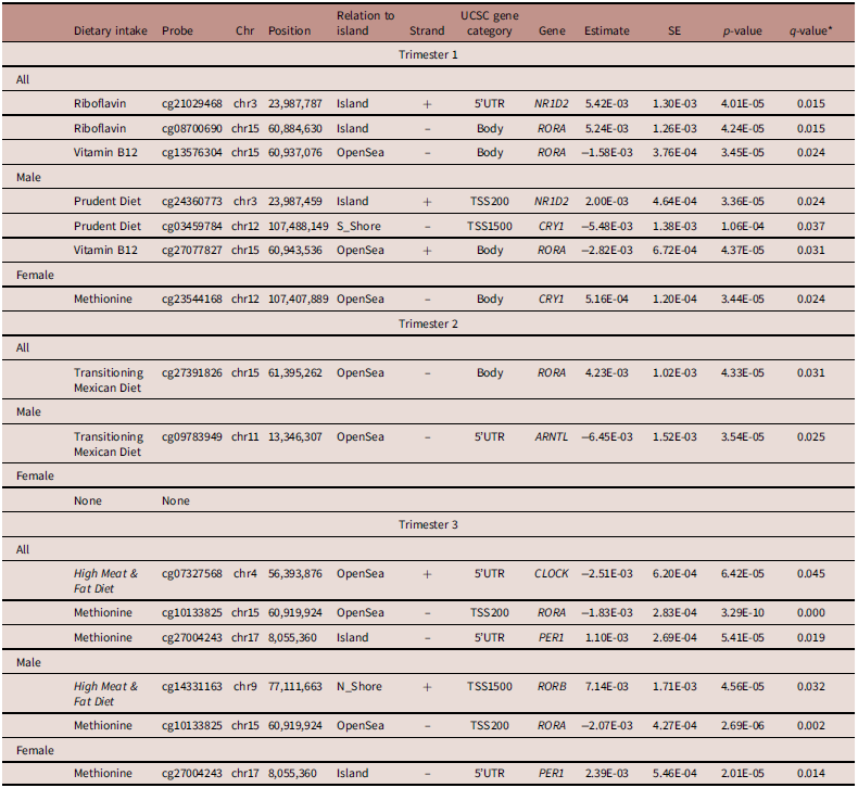
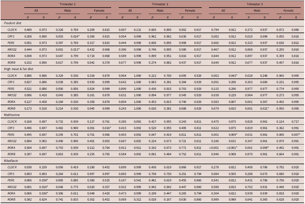
While adherence to prenatal dietary patterns in the first trimester was not associated with circadian gene DNA methylation in adolescents (Table 2), riboflavin intake was positively associated with DNA methylation at cg21029468 [NR1D2, β = 5.42 × 10⁻³, standard error (SE) = 1.30 × 10⁻³, q = 0.015] and cg08700690 (RORA, β = 5.24 × 10⁻³, SE = 1.26 × 10⁻³, q = 0.015), both annotated to CpG island regions. Additionally, vitamin B12 intake was negatively associated with DNA methylation at cg13576304 (β = −1.58 × 10⁻³, SE = 3.76 × 10⁻⁴, q = 0.024) located in the 3’ untranslated region (UTR) of RORA within an open sea region. Sex-stratified analysis showed that, in male adolescents, first-trimester adherence to the Prudent Diet was positively associated with DNA methylation at cg24360773 (β = 2.00 × 10⁻³, SE = 4.64 × 10⁻⁴, q = 0.024) annotated to a CpG island of NR1D2, and negatively associated with cg03459784 (β = −5.48 × 10⁻³, SE = 1.38 × 10⁻⁴, q = 0.037) near the promoter region of CRY1. Vitamin B12 intake was negatively associated with DNA methylation at cg27077827 (β = −2.82 × 10⁻³, SE = 6.72 × 10⁻⁴, q = 0.031) located in the gene body (3’ UTR) of RORA in male adolescents. ACAT showed a significant association between riboflavin intake with aggregated p-values for PER1 (q = 0.036), NR1D2 (p = 0.016), and RORA (q = 0.036) in unstratified analysis (Table 3). When stratified by sex, the Prudent Diet was associated with CRY1 (p = 0.024) and NR1D2 (p = 0.012) in males, while methionine intake was associated with CRY1 (p = 0.016) in females.
Associations between prenatal dietary patterns and methyl-donor nutrient intake with adolescent circadian gene DNA methylation, unstratified and stratified by sex
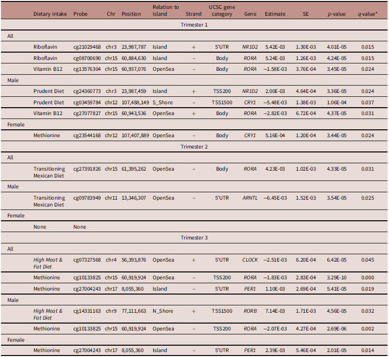
Table 2. Long description
A table with 15 rows and 11 columns. The columns are Dietary intake, Probe, Chr, Position, Relation to island, Strand, UCSC gene category, Gene, Estimate, SE, p-value, and q-value. The table is divided into three sections based on trimesters and further stratified by sex. Each row lists specific dietary intakes, probes, chromosome positions, and their relation to gene islands, along with corresponding gene categories, genes, estimates, standard errors, p-values, and q-values. Notable associations include riboflavin intake with DNA methylation at specific probes in the first trimester, and sex-stratified associations with various dietary patterns and nutrient intakes.
Chr, Chromosome; SE, standard error; Linear regression between dietary intake and 707 CpG sites annotated to 18 circadian genes, adjusting for maternal age, household socioeconomic status, maternal smoking, adolescent age, adolescent sex, calcium treatment, gestational weeks at each trimester, five batch effects, % monocytes, granulocytes, natural killer cells, CD8 T cells, CD4 T cells, memory and effector T cells, plasma blasts, naïve CD8 T cells, and naïve CD4 T cells. *False discovery rate adjustment using Benjamini–Hochberg method significant at q ≤ 0.05.
Significantly associated circadian genes with prenatal dietary patterns and methyl-donor nutrient intake identified via the Aggregated Cauchy Association Test, unstratified and stratified by sex

Table 3. Long description
A table with 12 rows and 21 columns comparing circadian genes with prenatal dietary patterns and nutrient intake across trimesters and sexes. The columns are divided into three main sections: Trimester 1, Trimester 2, and Trimester 3, each further divided into All, Male, and Female categories. The rows are labeled with different genes (CLOCK, CRY1, PER1, NR1D2, RORA, RORB) and dietary patterns (Prudent diet, High meat & fat diet, Methionine, Riboflavin). Each cell contains p and q values representing the statistical significance of the associations. Notable trends include specific associations between dietary patterns and gene methylation, with variations observed between males and females and across different trimesters.
Model adjusting for maternal age, household socioeconomic status, maternal smoking, adolescent age, adolescent sex, calcium treatment, gestational weeks at each trimester, five batch effects, % monocytes, granulocytes, natural killer cells, CD8 T cells, CD4 T cells, memory and effector T cells, plasma blasts, naïve CD8 T cells, and naïve CD4 T cells. *False discovery rate adjustment using Benjamini–Hochberg method.
Significant at q < 0.05.
Second-trimester adherence to the Transitioning Mexican Diet was positively associated with DNA methylation at cg27391826 (β = 4.23 × 10⁻³, SE = 1.02 × 10⁻³, q = 0.031) located in the gene body (3’ UTR) of RORA. No significant associations were found between adherence to the Prudent Diet, the High Meat & Fat Diet, or intake of any methyl-donor nutrients in the second trimester with adolescent circadian gene DNA methylation. Sex-stratified results showed that second-trimester adherence to the Transitioning Mexican Diet was negatively associated with DNA methylation at cg09783949 (β = −6.45 × 10⁻³, SE = 1.52 × 10⁻³, q = 0.025) annotated to the 5’ UTR of ARNTL in male adolescents. This negative association remained significant in the third trimester. No significant associations were observed with female adolescents. Additionally, no significant associations were observed between second-trimester dietary intake with aggregated p-values for circadian genes in the ACAT analysis.
Third-trimester adherence to the High Meat & Fat Diet was inversely associated with DNA methylation at cg07327568 (β = −2.51 × 10⁻³, SE = 6.20 × 10⁻⁴, q = 0.045) annotated to the 5’ and 3’ UTRs of CLOCK. Third-trimester methionine intake was negatively associated with DNA methylation at cg10133825 (β = −1.83 × 10⁻³, SE = 2.83 × 10⁻⁴, q< 0.001) near transcription start site of RORA, and positively associated with DNA methylation at cg27004243 (β = 1.10 × 10⁻³, SE = 2.69 × 10⁻⁴, q = 0.019) annotated to a CpG island of PER1. When stratified by sex, methionine intake was also negatively associated with DNA methylation at cg10133825 (β = −2.06 × 10⁻³, SE = 4.30 × 10⁻⁴, q = 0.002) located in the TSS200 region of RORA in male adolescents, and was also positively associated with cg27004243 (β = 2.39 × 10⁻³, SE = 5.42 × 10⁻⁴, q = 0.013) annotated to the CpG island region of PER1 in female adolescents. Additionally, the High Meat & Fat Diet was associated with DNA methylation at cg14331163 (β = 7.14 × 10⁻³, SE = 1.71 × 10⁻⁴, q = 0.032) annotated to the TSS1500 region of RORB in males. ACAT revealed significant associations between adherence to the High Meat & Fat Diet with aggregated p-values for CLOCK (q = 0.043) and between methionine intake with PER1 (q = 0.009) and RORA (q< 0.001). When stratified by sex, significant association was found between adherence to the Meat & Fat Diet with RORB (q = 0.021) in male adolescents, and between methionine intake with PER1 (q = 0.006) in female adolescents. Sensitivity and complete case analyses revealed some variation, but key significant and sex-specific associations remained largely consistent. The associations between dietary exposures and DNA methylation at CpG sites in circadian genes are summarized in Supplementary Figure 1. VIFs indicated generally low multicollinearity among covariates, with elevated values observed for adolescent’s sex, one principal component, and selected immune cell composition variables (granulocytes and CD4 T cells).
Post hoc EWAS analysis
EWAS showed several significant associations in prenatal methionine and vitamin B6 intake throughout pregnancy, but not in prenatal dietary patterns, folate, and vitamin B12 intakes in any trimester (Supplemental Table 1). First-trimester methionine intake was significantly associated with DNA methylation at 36 CpG sites. While most showed an inverse association (Supplemental Table 2), positive associations were found in five sites annotated to the island regions of genes. First-trimester vitamin B6 intake was associated with DNA methylation at 22 CpG sites, including 11 sites annotated to the island region of genes, mostly in a positive direction of association. Notably, a positive association was observed with DNA methylation at cg18109673 (β = 5.21 × 10⁻³, SE = 1.05 × 10⁻³, q = 0.040) annotated to the first exon of BHLHE40, a known circadian gene.
Second-trimester methionine intake was associated with DNA methylation at 313 CpG sites, with 45 sites in the promoter region (within 1,500 base pairs of the transcription start site of a gene of interest). Additionally, vitamin B6 intake in the second trimester was inversely associated with DNA methylation at four CpG sites annotated to genes located in the body region. Third-trimester methionine intake was significantly associated with DNA methylation at 233 CpG sites, including 38 sites located in the promoter region. Among them, an inverse association was found with DNA methylation at cg10133825 (β = −1.83 × 10⁻³, SE = 2.83 × 10⁻⁴, q<0.001) near the transcription start site of the circadian gene, RORA. Additionally, a positive association was found with DNA methylation at cg00046625 (β = 2.94 × 10⁻⁴, SE = 6.15 × 10⁻⁵, q = 0.015) annotated to a CpG island of PDE4B, a circadian-response gene.
Several significant CpG sites were identified as overlapping between methionine intakes during the second and third trimesters, including an inverse association with DNA methylation at cg23894919 annotated to the 3’UTR of GRIN2A, cg26220781 annotated to the gene body of DENND5B, and cg14454798 near the transcription start site of CPEB4. Stratifying by sex revealed similar results, with no significant associations observed between dietary patterns and trimester. While fewer associations were found in the first trimester, methionine intake showed multiple associations across all trimesters in both sexes. Volcano plots of the EWAS results are presented in Supplementary Figure 2. Across the EWAS models, λ ranged from 0.83 to 1.28, with most values close to one.
Discussion
Our study revealed several trimester-specific associations between gestational dietary intake and circadian gene DNA methylation during peri-adolescence. In the first trimester, riboflavin intake was positively associated with DNA methylation of CpG sites on NR1D2 and RORA, while vitamin B12 intake showed an inverse relationship with methylation at RORA-related sites. The second trimester showed the fewest associations, with adherence to the Transitioning Mexican Diet being positively associated with DNA methylation at a CpG site annotated to RORA. In the third trimester, adherence to the High Meat & Fat Diet was inversely associated with DNA methylation at a CpG site annotated to CLOCK, while methionine intake was linked to sites on both RORA and PER1. Sex-stratified results revealed differential DNA methylation between male and female adolescents, with more associations observed in males. Specifically, first-trimester adherence to the Prudent Diet was linked to with DNA methylation of CpG sites in the promoter regions of NR1D2 and CRY1, and second-trimester adherence to the Transitioning Mexican Diet was inversely associated with DNA methylation of a CpG site annotated to ARNTL, both in males. In the third trimester, adherence to the High Meat & Fat Diet was positively associated with DNA methylation at a CpG site in RORB in males, and methionine intake was inversely associated with RORA in males, while being positively related to PER1 in females. EWAS found that prenatal methionine and vitamin B6 intake were associated with DNA methylation changes, including at circadian genes BHLHE40 (first trimester, vitamin B6) and RORA (third trimester, methionine), while no significant associations were seen for folate, vitamin B12, or dietary patterns.
These results provide evidence that gestational diet may be linked with offspring circadian gene DNA methylation. The circadian-related regulatory system undergo epigenetic programming in utero, where maternal signals, including nutrients, hormones, and external cues, may influence the developing biological clock. Reference Crew, Mark, Waddell and Ferguson43 These early stages contribute to the formation of circadian-related physiological regulation systems, although their developmental trajectories remain to be fully understood. The biological clock is involved in physiological processes such as sleep–wake regulation, metabolism, and other physiological functions. Reference Wong, Wright and Spencer4 Disruptions in the prenatal environment, such as an imbalanced or suboptimal maternal diet, may be associated with differences in timing and function of the fetal circadian system, potentially impacting sleep–wake patterns and metabolic processes later in life. Further in support, a previous ELEMENT study linked prenatal adherence to the High Meat & Fat Diet in the first trimester with later sleep timing in adolescents, Reference Zamora, Peterson and Téllez-Rojo23 suggesting that maternal diet during pregnancy may be associated with regulation of sleep–wake patterns in offspring later in life. However, the extent to which the modest DNA methylation differences observed at individual CpG sites in this study are associated with meaningful differences in circadian-related physiological processes remain to be determined.
Overall, our findings align with the broader understanding that maternal nutrition during pregnancy can have lasting epigenetic consequences in the offspring. Yet, our findings add to existing literature by examining associations of gestational dietary intake on DNA methylation of CpG sites in circadian genes during adolescence. This study found that methionine, riboflavin, and vitamin B12 intakes were generally positively associated with DNA methylation, consistent with their involvement in one-carbon metabolism and methylation pathways. However, some associations were in the opposite direction. This observed heterogeneity in directionality suggests that the associations between prenatal nutrient intake and DNA methylation may be context dependent, potentially varying across developmental timing and genomic context rather than following a uniform directional pattern. Reference Anderson, Sant and Dolinoy44 While methyl-donor nutrients are biologically involved in one-carbon metabolism, their associations with site-specific DNA methylation may vary across genomic contexts and developmental stages, and some observed associations may also represent chance findings. Few studies have examined DNA methylation during adolescence, with one study finding no significant association between folic acid supplementation during the peri-conceptional period and offspring DNA methylation in 179 adolescents aged 14 to 17 years. Reference Crider, Wang and Ling18 In another study, a cross-sectional meta-analysis of data from six cohorts found a positive association between dietary glycemic load and DNA methylation at a CpG site within the WDR27 gene in 1187 overweight or obese children aged 4.5 to 17 years. Reference Ott, Stein and Hauta-alus19 However, these studies did not investigate specific maternal dietary patterns nor did they look at circadian genes.
Various components of the diet beyond methyl-donor nutrients, consisting of a range of nutrients, food types, and bioactive components, may differentially affect DNA methylation of individual CpG sites. Given the complexity of prenatal diet, components in the diet could influence methylation in distinct ways, contributing to the mixed associations observed in males with the first-trimester Prudent Diet. For instance, the presence of certain dietary polyphenols may have distinct effects on DNA methylation through inhibition of DNA methyltransferases. Reference Fang, Chen and Yang45 Antioxidants, such as vitamin C and E, may influence oxidative stress pathways, and they were related to DNA methylation in a recent meta-analysis. Reference Keshawarz, Joehanes and Ma46 Similarly, a randomized-controlled trial showed maternal vitamin C supplementation reduced changes in cord blood DNA methylation in response to maternal smoking. Reference Shorey-Kendrick, McEvoy and Ferguson47 Omega-3 fatty acids, found in nuts, have been shown to affect epigenetic mechanisms by modulating enzymatic activities that control DNA methylation. Reference Frankhouser, Steck and Sovic48,Reference Arpón, Milagro and Razquin49 A cross-sectional study also found significant associations between dietary fiber intake with DNA methylation in African American adolescents. Reference Chen, Dong and Wang50
Our study offered several notable strengths. The ELEMENT study included comprehensive information on various covariates, allowing for rigorous sensitivity analyses and accounting for potential confounders. The prospective design enabled a robust investigation into how maternal dietary intake during each trimester influenced their child’s circadian gene DNA methylation, providing important insights into critical windows of exposure and potential long-term effects. Repeated measures of maternal diet, combined with specific data on methyl-donor nutrient intake, provided a detailed and dynamic view of how dietary intake in different trimesters may be associated with epigenetic differences over time. The use of a validated FFQ tailored to Mexican women enhanced the accuracy of dietary assessments and the overall reliability of our data. Additionally, the use of PCA to derive dietary patterns specific to our study population ensured that the identified patterns directly reflected the unique dietary habits and cultural contexts. Furthermore, using genomic DNA from whole blood leukocytes provided a practical method for sample collection, as the extraction methods for leukocyte DNA were well-established and standardized. Whole blood reflected systemic biological changes and was particularly useful for studying overall health impacts, immune responses, and the ways in which various factors, such as diet and stress, influenced the body as a whole.
There were limitations that warrant consideration. While we captured a wide array of covariates, the long follow-up period may have precluded the consideration of subsequent factors, including childhood and adolescent lifestyle and environmental exposures, that could have influenced epigenetic changes. Additionally, the timing of recruitment during pregnancy restricted our ability to assess maternal dietary impacts at conception or during early childhood. Our study also did not compare associations between specific food groups or analyze variations in dietary intake across trimesters, which may have obscured critical insights into the nature of the maternal dietary influences. Although our sample size was substantial, the presence of potentially non-random missing data across trimesters, along with the use of mean substitution and indicator coding for missing values, could have introduced selection bias and affected the robustness of our findings. Analyses were stratified by sex, but formal tests of interaction between sex and maternal dietary exposures were not performed. Another limitation is that nutrient intakes did not account for dietary supplement use, which may have limited our ability to capture total nutrient exposure during pregnancy. Additionally, DNA methylation at individual CpG sites may not reflect gene expression or downstream physiological function, and some identified loci were located in intergenic or uncharacterized regions. The modest magnitude of the observed methylation differences further limits inference regarding their biological relevance, particularly in relation to transcriptional activity and downstream phenotypic outcomes. In addition, gene expression and other molecular measures of circadian activity were not examined in the present analyses, limiting the ability to infer functional regulation of circadian genes. We also did not account for the high correlation among circadian genes in our analysis, which could have oversimplified our findings and limited the interpretation of how individual genes interacted, and additional circadian-related genes may not have been captured by our candidate-gene approach. Given the large number of statistical tests performed, it is also possible that some observed associations may be chance findings due to multiple testing. Lastly, while we collected repeated measures of maternal diet, incorporating similar longitudinal measures for DNA methylation would have enhanced our understanding of temporal trends and dynamics in epigenetic modifications over the study period. The single time-point measurement of DNA methylation and the absence of functional validation limit the interpretability of the findings.
This study identified associations between gestational dietary intake and DNA methylation at circadian genes during peri-adolescence, with notable trimester-specific differences. The intake of methyl-donor nutrients, such as methionine, vitamin B6, and riboflavin, were associated with circadian gene methylation. Additionally, the impact of maternal diet varied by sex, with more associations observed in male adolescents. While these findings contribute to the growing body of research on the role of prenatal diet in circadian system development, further research is needed to explore the underlying mechanisms and long-term implications for circadian regulation in later stages of life.
Supplementary material
The supplementary material for this article can be found at https://doi.org/10.1017/S2040174426100786
Acknowledgements
We gratefully recognize the mothers and children who participated in the ELEMENT study and appreciate the ABC Medical Center for their support in providing research facilities.
Author contribution
JMG, DCD, KEP, MMT-R, AC, and LAT-O provided data; JTL and ECJ conceptualized the study; JTL, JMG, and ECJ analyzed and interpreted the data; JTL wrote the original draft; JTL, JMG, DCD, KEP, and ECJ reviewed and edited the manuscript; JMG and ECJ supervised the work. JTL had primary responsibility for final content. All authors critically revised the manuscript for important intellectual content and approved the final version
Financial support
The ELEMENT study was supported by the U.S. Environmental Protection Agency (RD83543601) and the National Institute of Environmental Health Sciences through the following awards: P01ES022844, R01ES007821, R01ES014930, R01ES013744, P30ES017885, 1U2CES026553, K01HL151673, and R35ES031686. Additional support was provided by the National Institute of Public Health/Ministry of Health of Mexico and the American British Cowdray Medical Center, which provided facilities for data collection.
Competing interests
None



 Open access

