Introduction
Worldwide, long-term exposure to opioids such as morphine, codeine, heroin and methadone is common, both in the substance misuse and other clinical populations. Chronic use of opioids leads to long-lasting changes, including tolerance, sensitization and physical dependence (Trujillo, Reference Trujillo2002). One of the most common opioid drugs to be misused is heroin, which is also prescribed in the UK as ‘diamorphine’ as an analgesic (Strang et al. Reference Strang, Babor, Caulkins, Fischer, Foxcroft and Humphreys2012). For those seeking treatment after developing dependence following repeated exposure to illicit heroin, methadone maintenance treatment (MMT) is the most commonly offered therapeutic intervention. In Scotland alone, 30 000 patients receive MMT as part of a ‘harm reduction programme’ to reduce or cease heroin use, diminish associated criminality, and reduce the risk of blood-borne virus infection (McKeganey et al. Reference McKeganey, Bloor, Robertson, Neale and MacDougall2006). However, an MMT programme is associated with low rates of sustained abstinence with as few as 17–28% of those dependent on MMT achieving abstinence (The Scottish Government, 2010). One of the core features of opioid dependence is the dysregulation of dopaminergic neurotransmission, including decreased dopamine receptor availability and release (Gradin et al. Reference Gradin, Baldacchino, Balfour, Matthews and Steele2014), associated with functional impairments in frontal brain regions innervated by dopamine. However, it is not known whether the effects of opioids could lead mechanistically to long-lasting changes, or could be reversible following a period of abstinence (Ersche et al. Reference Ersche, Roiser, Clark, London, Robbins and Sahakian2005; Baldacchino et al. Reference Baldacchino, Balfour and Matthews2015).
Chronic misuse of drugs appears to be associated with a variety of cognitive abnormalities. Such deficits may have important clinical consequences: e.g. increasing drug-seeking, presumably due to a failure of impulse control and impaired engagement in therapeutic programmes, with increased rates of relapse following treatment (Rogers & Robbins, Reference Rogers and Robbins2001). In comparison with work on stimulants and cannabis, there has been less research on cognitive impairments associated with opioid dependence (Baldacchino et al. Reference Baldacchino, Balfour, Passetti, Humphris and Matthews2012). Opioid-dependent subjects discounted the value of delayed money rewards more than non-drug-using participants, and discounted the value of delayed heroin more than delayed money (Madden et al. Reference Madden, Petry, Badger and Bickel1997). Whilst some researchers concluded that opioid misuse is not associated with frontal lobe impairments, others reported that it is associated with a broad range of deficits such as attentional control, planning and spatial working memory (Ornstein et al. Reference Ornstein, Iddon, Baldacchino, Sahakian, London, Everitt and Robbins2000). More recently, ‘reflection impulsivity’, the extent to which subjects sample information before responding, has been reported as decreased in currently opioid-dependent and former substance misusers (Clark et al. Reference Clark, Robbins, Ersche and Sahakian2006). A variety of cognitive deficits associated with opioid misuse has, therefore, been reported, ranging from general deficits, to selective memory impairment, increased discounting and impulsivity, to no prefrontal-linked cognitive impairments. Importantly, these studies have revealed significant overlaps in candidate markers of impulsivity (Dalley et al. Reference Dalley, Everitt and Robbins2011; Robbins et al. Reference Robbins, Gillan, Smith, de Wit and Ersche2012).
A post-mortem study reported an increased prevalence of ischaemic brain lesions in the globus pallidus in opioid-dependent individuals (Andersen & Skullerud, Reference Andersen and Skullerud1999). Structural magnetic resonance imaging (MRI) studies of human opioid-dependent populations have revealed significant grey matter reductions in the prefrontal and temporal cortices (Lyoo et al. Reference Lyoo, Pollack, Silveri, Ahn, Diaz, Hwang, Kim, Yurgelun-Todd, Kaufman and Renshaw2006; Liu et al. Reference Liu, Hao, Kaneko, Ouyang, Zhang, Xu, Xue and Liu2009; Yuan et al. Reference Yuan, Zhu, Shi, Zou, Yuan, Liu, Lee and Weng2009) and increased white matter hyper-intensities, mainly in frontal areas (Lyoo et al. Reference Lyoo, Streeter, Ahn, Lee, Pollak, Silveri, Nassar, Levin, Sarid-Segal, Ciraulo, Renshaw and Kaufman2004). Indeed, overall results show that the neuroanatomical impairments are located in neural networks linked to executive and attentional function, such as the dorsolateral prefrontal cortex, anterior cingulate cortex and medio-temporal cortices. Abstinent previously heroin-dependent individuals have shown, after 3 days of abstinence, significant grey matter reductions in the left frontal gyrus and cingulate gyrus. However, after 1 month of abstinence there were no significant differences between patients and controls in any structural brain region (Wang et al. Reference Wang, Li, Li, Yang, Zhu and Wang2011).
Previous studies investigating the chronic effects of opioid use either due to heroin misuse or as part of MTT have largely focused on the evaluation of cognitive impulsivity and non-planning impulsivity using Barratt's Impulsivity Scale (Patton et al. Reference Patton, Stanford and Barratt1995; Ersche et al. Reference Ersche, Roiser, Clark, London, Robbins and Sahakian2005; Baldacchino et al. Reference Baldacchino, Balfour and Matthews2015). The prefrontal cortex may play a key role in processing such information (Rogers et al. Reference Rogers, Everitt, Baldacchino, Blackshaw, Swainson, Wynne, Baker, Hunter, Carthy, Booker and London1999). However, to our knowledge, only one study has reported significant orbitofrontal structural abnormalities associated with heroin and methadone use (Ersche et al. Reference Ersche, Fletcher, Roiser, Fryer, London, Robbins and Sahakian2006).
The aim of this study was to test the following hypotheses:
-
(i) Behavioural impairment in cognitive impulsivity and non-planning impulsivity are related to different stages of treatment (stable MMT use v. abstinent).
-
(ii) Stable MMT patients exhibit more reductions in grey matter volume in a priori regions of interest, such as the prefrontal cortex and basal ganglia, because these regions have been linked to impulsivity and reported as abnormal in structure in opioid misuse (Robbins et al. Reference Robbins, Gillan, Smith, de Wit and Ersche2012), relative to controls and abstinent patients. MMT patients may be most vulnerable to the effects of current opioid exposure associated with cognitive impulsivity and non-planning impulsivity (Robbins et al. Reference Robbins, Gillan, Smith, de Wit and Ersche2012). We therefore predicted that behavioural measures of impulsivity correlate with brain structure measures.
Method
Participants
Study approval was granted by the East of Scotland, the Lothian and Glasgow Research Ethics Committees and written informed consent obtained from all participants. National Health Service (NHS) Scotland Research Governance approvals were provided by the NHS Fife Research and Development Department. The University of Dundee was the sponsor for this study.
A total of 122 male individuals were enrolled in the study. They underwent detailed clinical screening assessed by a clinician (A.B. or S.G.) that included the collection of sociodemographic information, and semi-structured interviews to ascertain detailed histories of drug and alcohol use and opioid-dependence status (Marsden et al. Reference Marsden, Gossop, Stewart, Best, Farrell, Lehmann, Edwards and Strang1998). Mental health status and history were assessed using the Mini International Neuropsychiatric Interview (MINI Plus, version 5.0; Sheehan et al. Reference Sheehan, Lecrubier, Sheehan, Amorim, Janavs, Weiller, Hergueta, Baker and Dunbar1998). Current and pre-morbid intelligence was estimated using the Wechsler Abbreviated Scale of Intelligence and National Adult Reading Test (NART) (Nelson & Willison, Reference Nelson and Willison1991; Woerner & Overstreet, Reference Woerner and Overstreet1999). The Fagerström Test for Nicotine Dependence (Fagerstrom & Schneider, Reference Fagerstrom and Schneider1989) was also used.
Case records from the addiction, psychiatric and general practitioners’ services helped in the identification of overdose episodes, confirmed the absence of a history of epilepsy and other neurological phenomena, hepatitis B, C and HIV status and diagnoses of personality disorders (e.g. borderline). These records also allowed us to validate medical and psychiatric histories, substance misuse career timelines and quantify current drug and alcohol use. Exclusion criteria were past or current histories of psychotic disorder, post-traumatic stress disorder, neurological and neurodevelopmental disorders, head injury, confirmed history of non-fatal overdose episodes and co-occurring benzodiazepine, stimulant and alcohol dependence, and antisocial and borderline personality disorder.
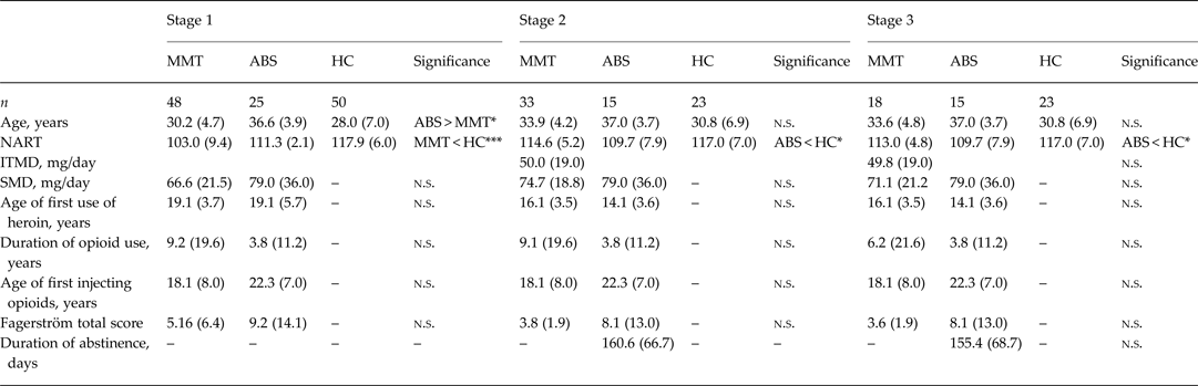
The methadone group (n = 48) had a diagnosis of Diagnostic and Statistical Manual of Mental Disorders, fourth edition (DSM-IV) opioid dependence and were engaged in a methadone maintenance programme (MMT) with objective confirmation of the absence of illicit drug use for more than 6 months. The previously dependent opioid (abstinent; ABS) group (n = 24) had a history of chronic opioid use with an abstinence duration exceeding 6 weeks. As patients were either stably maintained on methadone or were stably abstinent, no acute intoxication or withdrawal effects were observed. A representative subset of these volunteers took part in stages 2 and 3. Both MMT and ABS groups had been taking between 30 and 120 mg of methadone daily on an MMT programme and presented with more than 3 years of continuous daily illicit heroin use. The two groups were matched by lifetime drug use and methadone doses [initial titration of methadone dose (ITMD), current methadone treatment dose (CMTD) and/or last stable methadone dose (LSMD)]. The ITMD included the initial titration dose of methadone that each patient received, defined as the dose of methadone required to reduce heroin withdrawal symptoms at the commencement of MMT and was between 10 and 30 mg daily, depending on objective evidence of opioid withdrawal symptoms. Ongoing abstinence from illicit drug use was confirmed just prior to both neuropsychological testing, and scanning, using an onsite multidrug urine test (Armbruster & Krolak, Reference Armbruster and Krolak1992). Some participants withdrew from scanning and from neuropsychological assessment because they were not contactable or they were not able to take part. Details of participants included in the analysis are presented in Table 1. Fig. 1 describes the cohorts who participated in the stages of the study as described below.
Flowchart of study stages and participants. MMT, Methadone maintenance treatment group; ABS, abstinent group; HC, healthy controls.

Demographic and clinical characteristics

Data are given as mean (standard deviation).
MMT, Methadone maintenance group; ABS, abstinent group; HC, healthy control group; n.s., not significant; NART, National Adult Reading Test; ITMD, initial methadone titration dose; SMD, stable methadone dose.
* p < 0.05, *** p < 0.001.
Stage 1 – behavioural outcomes
Each participant was assessed using the standardized Cambridge Neuropsychological Test Automated Battery (CANTAB; http://www.cambridgecognition.com). Executive functions were assessed using the Cambridge Gambling Task (CGT) and Stockings of Cambridge (SOC) tests. These tasks assessed cognitive impulsivity and non-planning impulsivity, respectively (Patton et al. Reference Patton, Stanford and Barratt1995; Ersche et al. Reference Ersche, Roiser, Clark, London, Robbins and Sahakian2005; Baldacchino et al. Reference Baldacchino, Balfour and Matthews2015).
Statistical analysis of the behavioural outcomes
Data meeting assumptions of normality and homogeneity of variance were analysed using analysis of variance (ANOVA) and analysis of covariance (ANCOVA). All other data were compared using appropriate non-parametric tests (e.g. Kruskal–Wallis and Mann–Whitney tests). ANOVA was used to test for group differences. To control for family-wise error, we used post-hoc Bonferroni-corrected pairwise comparisons. After correction, results with p < 0.05 were considered significant. ANOVA was used to test for group differences. Effect sizes were calculated as Cohen's d statistics and analyses conducted using SPSS v. 20 (SPSS Inc., USA).
Stage 2 – neuroimaging
Scanning
Data were acquired with a Siemens 3 T Tim Trio at the Clinical Research Centre, Ninewells Hospital and Medical School, Dundee, UK. ‘Structural’ T1 weighted images were acquired with a voxel size 0.8 × 0.8 × 1.0 mm3 with whole brain coverage, repetition time (TR) = 1.9 s and echo time (TE) = 2.64 ms and reported by a consultant radiologist for incidental findings.
Image analyses
Voxel-based morphometry was done using SPM8 (http://www.fil.ion.ucl.ac.uk/spm/). For pre-processing, T1 weighted images for each participant were segmented into grey and white matter probability maps, spatially normalized with modulation to preserve the total amount of grey matter and smoothed with an 8 mm Gaussian kernel (Ashburner & Friston, Reference Ashburner and Friston2005). We used t tests to test the null hypothesis of no difference between MMT patients and controls.
The threshold of significance was defined as p < 0.05 at a whole-brain corrected level using a customized version of a popular Monte Carlo technique (Slotnick et al. Reference Slotnick, Moo, Segal and Hart2003) (http://www2.bc.edu/~slotnics/scripts.htm). Brain regions were identified by converting Montreal Neurological Institute coordinates into Talairach coordinates using the Yale conversion calculation (http://bioimagesuite.yale.edu/mni2tal/index.aspx) and inspection of the Talairach Atlas (Talairach & Tournoux, Reference Talairach and Tournoux1988).
Stage 3 – behaviour and neuroimaging and correlations with opioid exposure
Correlation analyses were similarly used to test null hypotheses of no relationship between grey matter indices and measures of opioid exposure: ITMD and CMTD. Correlation analyses were also used to test null hypotheses of no relationship between grey matter abnormalities and CGT or SOC behavioural measures.
Statistical analysis for treatment outcome
An exploratory analysis to investigate potential predictors of outcome was also done. Logistic regression with 6-fold cross-validation was used to generate an unbiased prediction of treatment outcome (MMT v. ABS) in the experimental groups using the following predictors: frequency use, withdrawal exposure and treatment. We were interested in particular in two measures: (1) ITMD and (2) stable methadone dose (SMD) for the MMT group and the LSMD for the ABS group. Analyses were conducted using SPSS for Windows v. 20 (SPSS Inc., USA).
Results
Participants
MMT and ABS groups were matched for clinical characteristics: SMD, age of first use of heroin, years of opioid use, age of first injecting opioids and Fagerström total score in the first stage of the study. The ABS group were on average older than the MMT group (p < 0.05) and the MMT group had lower intelligence quotient scores than the healthy control group (p < 0.001). In the second and third stages, the ABS group differed significantly in NART scores from the healthy control group (p < 0.05) but the clinical characteristics remained matched (p > 0.05). Table 1 shows the demographic and the substance use data of the participants.
Behavioural results
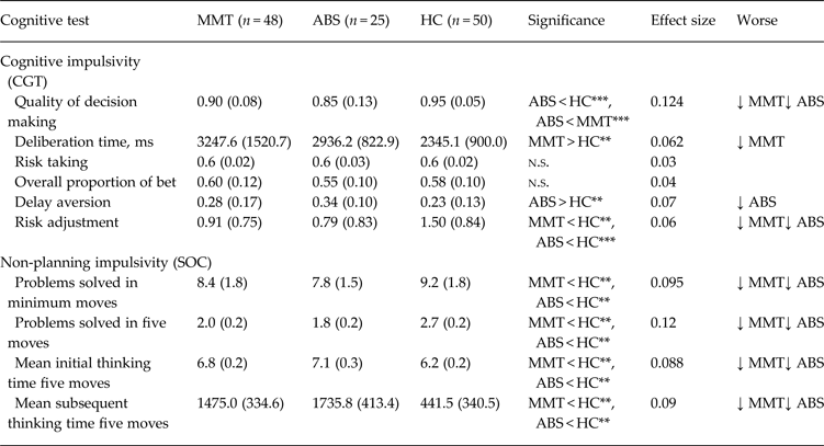
Significant decision-making abnormalities for the MMT and ABS groups relative to the control group were identified for cognitive impulsivity (CGT) (p < 0.01) and non-planning impulsivity (SOC) (p < 0.01) as summarized in Table 2.
Between-group comparisons of the neuropsychological domains

Data are given as mean (standard deviation).
MMT, Methadone maintenance group; ABS, abstinent group; HC, healthy control group; CGT, Cambridge Gambling Task; ↓, worse; n.s., not significant; SOC, Stockings of Cambridge.
** p < 0.01, *** p < 0.001.
Neuroimaging results
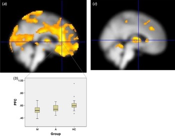
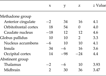
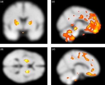
As shown in Fig. 2, significant reductions in grey matter probability were present in the MMT group relative to controls: medial-orbital (18, 54, 0) cortices, bilateral caudate (18, 12, 12; −18, 12, 12), bilateral globus pallidus (10, 10, 2; −10, 10, 2), nucleus accumbens (−6, 10, 10), anterior cingulate (−1, 34, 16) and bilateral insula (34, −6, 16; −38, −4, −0). Significant reductions in grey matter were present in the ABS group relative to controls: thalamus (−2, −6, 10) and midbrain (2, 30, 36). This is summarized in Table 3. In the prefrontal cortex, the region of interest was centred on −2, 34, 18. The MMT group had significantly reduced grey matter probability (p < 0.001) in comparison with controls.
Grey matter volume reductions. (a) Grey matter volume reductions in methadone maintenance treatment (MMT) patients relative to controls. Regions are significant (p < 0.01 whole-brain corrected level). (b) MMT patient (M), abstinent patient (A) and healthy control (HC) subgroups within the prefrontal cortex (PFC) (Montreal Neurological Institute coordinates −2, 34, 18). The central lines are medians; the boxes represent interquartile ranges; the whiskers represent ranges; circles outliers; the asterisk is an extreme outlier. Median value of the MMT group was significantly different (p < 0.01) from those of the abstinent and control groups. (c) Grey matter volume reductions in abstinent patients relative to controls. Regions are significant (p < 0.01 whole-brain corrected level).

Between-group comparisonsa

Grey matter reduction in methadone patients and abstinent patients in comparison with controls. Coordinates (x, y, z) reported in Montreal Neurological Institute space. All results significant at p < 0.01, cluster extent corrected across the whole brain.
Fig. 3 shows that current SMD for the MMT group correlated negatively with bilateral globus pallidus, grey matter probability (20, 2, 6; −20, 4, 6). Increased CGT risk adjustment was associated in the MMT group with bilateral globus pallidus grey matter reductions (20, −4, 6; −24, −6, 10) and increased CGT risk taking was associated with significantly decreased grey matter in the orbitofrontal and inferior medial prefrontal cortices (10, 62, 8; 10, 48, −20) in the MMT group. Increased mean subsequent moves in the SOC were correlated with grey matter reductions in the rostromedial prefrontal cortex (10, 26, −16) in MMT patients, which also correlated with longer times to solve complex problems (such as ‘5-moves’).
Grey matter volume and methadone maintenance treatment (MMT). (a) Grey matter reductions in the bilateral globus pallidus in MMT patients correlating with stable methadone dose. (b) Grey matter reductions in the bilateral globus pallidus in MMT patients correlating with Cambridge Gambling Task (CGT) risk adjustment scores. (c) Grey matter reductions in the orbito-medial prefrontal cortex correlating with CGT risk-taking scores. (d) Grey matter volume reductions in the rostral prefrontal cortex correlating with mean subsequent five moves from Stockings of Cambridge.

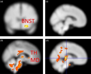
In Fig. 4 the LSMD for the ABS group correlated negatively with bilateral: bed nucleus of stria terminalis (BNST) (20, 18, −14; −14, 18, −14), midbrain (−2, −22, 14) and thalamus (−6, −18, 0) grey matter probability. Greater delay aversion on the CGT in the ABS group was associated with periaqueductal grey matter reductions (8, −26, 2). Increased mean subsequent moves of the SOC correlated negatively with grey matter probability in the putamen (−28, 16, 2) and bilateral BNST (24, 18, −24; −22, 16, −24) in ABS patients.
Grey matter volume and abstinent (ABS) patients. (a, b) Grey matter reductions in the bed nucleus of the stria terminalis (BNST), thalamus (TH) and midbrain (MD) in ABS patients correlating with the last stable methadone dose. (c) No grey matter reductions in the ABS group correlating with Cambridge Gambling Task risk taking. (d) Grey matter reductions in the putamen and BNST in ABS patients correlating with mean subsequent five moves from Stockings of Cambridge.

Summary of the neuroimaging and behavioural results
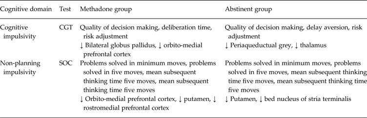
Regions of grey matter reduction in the MMT and ABS participants where the variance in grey matter probability was significantly explained by SMD and LSMD and CGT and SOC behavioural measures are summarized in Table 4 and Fig. 5.
Summary. Brain abnormalities summarized according to the cortico-basal ganglia-thalamocortical loop. This circuit originates from grey matter volume reductions in methadone maintenance treatment and abstinent patients significantly explained by opioid exposure (stable methadone dose, last stable methadone dose) and behavioural measures (Cambridge Gambling Task and Stockings of Cambridge). GP, Globus pallidus.

Summary of the cognitive and neuroimaging outcomes

CGT, Cambridge Gambling Task; ↓, reductions in grey matter; SOC, Stockings of Cambridge.
Predictions of treatment outcome
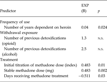
Using logistic regression with cross-validation (within study replication), SMD and LSMD predicted treatment status at the time of the present study (being on MMT treatment v. abstinence) with 92% accuracy (98% sensitivity and 72% specificity). In addition, ITMD, which reflected extent of heroin use prior to commencing MMT, predicted treatment status at the time of the present study with 86.2% accuracy (85.7% sensitivity and 86.2% specificity). This indicates that in both ITMD, when commencing MMT, and SMD or LSMD were significant predictors for much later treatment outcomes (Table 5).
Logistic regression model a

n.s., Not significant.
a Results of the binary logistic regression model with exponential (β) = EXP(B), using as the response at the treatment outcome for the methadone group and abstinent group.
Discussion
This study tested the hypotheses that both behavioural and structural grey matter changes were related to different stages of the treatment programme (stable MMT v. abstinence in former MMT patients). We also tested for differences in cognitive impulsivity and non-planning impulsivity comparing MMT, ABS and healthy control groups. Stable MMT participants deliberated longer and placed higher bets earlier on the CGT and showed impaired strategic planning compared with healthy controls. ABS participants showed impairment in quality of decision-making, delay aversion and risk adjustment on the CGT and non-planning impulsivity compared with healthy controls. MMT participants had widespread grey matter reductions in the orbitomedial prefrontal cortex, caudate, putamen and globus pallidus. In contrast, ABS participants showed midbrain-thalamic grey matter reductions. The behavioural abnormalities correlate with structural grey matter abnormalities, providing evidence that the ABS group represent an intermediate group, with regard to behavioural abnormalities and brain structure abnormalities, between MMT participants and healthy controls.
Several studies have suggested impairment in cognitive impulsivity and non-planning impulsivity in opioid dependence (Ersche et al. Reference Ersche, Roiser, Clark, London, Robbins and Sahakian2005; Baldacchino et al. Reference Baldacchino, Balfour and Matthews2015). However, these studies did not address the issue of possible long-lasting changes as a result of chronic opioid use, and not address the issue of possible reversibility with abstinence. Our findings provide evidence that cognitive processes that are particularly associated with the prefrontal cortex are disrupted during chronic opioid use, but not during abstinence. MMT patients differed from healthy controls in that they took longer to make decisions during the CGT. However, the ABS group differed in delay aversion outcomes for cognitive impulsivity. It is possible that these changes are related to opioid dependence or the effects of subacute opioid withdrawal.
Our results show that MMT patients exhibited grey matter reductions in the orbito-medial prefrontal cortex, bilateral caudate nucleus and globus pallidus. Notably, the orbito-medial prefrontal cortex is implicated in processing drug-related information in addiction (Volkow et al. Reference Volkow, Wang, Fowler, Logan, Angrist, Hitzemann, Lieberman and Pappas1997; Volkow & Fowler, Reference Volkow and Fowler2000) and the dorsal caudate in compulsive drug seeking and habit formation (Everitt et al. Reference Everitt, Belin, Economidou, Pelloux, Dalley and Robbins2008; Koob & Volkow, Reference Koob and Volkow2010). Abnormally reduced frontal cortex activity has been linked with a reduction in dopamine striatal D2 receptor availability (Volkow et al. Reference Volkow, Wang, Fowler, Logan, Angrist, Hitzemann, Lieberman and Pappas1997). Consistent with the latter, increased cognitive impulsivity correlated with grey matter reductions in the orbito-medial prefrontal cortex, but did not similarly correlate in the ABS group. This suggests that orbitofrontal cortex abnormalities are not linked to opioid exposure and may, alternatively, be linked to vulnerability to develop drug dependence (Adinoff et al. Reference Adinoff, Devous, Best, George, Alexander and Payne2001), or an effect of non-opioid drug exposure prior to MMT (Volkow et al. Reference Volkow, Wang, Fowler, Tomasi and Telang2011). Consistent with the latter interpretation, orbitofrontal grey matter reductions have been reported to be associated with increased risk taking in cocaine-, amphetamine- and alcohol-dependent individuals, using the Iowa Gambling Task (Tanabe et al. Reference Tanabe, Tregellas, Dalwani, Thompson, Owens, Crowley and Banich2009). In animal studies, there is evidence that morphine administration increases impulsivity (Pattij et al. Reference Pattij, Schetters, Janssen, Wiskerke and Schoffelmeer2009) which normalizes when administration is stopped (Harvey-Lewis et al. Reference Harvey-Lewis, Perdrizet and Franklin2012). In the present study, a negative correlation between methadone dose and CGT risk adjustment was present. This is consistent with animal studies reporting pallidal changes as a result of chronic opioid use (Gurwell et al. Reference Gurwell, Nath, Sun, Zhang, Martin, Chen and Hauser2001). As reported decades ago, bilateral gross symmetric lesions of the globus pallidus were attributable to heroin intoxication (Strassmann et al. Reference Strassmann, Sturner and Helpern1969) and subsequently more subtle lesions were observed in 5–10% of heroin addicts at post-mortem (Andersen & Skullerud, Reference Andersen and Skullerud1999). As with motor impairments as a consequence of globus pallidus lesions (Andersen & Skullerud, Reference Andersen and Skullerud1999), neuropsychological impairments may depend on which part of the globus pallidus that has been damaged. Furthermore, it has been reported that hypoventilation caused by opioid use with paradoxical vasoconstriction results in bilateral, symmetrical infarcts of the globus pallidus (Daras et al. Reference Daras, Orrego, Akfirat, Samkoff and Koppel2001). Notably, sleep apnoea (Egan et al. Reference Egan, Becker and Bundt2005) has been reported to occur in 30% of patients receiving MMT (Wang & Teichtahl, Reference Wang and Teichtahl2007). The incidence of sleep apnoea and ataxic breathing correlates with plasma methadone levels (Wang et al. Reference Wang, Teichtahl, Drummer, Goodman, Cherry, Cunnington and Kronborg2005) and opioid dose (Walker et al. Reference Walker, Farney, Rhondeau, Boyle, Valentine, Cloward and Shilling2007). Consequently, there may be a link between higher doses of methadone, sleep apnoea and globus pallidus grey matter reductions. However, it has been reported that changes observed in MRI may be related to shifts in water content from and to glia cells because of the effects of steroid hormones or cytokines (Lucassen et al. Reference Lucassen, Pruessner, Sousa, Almeida, Van Dam, Rajkowska, Swaab and Czéh2014). This could be linked to stress responses due to short-term abstinence (6 months) or as a result of methadone intake.
Abstinent patients exhibited grey matter reductions in the thalamus and midbrain consistent with a previous study (Williams et al. Reference Williams, Daglish, Lingford-Hughes, Taylor, Hammers, Brooks, Grasby, Myles and Nutt2007). Our study also found grey matter reductions in the BNST associated with increased LSMD. The BNST is a powerful modulator of addictive behaviour, with a major input from noradrenergic innervation and is a dense centre of neurones expressing corticotropin-releasing factor with cell bodies and terminals which could also be linked with impairment in cognitive impulsivity and non-planning impulsivity (Koob & Volkow, Reference Koob and Volkow2010). The reduction in the BNST may be specific to the abstinent group because it is involved in stress responses or it could be associated with decreased stress in recovery. Deep brain stimulation has been attempted previously in the BNST in patients and there is some evidence for drug dependence reduction (Langevin, Reference Langevin2012). Consistent with this, we found that grey matter volume reductions in thalamus and midbrain grey matter correlated negatively with measures of impaired cognitive impulsivity and non-planning impulsivity.
In conclusion, as hypothesized, MMT maintained patients exhibited abnormal behaviour linked to abnormal brain structure, and behavioural and brain abnormalities were least in ABS patients. However the extent to which these differences reflect a coincidence of MMT cessation or factors present during MMT promoting cessation remain unclear. We therefore suggest that a longitudinal study is required to determine whether abstinence improves abnormalities, or patients who achieve abstinence have reduced abnormalities before methadone cessation.
Acknowledgements
We thank the NHS Fife Research and Development Department and NHS Fife Addiction Services for their support in the recruitment, especially Angela Swift, Dawn Stewart, Michelle Hyslop, Anthony Robb and Fiona Boyce. We thank the Lothian and Edinburgh Abstinence Programme (LEAP) and Phoenix Futures Scottish Residential Service. We are grateful to Victoria Gradin and Blair Johnston for initial analysis of some of the cohort data, to Mairi Stirling and Christine Matthews with data management and David Balfour for expert advice. Finally we thank all participants in this study. This study was part funded by an unrestricted educational grant provided by Schering-Plough, a grant by an anonymous trust and the Scottish Mental Health Research Network. The funding sources had no role in the design, conduct of the study and interpretation of the data.
Declaration of Interest
S.T. has received funding from Merck Serono and Lundbeck, and K.M. has chaired advisory boards for studies of Deep Brain Stimulation for Obsessive–Compulsive Disorder sponsored by Medtronic. He has received educational grants from Cyberonics Inc. & Schering Plough, and he has received research project funding from Schering-Plough, Merck Serono, and Indivior and also from St Jude Medical for a multi-centre clinical trial of Deep Brain Stimulation for depression. He has received travel and accommodation support to attend meetings from Medtronic and St Jude Medical. J.D.S. has received research funding via an honorarium associated with a lecture from Wyeth and an unrestricted educational grant from Schering-Plough. A.B. has received educational grants from Schering Plough and he has received research project funding from Schering-Plough, Merck Serono, Lundbeck, and Indivior. S.G. reported no biomedical financial interests or potential conflicts of interest.